SciELO - Scientific Electronic Library Online

 
vol.36 número1Perforación gástrica por espina de pescado. Reporte de un caso y revisión de literaturaMucosectomía endoscópica para cáncer gástrico temprano: una alternativa vigente índice de autoresíndice de materiabúsqueda de artículos
Home Pagelista alfabética de revistas  

Servicios Personalizados

Revista

Articulo

Indicadores

Links relacionados

  • En proceso de indezaciónCitado por Google
  • No hay articulos similaresSimilares en SciELO
  • En proceso de indezaciónSimilares en Google

Compartir


Revista colombiana de Gastroenterología

versión impresa ISSN 0120-9957versión On-line ISSN 2500-7440

Rev. colomb. Gastroenterol. vol.36 no.1 Bogotá ene./mar. 2021  Epub 21-Oct-2021

https://doi.org/10.22516/25007440.490 

Report of cases

Biliodigestive shunt guided by endoscopic ultrasonography in patient with stage IV pancreatic adenocarcinoma and obstruction of the bile duct. A case report

María Manrique1 
http://orcid.org/0000-0002-8262-8752

Wilmar Martín-M2  * 

1 Gastrointestinal surgery, echoendoscopy and digestive endoscopy, Instituto Nacional de Cancerología. Bogotá D. C., Colombia.

2 Gastrointestinal and digestive endoscopy fellow, Instituto Nacional de Cancerología. Bogotá D. C., Colombia.


Abstract

Endoscopic endosonography is an alternative to guide biliodigestive shunting in cases where endoscopic retrograde cholangiopancreatography (ERCP) has failed. Techniques such as choledoduodenostomy or choledochoantrostomy guided by endoscopic ultrasonography have been described.

This is the case of a 72-year-old patient with pancreatic adenocarcinoma, portal vein and duodenal wall involvement, in whom ERCP to try a palliative shunt failed. Therefore, a metal stent was placed using endoscopic endosonography, with adequate clinical response to the procedure and treatment. It is concluded that the procedure is safe and that endosonography is an effective alternative in cases of ERCP failure to achieve biliary bypass in cases of malignant biliary obstruction.

Keywords: Bile duct obstruction; Retrograde endoscopic cholangiopancreatography (ERCP); Endoscopic ultrasonography (EUS)

Resumen

La endosonografía endoscópica es una alternativa que sirve como guía para la realización de derivaciones biliodigestivas en los casos en los que la colangiopancreatografía retrógrada endoscópica (CPRE) ha fallado. Se han descrito técnicas como la coledocoduodenostomía o coledocoantrostomía guiadas por ultrasonografía endoscópica (USE).

Se describe el caso de una paciente de 72 años con adenocarcinoma de páncreas, compromiso portal y de paredes duodenales, en quien se usó la CPRE para intentar una derivación paliativa y fue fallida, por lo cual se realizó la colocación de un stent metálico guiado por endosonografía endoscópica, con adecuada respuesta clínica al tratamiento. En conclusión, el procedimiento es seguro y la endosonografía es una vía alterna efectiva en los casos de CPRE fallidas para lograr derivaciones biliares en casos de obstrucción de la vía biliar de origen maligno.

Palabras clave: Obstrucción del conducto biliar; colangiopancreatografía endoscópica retrógrada (CPRE); ultrasonografía endoscópica (USE)

Introducción

Endoscopic guided biliary stent placement is a procedure used in patients with stenosis of the biliopancreatic confluent secondary to unresectable neoplasias, and ERCP is the technique of choice to perform such procedure 1. Even being the method of choice, ERCP fails in 5% to 10% of cases 2 due to situations such as anatomical alterations, gastroduodenal obstruction, obstructed segment of considerable length, distortion of the papilla, periampullary diverticulum, and duodenal stents 2. Traditionally, if ERCP fails, percutaneous transhepatic cholangiography is performed, a procedure in which complication and morbidity rates can be up to 33% 2, including catheter dysfunction and associated cholangitis, bile leak or bleeding to the peritoneal cavity. In case of failure with the two abovementioned techniques, surgery is the final treatment option; however, this approach implies the morbidity of undergoing any surgical intervention. For these reasons, endoscopic ultrasound (EUS)-guided biliary stent placement has emerged as a drainage alternative.

The clinical case of a female older adult with a biliary obstruction secondary to a malignancy who underwent an EUS-guided metal stent placement after undergoing a failed biliary stent placement using ERCP is presented.

CASE DESCRIPTION

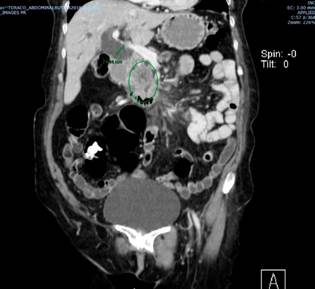
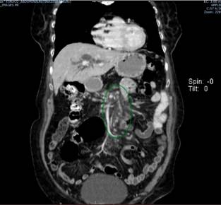
This is the case of a 72-year-old female patient with a biliary obstruction secondary to a mass located in the head of the pancreas. The following findings were reported in her paraclinical tests: laboratory tests: initial total bilirubin levels of 12.32 mg/dL at the expense of direct bilirubin levels (10.8 mg/dL); abdominal CT scan: presence of a neoplasm in the head of the pancreas surrounding in 180° the superior mesenteric vein; patent splenoportal confluence; patent hepatic artery; dilatation of the intrahepatic and extrahepatic biliary ducts, and of the main pancreatic duct; increased density of mesenteric fat with lymph nodes with short axes (between 5 mm and 6 mm) suspicious due to their enhancement; and focal hepatic lesion in the segment 6 with an appearance suggestive of suspected neoplastic infiltration, without presence of free fluid (Figures 1 and 2).

Figure 1 Mass located in the head of the pancreas, distal bile duct obstruction. 

Figure 2 Increased density of mesenteric fat with underlying adenopathies. 

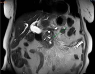
An abdominal MRI showed a dilatation of the intrahepatic and extrahepatic biliary ducts associated with a mass located in the head of the pancreas, with signs of involvement of the root of the mesentery and the presence of a focal hepatic lesion in the sixth segment of suspicious appearance (Figure 3).

Figure 3 Mass located in the head of the pancreas, intra- and extrahepatic bile duct dilatation. 

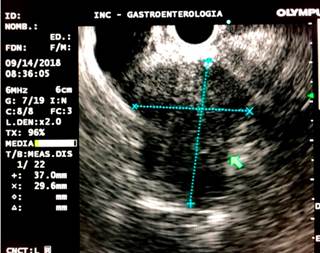
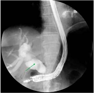
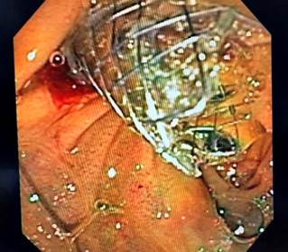
Based on these findings, palliative care was decided by performing a biliary stent placement by means of an ERCP. During the procedure, the papilla was found to be distorted with local retraction of the duodenal walls, and despite cannulation of the distal bile duct was achieved, cannulation of the proximal bile duct was unsuccessful. EUS-guided biliodigestive shunt was then performed, in which a neoplastic infiltration of the duodenal papilla preventing its cannulation and a 23 mm heterogeneous hypoechoic lesion in the head of the pancreas with an up to 18 mm retrograde bile duct dilation were found (Figure 4). An initial puncture of the mass was performed for histological study purposes and then of the transduodenal bile duct with a 19 Fr. needle, which allowed the advancement of the 0.035” hydrophilic guide wire; dilatation was achieved using a biliary balloon dilator under fluoroscopic control and subsequent placement of the 10 x 80 mm fully covered metal stent was performed, which was released under endoscopic and fluoroscopic control achieving adequate opening and drainage of the contrast medium and clear bile, without subsequent complications (Figures 5 and 6). The cytology report of the samples obtained through endosonography confirmed adenocarcinoma involvement. Two months later, a total bilirubin control was performed obtaining the following results: total bilirubin: 2.42 mg/dL; direct bilirubin: 1.85 mg/dL, and indirect bilirubin: 0.58 mg/dL (Figure 7). Palliative care was continued in the oncology service.
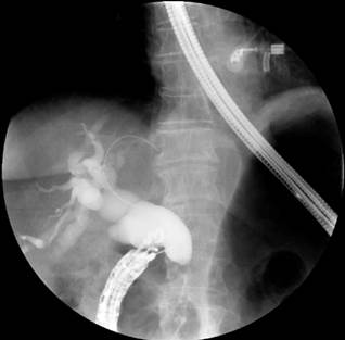
Figure 4 Mass in the head of the pancreas and dilatation of the bile duct in the endosonography. 

Figure 5 Endoscopic-guided bile duct cannulation and fluoroscopy in which dilatation with distal amputation is observed. 

Figure 6 Metal stent released under endoscopic ultrasound control and fluoroscopic guidance. 

Figure 7 Final result of the EUS-guided biliodigestive shunt. 

Discussion

This is the case of a patient with an obstruction of the distal common bile duct secondary to an inoperable adenocarcinoma located in the head of the pancreas, who required undergoing biliary stent placement due to the effects of compression. In this regard, similar cases describing the inability to cannulate the biliary duct due to the compressive nature of malignant tumors have been documented, but this situation has also been described in obstructions caused by benign tumors3-6.

EUS-guided biliodigestive shunt is an alternative procedure when failure to access the papilla or the bile duct occurs using ERCP (5% to 10% of cases)7; when the failure is caused by anatomical alteration, as it happened in the case presented here; or when ampullary distortion, periampullary diverticula, gastric outlet obstruction or in situ duodenal stents are present2.

When ERCP fails, biliary stent placement is then performed using percutaneous transhepatic cholangiography, but adverse events occur in up to 33% of cases, including hemorrhage, bilioperitoneum, infection and acute cholangitis, among others8. On the contrary, EUS-guided biliodigestive shunt has a lower risk of adverse events.

Transhepatic drainage is associated with significant morbidity. There are reports of complications in up to 60% of biliodigestive shunts performed using transpariticohepatic cholangiography, compared to 35% of those performed using EUS3.

EUS-guided biliodigestive shunt was first described by Giovannini et al. in 20019, and its safety and efficacy are currently being demonstrated given its approach2; in addition, it offers the following advantages: it is a minimally invasive procedure, it can be performed immediately after a failed ERCP and by the same specialist, it allows the drainage of both intrahepatic and extrahepatic bile ducts, it is a procedure that only causes mild pain to the patient (and even it does not cause any pain at all in some cases), it does not involve external drainage or dislocating the drains, it does not restrict the patient’s ability to engage in their daily activities as it only requires a very short hospitalization time, and it has a low complication rate3,10,11.

Regarding its complications, a 3.4 % to 38.6 % complication rate with an average complication rate between 17 % and 18.9 % has been described3,12,13; however, the rate of complications has shown a decreasing trend over time10,13-15. Complications include bile leak (3 %), hemorrhage (2.7 %), cholangitis (0.3 %), sepsis and peritonitis (3.5 %)10. Serious complications include the migration of the stent into the peritoneal cavity and fatal perforations, but their frequency is very low10,16,17; however, a consensus regarding the complications of this procedure has not yet been defined2.

As mentioned before, the performance of biliodigestive shunts through percutaneous transhepatic cholangiography is associated with a lower clinical success rare and, therefore, a higher cost. ERCP and EUS have the same efficacy to perform a biledigestive shunt, but there are no prospective studies comparing EUS as a primary procedure to perform biliodigestive shunts2.

Cannulation of the papilla can be quite complex in cases where the obstruction is caused by a malignant tumor, and, therefore, cannulation time increases, which can trigger the development of adverse events, being post-ERCP pancreatitis the main adverse event, which can occur in up to 10% of cases in which cannulation time exceeds 10 minutes18.

In this sense, the last multi-institutional consensus determined that EUS-guided biliodigestive shunt is the technique of choice in patients with a bile duct obstruction secondary a malignancy and in which bile stent placement was not possible using ERCP. In this regard, the following management algorithm has been proposed (Figure 8)1.
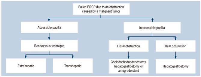
Figure 8 Management algorithm for patients with failed ERCP due to an obstruction caused by a malignancy1. Taken from: Guo J et al. Endosc Ultrasound. 2018;7(6):356-365. 

It has been demonstrated that this is a safe and effective procedure for the management of obstructions secondary to malignant tumors with very similar results when compared to ERCP. In fact, it can be thought that it is not only the method of choice in cases of failed ERCP, but also in distal intestinal obstruction cases. In addition, due to the fact that it is performed under direct observation, the risk of acute pancreatitis can be avoided.

Conclusion

EUS-guided biliodigestive shunt is an emerging technique that is considered a very good alternative in patients in which initial ERCP was unsuccessful. It is a practical, effective and efficient procedure with an adequate safety margin and clinical response. As it is an emerging technique, further studies are required to consolidate such advantages.

REFERENCES

1. Guo J, Giovannini M, Sahai AV, Saftoiu A, Dietrich CF, Santo E, Fusaroli P, Siddiqui AA, Bhutani MS, Bun Teoh AY, Irisawa A, Arturo Arias BL, Achanta CR, Jenssen C, Seo DW, Adler DG, Kalaitzakis E, Artifon E, Itokawa F, Poley JW, Mishra G, Ho KY, Wang HP, Okasha HH, Lachter J, Vila JJ, Iglesias-Garcia J, Yamao K, Yasuda K, Kubota K, Palazzo L, Sabbagh LC, Sharma M, Kida M, El-Nady M, Nguyen NQ, Vilmann P, Garg PK, Rai P, Mukai S, Carrara S, Parupudi S, Sridhar S, Lakhtakia S, Rana SS, Ogura T, Baron TH, Dhir V, Sun S. A multi-institution consensus on how to perform EUS-guided biliary drainage for malignant biliary obstruction. Endosc Ultrasound. 2018;7(6):356-365. https://doi.org/10.4103/eus.eus_53_18Links ]

2. Baars JE, Kaffes AJ, Saxena P. EUS-guided biliary drainage: A comprehensive review of the literature. Endosc Ultrasound. 2018;7(1):4-9. https://doi.org/10.4103/eus.eus_105_17Links ]

3. Giovannini M, Bories E, Napoleon B, Barthet M, Caillol F, Pesenti C. Multicenter randomized phase II study: Percutaneous biliary drainage vs. EUS guided biliary drainage: Results of intermediate analysis. Gastrointest Endosc 2015;81(5 Suppl):AB174. https://doi.org/10.1016/j.gie.2015.03.1926Links ]

4. Du L, D’Souza P, Thiesen A, Girgis S, Owen R, McNally D, Sarlieve P, Sandha G. Percutaneous transhepatic cholangioscopy for indeterminate biliary strictures using the SpyGlass system: a case series. Endoscopy. 2015;47(11):1054-6. https://doi.org/10.1055/s-0034-1392527Links ]

5. Franzini T, Cardarelli-Leite L, Figueira ERR, Morita F, Domingos FUG, Carnevale FC, de Moura EGH. SpyGlass percutaneous transhepatic cholangioscopy-guided lithotripsy of a large intrahepatic stone. Endoscopy. 2017;49(12):E292-E293. https://doi.org/10.1055/s-0043-117943Links ]

6. Mendonça EQ, de Oliveira JF, Baba ER, Dias AR, Maluf-Filho F. Percutaneous transhepatic cholangioscopy for the diagnosis of biliary obstruction in a patient with Roux-en-Y partial gastrectomy. Gastrointest Endosc. 2017;85(1):255-256. https://doi.org/10.1016/j.gie.2016.07.057Links ]

7. Enochsson L, Swahn F, Arnelo U, Nilsson M, Löhr M, Persson G. Nationwide, population-based data from 11,074 ERCP procedures from the Swedish Registry for Gallstone Surgery and ERCP. Gastrointest Endosc. 2010;72(6):1175-84, 1184.e1-3. https://doi.org/10.1016/j.gie.2010.07.047Links ]

8. Nennstiel S, Weber A, Frick G, Haller B, Meining A, Schmid RM, Neu B. Drainage-related Complications in Percutaneous Transhepatic Biliary Drainage: An Analysis Over 10 Years. J Clin Gastroenterol. 2015;49(9):764-70. https://doi.org/10.1097/MCG.0000000000000275Links ]

9. Giovannini M, Moutardier V, Pesenti C, Bories E, Lelong B, Delpero JR. Endoscopic ultrasound-guided bilioduodenal anastomosis: a new technique for biliary drainage. Endoscopy. 2001;33(10):898-900. https://doi.org/10.1055/s-2001-17324Links ]

10. Sharaiha RZ, Khan MA, Kamal F, Tyberg A, Tombazzi CR, Ali B, Tombazzi C, Kahaleh M. Efficacy and safety of EUS-guided biliary drainage in comparison with percutaneous biliary drainage when ERCP fails: a systematic review and meta-analysis. Gastrointest Endosc. 2017;85(5):904-914. https://doi.org/10.1016/j.gie.2016.12.023Links ]

11. Khashab MA, Valeshabad AK, Afghani E, Singh VK, Kumbhari V, Messallam A, Saxena P, El Zein M, Lennon AM, Canto MI, Kalloo AN. A comparative evaluation of EUS-guided biliary drainage and percutaneous drainage in patients with distal malignant biliary obstruction and failed ERCP. Dig Dis Sci. 2015;60(2):557-65. https://doi.org/10.1007/s10620-014-3300-6Links ]

12. Khan MA, Akbar A, Baron TH, Khan S, Kocak M, Alastal Y, Hammad T, Lee WM, Sofi A, Artifon EL, Nawras A, Ismail MK. Endoscopic Ultrasound-Guided Biliary Drainage: A Systematic Review and Meta-Analysis. Dig Dis Sci. 2016;61(3):684-703. https://doi.org/10.1007/s10620-015-3933-0Links ]

13. Dhir V, Isayama H, Itoi T, Almadi M, Siripun A, Teoh AYB, Ho KY. Endoscopic ultrasonography-guided biliary and pancreatic duct interventions. Dig Endosc. 2017;29(4):472-485. https://doi.org/10.1111/den.12818Links ]

14. Tyberg A, Desai AP, Kumta NA, Brown E, Gaidhane M, Sharaiha RZ, Kahaleh M. EUS-guided biliary drainage after failed ERCP: a novel algorithm individualized based on patient anatomy. Gastrointest Endosc. 2016;84(6):941-946. https://doi.org/10.1016/j.gie.2016.05.035Links ]

15. Kunda R, Pérez-Miranda M, Will U, Ullrich S, Brenke D, Dollhopf M, Meier M, Larghi A. EUS-guided choledochoduodenostomy for malignant distal biliary obstruction using a lumen-apposing fully covered metal stent after failed ERCP. Surg Endosc. 2016;30(11):5002-5008. https://doi.org/10.1007/s00464-016-4845-6Links ]

16. Minaga K, Kitano M, Yamashita Y, Nakatani Y, Kudo M. Stent migration into the abdominal cavity after EUS-guided hepaticogastrostomy. Gastrointest Endosc. 2017;85(1):263-264. https://doi.org/10.1016/j.gie.2016.03.016Links ]

17. Martins FP, Rossini LG, Ferrari AP. Migration of a covered metallic stent following endoscopic ultrasound-guided hepaticogastrostomy: fatal complication. Endoscopy. 2010;42 Suppl 2:E126-7. https://doi.org/10.1055/s-0029-1243911Links ]

18. Bailey AA, Bourke MJ, Williams SJ, Walsh PR, Murray MA, Lee EY, Kwan V, Lynch PM. A prospective randomized trial of cannulation technique in ERCP: effects on technical success and post-ERCP pancreatitis. Endoscopy. 2008;40(4):296-301. https://doi.org/10.1055/s-2007-995566Links ]

Citation: Manrique M, Martín W. Biliodigestive shunt guided by endoscopic ultrasonography in patient with stage IV pancreatic adenocarcinoma and obstruction of the bile duct. A case report. Rev Colomb Gastroenterol. 2021;36(1):120-125. https://doi.org/10.22516/25007440.490

Funding

The authors state that this study was funded with their own resources and did not involve external contributions of any kind.

Received: December 11, 2019; Accepted: February 13, 2020

*Correspondence: Wilmar Martín. wilmartin16@yahoo.com

Conflicts of interest

None declared by the authors.

Creative Commons License This is an open-access article distributed under the terms of the Creative Commons Attribution License