


 text in
  English (pdf)
 Article in xml format
 Article references
 Send this article by e-mail
 Cited by SciELO
  Cited by Google
  Similars in
SciELO
Similars in
SciELO  Similars in Google
 Permalink
Introduction
Cancer entails a global health problem and is one of the most important causes of mortality 1. Oral cancer represents 2% of all cancers, almost 30% of head and neck tumors, and 90% are squamous or epidermoid cell carcinomas; the remaining 10% consist of salivary gland tumors, melanomas, sarcomas, basal carcinomas, lymphomas, odontogenic tumors and metastatic lesions 2.
There is a wide geographical variation in the incidence of oral cancer (OCa). The highest risk in males is in France, India, Brazil and some South Asian countries 3. It is more common after the age of 50, and in many countries, is more common in men than women, due to risky habits or the risk of sun exposure in some professions. It is a multifactorial disease, in which tobacco and alcohol play a very important role, but it can be prevented by avoiding the pertinent risk factors, as well as having annual dental exams 1.
Unlike other locations, oral lesions can be analyzed visually and various types of noninvasive and invasive methods, such as biopsies, molecular markers, toluidine blue and oral cytology can be used. These are adjunctive techniques to the clinical exam which allow greater effectiveness in early diagnosis 4.
Despite everything, oral cancer detection continues to be a significant and challenging problem for physicians and dentists, due to many lesions being asymptomatic in early stages, as well as due to their lack of training 4.
The detection methods include toluidine blue, which is a thiazide, acidophilic and metachromatic dye. Its main characteristic is that it selectively stains acidic tissue components, such as sulphates and radical phosphates incorporated in the cell DNA and RNA. Therefore, it is used for in vivo nuclear staining, based on the fact that dysplastic and anaplastic cells have a quantitatively larger amount of nucleic acids and therefore retain the stain. The test has high sensitivity, and its drawbacks include its possible false positives 5.
Exfoliative cytology is another detection method; this is a simple, noninvasive process used to study the cells that are shed from the tissues, using conventional staining. This test, known as conventional smear cytology, was originally designed for early detection of cancerous cervical cells. Exfoliative cytology is defined as a morphological study based on the microscopic character of cells and extracellular components spontaneously or artificially (through procedures) detached from the organs, which are generally easily processed 6.
ViziLite TBlue and Microlux DL devices are light-based methods which use a blue-white light (430 and 580 nm spectral wavelengths) to evaluate the tissues. For ViziLite TBlue, the blue-white light is created by the reaction between acetylsalicylic acid and hydrogen peroxide (chemiluminescence), while Microlux DL's blue-white light is produced using a battery-operated light-emitting diode 4.
The objective of this study was to determine the effectiveness of four methods in detecting potentially malignant oral lesions, using an umbrella systematic review.
Data collection
An umbrella review was performed following the guidelines proposed by the Joanna Briggs Institute for umbrella reviews. A bibliographic search was carried out using the following key words: early detection of cancer, mouth neoplasms, and diagnostic techniques and procedures.
Two investigators, previously calibrated using a stomatology specialist as the gold standard, performed an electronic search in the following databases: PubMed and EBSCO-host. The use of only two databases is explained by a lack of access to others due to the financial limitations of the researchers and the institution to which they are affiliated.
The inclusion criteria for article selection were: systematic reviews whose objective was to evaluate the efficacy of at least one of the detection methods for potentially malignant lesions; specifically, exfoliative cytology, toluidine blue, or ViziLite or Microlux DL devices.
The following criteria were used to refine the search: systematic review articles, related to detection methods for potentially malignant oral lesions, published between 2013 and 2018 in English and Spanish, full text and the Boolean connector and.
The exclusion criteria were: studies which evaluated the efficacy of detection methods for potentially malignant lesions other than exfoliative cytology, toluidine blue, or ViziLite or Microlux DL devices. Studies which were not systematic reviews were also excluded.
The search produced a total of 90,819 references, of which 23 full text articles were selected, through title and abstract reading by the two investigators calibrated by the gold standard, to be evaluated for eligibility. Seventeen were excluded through a reading of the full text, due to insufficient relationship with the topic. Ultimately, six articles which discussed the methods of interest for detecting premalignant oral lesions were included, using a critical reading of the full text and the PRISMA format checklist (Figure 1). It should be noted that in cases where there was doubt regarding which articles to use, the gold standard was used for the final decision.
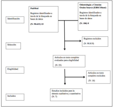
Figure 1 Flow chart of study selection. Data derived from the PubMed and EbscoHost databases identified, selected, chosen and included.
The following information was extracted from the selected studies: authors, title, journal, year, type of study, technique and result (Table 1), as well as specificity, sensitivity, positive predictive value (PPV) and negative predictive value (NPV) figures (Table 2). These processes were carried out by one of the investigators through critical reading of the full text.
Table 1 Characteristics of the included studies.
| Author | Title | Journal | Year |
|---|---|---|---|
| Marco Mascitti, Giovanna Orsini, Vincenzo Tosco, Riccardo Monterubbianesi, Andrea Balercia, Angelo Putignano, Maurizio Pro-caccini, Andrea Santarelli | An Overview on Current Non-invasive Diagnostic Devices in Oral Oncology 7 | Frontiers in Physiology | 2018 |
| Dongjuan Liu, Xin Zhao, Xin Zeng, Hongxia Dan, Qianming Chen | Non-Invasive Techniques for Detection and Diagnosis of Oral Potentially Malignant Disorders 8 | Tohoku Journal | 2016 |
| Carreras C, Cosme Gay E | Techniques for early diagnosis of oral squamous cell carcinoma: Systematic review 9 | Medicina oral Patología oral y Cirugía bucal | 2015 |
| Ravleen Nagi, Yashoda Bhoomi Reddy-Kantharaj, Nagaraju Rakesh, Sujatha Janardhan-Reddy, Shashikant Sahu | Efficacy of light based detection systems for early detection of oral cancer and oral potentially malignant disorders: Systematic review10 | Medicina oral Patología oral y Cirugía bucal | 2016 |
| Abdulhameed H. Alsarraf, Omar Kujan, Camile S. Farah | The utility of oral brush cytology in the early detection of oral cancer and oral potentially malignant disorders: A systematic review11 | Journal of Oral Pathology and Medicine | 2017 |
| A Rashid, S Warnakulasuriya | The use of light-based (optical) detection systems as adjuncts in the detection of oral cancer and oral potentially malignant disorders: a systematic review12 | Journal of Oral Pathology and Medicine | 2015 |
Table 2 Results
| Author | Title | DMPMOL | Number of articles | Sensitivity % | Specificity % | PPV % | NPV % |
|---|---|---|---|---|---|---|---|
| Marco Mascitti | An Overview on Current Non-invasive Diagnostic Devices in Oral Oncology 7 | -ViziLite | 15 (4 did not report sensitivity, sensibility, PPV or NPV; 1 did not report NPV; 1 did not report NPV or PPV) | 53 of the articles report more than 70 | 26.6 of the articles report more than 70 | 26.6 of the articles report more than 70 | 46.6 of the articles report more than 70 |
| Microlux/DL | 2 | 77.8 and 100 | 32.4 and 70.7 | 17.9 and 36.8 | 93.5 and 100 | ||
| Dongjuan Liu | Non-Invasive Techniques for Detection and Diagnosis of Oral Potentially Malignant Disorders 8 | -Toluidine blue staining | 10 | 38 - 100 | 9 - 100 | Not reported | Not reported |
| -ViziLite | 8 | 71 - 100 | 0 - 84.6 | Not reported | Not reported | ||
| Microlux/DL | 2 | 77.8 and 94.3 | 70.7 and 99.6 | Not reported | Not reported | ||
| Carreras C | Techniques for early diagnosis of oral squamous cell carcinoma: Systematic review 9 | -Toluidine blue staining | 2 | 92.5 and 97.8 | 63.2 and 92.9 | Not reported | Not reported |
| -ViziLite | 4 | 100 | 0 - 14 | 18-80 | Not reported | ||
| -Exfoliative cytology | 2 | 55 and 83.1 | 32.4 and 70.7 | 100 | 49 and 80 | ||
| Ravleen Nagi, et al. | Efficacy of light based detection systems for early detection of oral cancer and oral potentially malignant disorders: Systematic review10 | -ViziLite | 5 | 7.1- 100 | 0 - 27.8 | Not reported | Not reported |
| Microlux/DL | 1 | 77.8 | 70.7 | Not reported | Not reported | ||
| Abdulhameed H, et al. | The utility of oral brush cytology in the early detection of oral cancer and oral potentially malignant disorders: A systematic review11 | -Exfoliative cytology | 25 | 69 - 100 | 23.5-100 | Not reported | Not reported |
| Rashid A, et al | The use of light-based detection systems as adjuncts in the detection of oral cancer and oral potentially malignant disorders: a systematic review12 | -ViziLite | 9 (Four did not report sensitivity and five did not report specificity, six did not report PPV or NPV) | 77.1 - 100 | 0 - 27.8 | 18-80 | 11.1 of the articles reported more than 70 |
| DMPMOL: detection method for potentially malignant oral lesions. PPV: positive predictive value. NPV: negative predictive value. | |||||||
Results
To find the results, the number of articles reported in each review was extracted, and these were subsequently subdivided according to the detection method of potentially malignant oral lesions they analyzed. Data were also extracted on the sensitivity, specificity, PPV and NPV reported for each article (Table 2), analyzing these values with a 70% cut-off point for all data.
According to the findings of this overview of reviews and the analysis of the mean of the reported sensitivity and specificity, the Microlux/DL method was found to have the highest average sensitivity, at 82.63%, while exfoliative cytology had the lowest sensitivity, according to the reports, with 76.77%. For specificity, lower overall values were found than for sensitivity, with the exception of exfoliative cytology with an average specificity of 80.07%, which could be considered high, as opposed to the ViziLite method with 25.4%. Thus, it is fair to say that there may be a greater possibility of false positive results with this latter method, which is not very desirable in the diagnosis of diseases such as oral cancer (Figure 2).
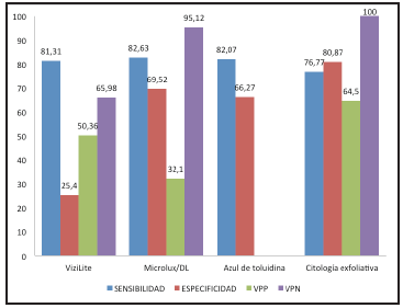
Figure 2 Bar graphs; sensitivity, specificity, positive predictive value and negative predic tive value results. Graphic presentation of the mean results in percentages of sensitivity, specificity, positive predictive value and negative predictive value.
In another vein, the PPV and NPV reported for Microlux/ DL were 32.1% and 95.12%, respectively, which are very distant from each other and, considering that PPV is the probability of having a disease when the test is positive, and NPV is the probability of not having the disease when the test is negative, it can be said that the use of Microlux/ DL may entail a greater risk of having false positive than false negative results. For their part, the values found for Vizilite were 50.36% PPV and 65.98% NPV, and for exfoliative cytology were 64.5% PPV and 100% NPV (Figure 2). It should be clarified that none of the reviewed articles reported PPV or NPV for toluidine blue (Tables 3 and 4 ).
Table 3 Summary of the sensitivity and specificity means.
| Title | DMPMOL | ||||
|---|---|---|---|---|---|
| ViziLite % | Microlux/DL % | Toluidene blue % | Exfoliative cytology % | ||
| An Overview on Current Non-invasive Diagnostic Devices in Oral Oncology 7 | Mean sensitivity | 78.99 | 88.9 | ---------- | ---------- |
| Mean specificity | 49.1 | 51.55 | ---------- | ---------- | |
| Non-Invasive Techniques for Detection and Diagnosis of Oral Potentially Malignant Disorders 8 | Mean sensitivity | 85.5 | 86.05 | 69 | ---------- |
| Mean specificity | 42.3 | 85.15 | 54.5 | ---------- | |
| Techniques for early diagnosis of oral squamous cell carcinoma: Systematic review 9 | Mean sensitivity | 100 | ---------- | 95.15 | 69.05 |
| Mean specificity | 7 | ---------- | 78.05 | 100 | |
| Efficacy of light based detection systems for early detection of oral cancer and oral potentially malignant disorders: Systematic review10 | Mean sensitivity | 53.55 | 77.8 | ---------- | ---------- |
| Mean specificity | 13.9 | 70.7 | ---------- | ---------- | |
| The utility of oral brush cytology in the early detection of oral cancer and oral potentially malignant disorders: A systematic review11 | Mean sensitivity | ---------- | ---------- | ---------- | 84.5 |
| Mean specificity | ---------- | ---------- | ---------- | 61.75 | |
| The use of light-based (optical) detection systems as adjuncts in the detection of oral cancer and oral potentially malignant disorders: a systematic review12 | Mean sensitivity | 88.55 | 77.8 | ---------- | ---------- |
| Mean specificity | 13.9 | 70.7 | ---------- | ---------- | |
| TOTAL | Mean sensitivity | 81.31 | 82.63 | 82.07 | 76.77 |
| Mean specificity | 25.4 | 69.52 | 66.27 | 80.87 | |
Table 4 Summary of the mean positive predictive value (PPV) and negative predictive value (NPV).
| Title | DMPMOL | ||||
|---|---|---|---|---|---|
| ViziLite % | Miciolux/DL % | Toluidene blue % | Exfoliative cytology % | ||
| An Overview on Current Non-invasive Diagnostic Devices in Oral Oncology 7 | Mean PPV | 50.5 | 27.4 | ---------- | ---------- |
| Mean NPV | 82.5 | 96.75 | ---------- | ---------- | |
| Non-Invasive Techniques for Detection and Diagnosis of Oral Potentially Malignant Disorders 8 | Mean PPV | Not reported | Not reported | Not reported | ---------- |
| Mean NPV | Not reported | Not reported | Not reported | ---------- | |
| Techniques for early diagnosis of oral squamous cell carcinoma: Systematic review 9 | Mean PPV | 49 | ---------- | Not reported | 64.5 |
| Mean NPV | Not reported | ---------- | Not reported | 100 | |
| Efficacy of light based detection systems for early detection of oral cancer and oral potentially malignant disorders: Systematic review10 | Mean PPV | Not reported | Not reported | ---------- | ---------- |
| Mean NPV | Not reported | Not reported | ---------- | ---------- | |
| The utility of oral brush cytology in the early detection of oral cancer and oral potentially malignant disorders: A systematic review11 | Mean PPV | ---------- | ---------- | ---------- | Not reported |
| Mean NPV | ---------- | ---------- | ---------- | Not reported | |
| The use of light-based (optical) detection systems as adjuncts in the detection of oral cancer and oral potentially malignant disorders: a systematic review12 | Mean PPV | 51.6 | 36.8 | ---------- | ---------- |
| Mean NPV | 49.46 | 93.5 | ---------- | ---------- | |
| Total | Mean PPV | 50.36 | 32.1 | Not reported | 64.5 |
| Mean NPV | 65.98 | 95.12 | Not reported | 100 | |
In order to determine the method with the greatest efficacy in diagnosing potentially malignant oral lesions, the sensitivity and specificity of each should be considered (the PPV and NPV were not used for the final comparison since these values were not available for each of the methods studied), clarifying that sensitivity takes precedence since it is the method's ability to detect the disease, while specificity is the ability to establish that an individual is healthy. Based on this and the results found (Table 3), it can be said that the method with the best-balanced sensitivity and specificity is Microlux DL, with 82.63% sensitivity and 69.52% specificity. Although it does not have the highest specificity it does have the highest sensitivity, which indicates a higher probability of an accurate positive result. Despite having a lower specificity, the values are not too distant from each other, and the possibility of a false positive is not alarming. This is not the case with ViziLite, which has 81.31% sensitivity and 25.4% specificity, leading to a high probability of obtaining a false positive result (Figure 2).
Discussion
This study found that the methods for detecting potentially malignant oral lesions are very promising and a very good option as an alternative to tissue biopsy and histopathological tests. For their part, in 2018, Yang E et al. 13 reported that although a biopsy is the gold standard for diagnosing cancer and dysplasia, it is limited by morbidity, the need for time and resources, and the risk of sampling bias. They also pointed out that the existing diagnostic complements which provide immediate feedback are limited by deficient diagnostic precision and that in vivo microscopy technologies are the most promising methods.
With regard to ViziLite, in 2019, Shashidara R 14 reported that, as a relatively recent technique, it does not have sufficient evidential support for its effectiveness in diagnosing oral precancer and cancer. This concurs with the present study, which shows that while this method is a good alternative to the conventional oral exam, it has some limitations and needs to be studied further.
With regard to toluidine blue, Awan K, in 2015 15 stated that it had better positive and negative predictive values (PPV = 50%, NPV = 71.2%) than the optic instruments VELscope and ViziLite, which were almost equal (VELscope, PPV = 37.8%, NPV = 61.1%; ViziLite, PPV = 39.5, NPV = 66.7%). Thus, this study concluded that the three complementary tests may be useful in detecting oral mucosal disorders seen in specialized oral medicine clinics, but their accuracy in detecting potentially malignant oral disorders and dysplastic lesions is questionable, and more well-designed studies are needed to examine their role in a primary care setting, which coincides with this paper.
In 2014, Ibrahim S et al. 16 reported that the sensitivity, specificity and PPV of Microlux DL for visualizing suspicious premalignant lesions, taking COE as a gold standard (that is, detection device), were 94.3, 99.6 and 96.2%, respectively, while when biopsy was considered a gold standard, the sensitivity, specificity and PPV were 100, 32.4 and 17.9%, respectively. These values are higher (when COE is considered the gold standard) than those reported in this study, where mean sensitivity, specificity and PPV were 82.63%, 69.52 and 32.1%, respectively. In both studies, Microlux DL was considered to be a promising device for detecting potentially malignant oral lesions, which does not replace biopsy, considered to be the gold standard.
In conclusion, the methods for detecting premalignant oral lesions evaluated in this study require greater scientific evidence to validate their effectiveness. According to the findings of this review, the method with the greatest effectiveness is Microlux/DL, due to its high sensitivity and specificity.

