SciELO - Scientific Electronic Library Online

 
vol.37 issue3Morphological subtypes of the duodenal papilla and their relationship with post-ERCP complicationsSedation Administered by General Practitioners for Low Complexity Endoscopic Procedures: Experience in an Endoscopy Unit of a Tertiary Referral Hospital in Cali author indexsubject indexarticles search
Home Pagealphabetic serial listing  

Services on Demand

Journal

Article

Indicators

Related links

  • On index processCited by Google
  • Have no similar articlesSimilars in SciELO
  • On index processSimilars in Google

Share


Revista colombiana de Gastroenterología

Print version ISSN 0120-9957On-line version ISSN 2500-7440

Rev. colomb. Gastroenterol. vol.37 no.3 Bogotá July/Sept. 2022  Epub Nov 29, 2022

https://doi.org/10.22516/25007440.769 

Original article

Association Between Body Mass Index and Liver Fibrosis Degree Measured Using Real-Time Elastography (Supersonic)

Diana Carolina Alfonso-Vergel,1  * 
http://orcid.org/0000-0003-4955-0700

Jhon Edison Prieto-Ortiz2 
http://orcid.org/0000-0002-8009-5999

1Surgeon, University Teaching Specialist, Master in Epidemiology, Resident of Internal Medicine, Pontificia Universidad Javeriana. Bogotá, Colombia.

2Surgeon, Internal Medicine, Gastroenterology, and Hepatology Specialist, Centro de Enfermedades Hepáticas y Digestivas (CEHYD). Bogotá, Colombia.


Abstract

Introduction:

Non-alcoholic fatty liver disease (NAFLD) or fatty liver, is characterized by an excessive accumulation of fat in the liver, is a metabolic disorder with a worldwide prevalence close to 25%, with a spectrum of liver damage that covers the steatosis without fibrosis, steatohepatitis with variable fibrosis and cirrhosis or maximum degree of fibrosis, this fibrosis determines prognosis and outcomes in the disease.

Objective:

To evaluate the association between body mass index and the degree of liver fibrosis in patients diagnosed with fatty liver in a hepatology center in the city of Bogotá, Colombia.

Patients and methods:

A case-control study is carried out with patients diagnosed with fatty liver, who have undergone real-time elastography (Supersonic). Information was taken from patients diagnosed with fatty liver who met the inclusion criteria. Continuous variables were described using measures of central tendency and standard deviation. Categorical variables were described with numbers and percentages. A 95% confidence interval was considered statistically significant.

Results:

361 patients were included, of which 95.2% (n=344) presented some degree of alteration (12% minimal fibrosis, 33% moderate fibrosis, 34% severe fibrosis and 16% cirrhosis) and only 5% showed a liver normal. Not having an adequate weight is related to severe fibrosis F3 OR 3.24 (1.03-10) and cirrhosis F4 OR 2.33 (2.33-42.99). No statistically significant differences were found between altered body mass index and any degree of fibrosis OR 2.74 (0.90-8.40). The presence of DM presents a 10-fold risk probability of ending in F4 cirrhosis, especially with poor disease control OR 5.16 (1.23-30.33).

Conclusion:

There is an association between abnormal body mass index and glycemic profile and the development of severe and advanced fibrosis. It is necessary in clinical practice, greater surveillance and evaluation of patients with fatty liver, in order to prevent the progression of fibrosis.

Keywords: Non-alcoholic fatty liver; hepatic fibrosis; cirrhosis; real-time elastography; supersonic

Resumen

Introducción:

La enfermedad del hígado graso no alcohólico (EHGNA) o hígado graso se caracteriza por una excesiva acumulación de grasa en el hígado, es un desorden metabólico con una prevalencia mundial cercana al 25 %, con un espectro de daño hepático que abarca la esteatosis sin fibrosis, esteatohepatitis con fibrosis variable y la cirrosis o grado máximo de fibrosis, dicha fibrosis determina el pronóstico y los desenlaces de la enfermedad.

Objetivo:

evaluar la asociación entre el índice de masa corporal (IMC) y el grado de fibrosis hepática en pacientes con diagnóstico de hígado graso en un centro de hepatología en la ciudad de Bogotá, Colombia.

Pacientes y métodos:

se realiza un estudio de casos y controles de pacientes con diagnóstico de hígado graso, a quienes se les haya realizado elastografía en tiempo real (Supersonic). Se tomó la información de pacientes con diagnóstico de hígado graso que cumplieron criterios de inclusión. Las variables continuas se describieron utilizando medidas de tendencia central y desviación estándar. Las variables categóricas se describieron con números y porcentajes. Se consideró un intervalo de confianza (IC) del 95 % como estadísticamente significativo.

Resultados:

se incluyeron 361 pacientes, de los cuales el 95,2 % (n = 344 pacientes) presentó algún grado de alteración (12 % fibrosis mínima, 33 % fibrosis moderada, 34 % fibrosis grave y 16 % cirrosis) y solo el 5 % mostró un hígado normal. No tener un adecuado peso se relaciona con fibrosis grave F3, odds ratio (OR): 3,24 (IC: 1,03-10) y cirrosis F4, OR: 2,33 (IC: 2,33-42,99). No se encontraron diferencias estadísticamente significativas entre la alteración del IMC y cualquier grado de fibrosis (OR: 2,74; IC: 0,90-8,40). La presencia de diabetes mellitus (DM) presenta una probabilidad de riesgo de 10 veces de terminar en cirrosis F4, en especial, con mal control de la enfermedad (OR: 5,16; IC: 1,23-30,33).

Conclusión:

existe una asociación entre el IMC, el perfil glicémico anormal y el desarrollo de fibrosis grave y avanzada. En la práctica clínica, son necesarias una mayor vigilancia y evaluación de los pacientes con hígado graso, con el fin de evitar la progresión de la fibrosis.

Palabras clave: Hígado graso no alcohólico; fibrosis hepática; cirrosis; elastografía en tiempo real; Supersonic

Introduction

Nonalcoholic fatty liver disease (NAFLD) or fatty liver disease is characterized by excessive fat accumulation in the liver and is associated with insulin resistance1. A histological analysis defines it as the presence of steatosis in 5% or more of the hepatocytes. Fatty liver disease is diagnosed through biopsy or imaging and radiology, usually after detecting fat through ultrasound and after ruling out secondary causes of hepatic steatosis such as alcohol consumption greater than 20 g/day for men and greater than 10 g/day for women, hepatotoxic drugs intake, hepatitis B and C virus, hemochromatosis, autoimmunity, and other chronic liver disease causes2.

Fatty liver disease is associated with obesity, diabetes mellitus (DM), dyslipidemia, and high blood pressure and is considered the hepatic manifestation of metabolic syndrome (MS)3. Most patients with fatty liver disease have hepatic steatosis without fibrosis in its initial phase and a good long-term prognosis4. Other patients have fibrosis and inflammation or steatohepatitis (intermediate phase) and may progress to the final stage of fibrosis or cirrhosis with an additional risk of developing hepatocellular carcinoma (HCC)5.

It is important to identify patients with fatty liver during the disease’s different stages and, according to findings, provide them with a treatment to prevent the progression of fibrosis. Given their poor prognosis, complications such as esophageal varicose veins, hepatic impairment, and HCC should be evaluated in patients with cirrhosis6,7. Currently, liver biopsy has remained the gold standard for assessing the degree of liver fibrosis8. However, since this is an invasive test, it can cause pain, bleeding, and even death9. Therefore, non-invasive methods to assess the degree of fibrosis in patients with fatty liver disease are becoming increasingly common due to the invasive nature of biopsy and its complications10.

There are several clinical scoring systems and non-invasive methods in medical practice. Some of these methods include the aspartate aminotransferase index (AST)/platelet count11, the relationship between AST/alanine aminotransferase (ALT)12, transient elastography (FibroScan), magnetic resonance elastography (MRE), and real-time elastography (RTE)13 or SuperSonic; the latter is a non-invasive test recently used in Colombia. The RTE test determines liver elasticity and calculates the grade of liver fibrosis. Sometimes, the RTE test supersedes liver biopsy and is useful for monitoring most patients with hepatopathies14.

The objective of this study is to evaluate the association between body mass index (BMI) and the degree of liver fibrosis in patients diagnosed with fatty liver in a hepatology center in Bogotá, Colombia.

Patients and methods

Population

Between January 1 and December 30, 2017, a case-control study of patients diagnosed with fatty liver disease was conducted through ultrasound or other imaging methods during hepatology consultation check-ups that would have undergone real-time elastography (SuperSonic) at Centro de Enfermedades Hepáticas y Digestivas (CEHYD) in Bogotá. We considered cases of patients who showed some degree of fibrosis and performed controls on participants with fatty liver without fibrosis determined as F0. We excluded patients who reported alcohol consumption (> 10 g in women and > 20 g in men per day), positive markers for hepatitis B virus (HBV) and hepatitis C virus (HCV), autoimmunity, hemochromatosis confirmed through a genetic study, or hepatotoxic drugs intake.

Variables

The variables included age, gender, metabolic syndrome condition, high blood pressure, BMI, transaminase level, AST/ALT ratio, dyslipidemia, glycemia, insulin, and degree of fibrosis.

Operational definitions were made according to the following criteria: the presence of metabolic syndrome and dyslipidemia, according to the Adult Treatment Panel III guidelines (ATP III)15; high blood pressure, according to the JNC8 guidelines; DM, according to the American Diabetes Association criteria (ADA)16; overweight and obesity, according to the World Health Organization criteria (WHO)17. Liver fibrosis determination was performed through real-time elastography (SuperSonic) using the Aixplorer ultrasound system (SuperSonic Imagine S. A. Aix-en-Provence, France) with a convex broadband probe (SC6-1). Values between 5.1 and 6.8 kPa, F2 between 7.2 and 8.3 kPa, F3 between 9.2 and 10.1, and F4 between 12.8 and 18.8 kPa were considered F0-F1.

Source of information

Project format: we used patients’ medical records as a secondary source in a consultation at a hepatology center in Bogotá.

Data collection plan and analysis

We used a format proposed for the data collection study that included the variables. Data was tabulated using the Stata 12 program and the Excel program database, creating the tables with their statistics and graphs, respectively.

Information was taken from patients diagnosed with fatty liver disease who met inclusion criteria to establish the degree of fibrosis and its relationship with BMI.

Continuous variables were described using measures of central tendency and standard deviation. Categorical variables were described with numbers and percentages. A 95% confidence interval (CI) was considered statistically significant.

Results

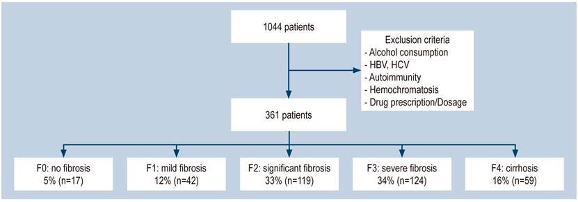
We excluded 683 patients from a group of 1044 seen between January and December 2017 because they did not meet the inclusion criteria or their information was incomplete. Included in the study were 361 patients (Figure 1), from whom 58% were women, with an average age of 57 years, a minimum age of 16 years, and a maximum of 90 years.

Figure 1 Patient flowchart. 

The overweight prevalence in this study was 49.2% (n = 177), with a BMI average of 26 ± 3.9, while the obesity prevalence was 20.7% (n = 75). The mean abdominal perimeter was 94 ± 10 cm. Twenty-seven percent (n = 99) had a complete metabolic syndrome. High blood pressure was observed in 28% (n = 101), DM, 17.7% (n = 64); impaired fasting glycemia (IFG), 27.9% (n = 101), and hyperinsulinemia in 56.9% (n = 205) (Table 1).

Table 1 Demographic, biochemical, metabolic, and diagnostic characteristics of liver fibrosis disease 

Variable Men Women
Age (years) 41.3 (149) 58.7 (212)
Mean BMI (kg/m2) 27 27
MS (%/n) 72.6 (262) 27.4 (99)
HBP (%/n) 72 (260) 28 (101)
DM (%/n) 82.2 (297) 17.7 (64)
Glycemia > 100 mg/dL 72 (260) 28 (101)
HDL< 40 mg/dl (%/n) 42.7 (154) 57.3 (207)
TGs>150 mgs/dl (%/n) 55.7 (201) 44.3 (160)
Degree of fibrosis
F0 41.2 (7) 58.8 (10)
F1 42.9 (18) 57.1 (24)
F2 39.5 (47) 60.5 (72)
F3 41.9 (52) 58.1 (72)
F4 42.4 (25) 57.6 (34)
Total 146 212
BMI classification
  % n
Healthy weight 30.1 109
Overweight 49.2 177
Obesity 20.7 75
Total 100 361

DM: diabetes mellitus; HDL: high-density lipoprotein; HBP: high blood pressure; BMI: body mass index; MS: metabolic syndrome; TGs: triglycerides.

We found 207 (57.3%) patients with a decreased HDL level and 160 (44%) of patients had > 150 mg/dL triglycerides levels. 81.5% (n = 294) of patients had a > 1 ALT/AST ratio, which is an indicator of liver fibrosis.

When measuring fibrosis with real-time elastography, 95.2% (n = 344) of the patients showed an alteration (11% mild fibrosis, 32% significant fibrosis, 34% severe fibrosis, and 16% cirrhosis), and only 4% of the patients showed a completely normal liver.

No statistically significant differences were found between the degree of fibrosis and gender (odds ratio (OR): 0.9; CI: 0.3-2.9). Unhealthy weight is associated with severe fibrosis F3 (OR: 3.24; CI: 1.03-10) and cirrhosis F4 (OR: 2.33; CI: 2.33-42.99) (Figure 2). No statistically significant differences were found between BMI impairment and any degree of fibrosis (OR: 2.74; CI: 0.90-8.40). Risk probability estimation between fibrosis and obesity was impossible because this group lacked fibrosis-free checks.
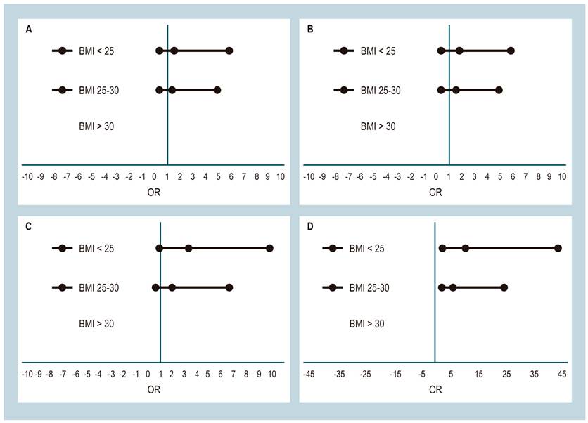
Figure 2 Association between liver fibrosis and BMI. A. Association between mild fibrosis and BMI. B. Association between significant fibrosis and BMI. C. Association between severe fibrosis and BMI. D. Association between cirrhosis and BMI. 

Women and men with larger abdominal circumference are 9.4-fold more likely to increase the risk of cirrhosis (CI: 2.41-39.64). High blood pressure was not associated with developing fibrosis to any degree, while the presence of DM has a 10-fold risk of ending in F4 cirrhosis, especially in patients with poor disease control (OR: 5.16; CI: 1.23-30.33). These values were unchanged with the logistic regression model (Table 2).

Table 2 Association between degrees of liver fibrosis and BMI 

Dependent/independent variable Adjusted OR 95%CI
Fibrosis/Gender 0.99 0.37-2.67
Fibrosis/BMI 2.74 1.02-7.31
Fibrosis/overweight 1.90 0.71-5.08
Fibrosis/obesity NA  
F1 fibrosis/BMI 1.64 0.53-5.14
F1 fibrosis/overweight 1.32 0.61-4.11
F1 fibrosis/obesity NA  
F2 fibrosis/BMI 1.85 0.66-5.14
F2 fibrosis/overweight 1.57 0.54-4.34
F2 fibrosis /obesity NA  
F3 fibrosis/BMI 3.24 1.15-9.09
F3 fibrosis/overweight 2.03 0.71-5.80
F3 fibrosis /obesity NA  
F4 cirrhosis/BMI 9.93 2.78-35.48
F4 cirrhosis/overweight 5.25 1.43-19.22
F4 cirrhosis/obesity NA  
Fibrosis/DM 3.58 0.46-27.55
F1 fibrosis/DM 2.16 0.23-20.02
F2 fibrosis/DM 1.74 0.21-14.74
F3 fibrosis/DM 3.64 0.45-28.8
F4 cirrhosis/DM 10.22 1.26-82.3
Fibrosis/Glyc > 100 mg/dL 1.85 0.52-6.61
F1 fibrosis/Glyc > 100 mg/dL 1.09 0.25-4.75
F2 fibrosis/Glyc > 100 mg/dL 1.24 0.33-4.55
F3 fibrosis/Glyc > 100 mg/dL 1.76 0.47-6.51
Cirrhosis F4/Glyc > 100 mg/dL 5.16 1.26-2.39

Discussion

Fatty liver disease is a metabolic disorder with a 20%-40% prevalence in Western countries18, 12%-30% in Asia19, and an overall prevalence between 20% and 25%20,21. The disease severity increases with risk factors, which is found in 10%-20% of individuals with healthy weight, 50% overweight, and 80% with obesity22.

Non-alcoholic fatty liver disease is a condition ranging across a spectrum of liver damage from steatosis to steatohepatitis with variable fibrosis and leading to cirrhosis with normal or elevated ALT values23. This disease is associated with some conditions, including metabolic syndrome, DM, obesity, high blood pressure, and dyslipidemia24. The metabolic syndrome is characterized by a group of risk factors that favor insulin resistance25; among these, BMI alteration is an important factor for developing NAFLD26. Additionally, we found that, among patients with fatty liver disease, 30.1% had a < 25 kg/m2 BMI; 49.2%, had between a 25 and 30 kg/m2 BMI, and 20.7%, had a > 30 kg/m2 BMI. Furthermore, this study showed an association between BMI alteration and the development of severe and advanced fibrosis, which was statistically significant (Table 2).

Several studies showed the association between fatty liver disease and DM27. An Italian study with 458 patients found that DM was the most important marker for fatty liver disease and a higher degree of fibrosis (OR: 1.97; 95%CI: 1.2-3.7)28. This study shows that 82.2% of men and 17.7% of women had DM. When adjusting the data for the association between severe fibrosis F3 and cirrhosis, we found an association between DM (OR: 10.22; 95 % CI: 1.36-44.6) and impaired fasting glycemia (OR: 5.16; 95 % CI: 1.23-30.33).

This study confirmed that fatty liver could occur at any age, though it is not a risk factor4 since no statistically significant association between age and sex was found in our patients.

In addition, 20% of patients with NAFLD develop cirrhosis29. In this study, cirrhosis was found in 16.3% of the entire series, with a slight predominance in women: 57.6% versus 42.4%.

However, this study is limited since it was a case-control study, and there was a selection bias in the check-ups group because all the participants were overweight or obese. In future studies, we can improve this aspect by increasing the number of participants. In summary, it was impossible to evaluate the association between abdominal perimeter and cirrhosis due to under-reporting in the medical histories.

Conclusions

The study found a statistically significant association between an abnormal BMI and glycemic profile and severe and advanced fibrosis development. Therefore, further surveillance and evaluation of patients with fatty liver disease are necessary for clinical practice to prevent fibrosis progression.

Acknowledgments

To our patients, the reason we continue researching, and to all the people who participated during the revision, writing, and translation process.

Referencias

1. Soresi M, Giannitrapani L, Noto D, Terranova A, Campagna ME, Cefalù AB, et al. Effects of steatosis on hepatic hemodynamics in patients with metabolic syndrome. Ultrasound Med Biol. 2015;41(6):1545-52. https://doi.org/10.1016/j.ultrasmedbio.2015.01.020Links ]

2. European Association for the Study of the Liver (EASL); European Association for the Study of Diabetes (EASD); European Association for the Study of Obesity (EASO). EASL-EASD-EASO Clinical Practice Guidelines for the Management of Non-Alcoholic Fatty Liver Disease. Obes Facts. 2016;9(2):65-90. https://doi.org/10.1159/000443344Links ]

3. Watanabe S, Hashimoto E, Ikejima K, Uto H, Ono M, Sumida Y, et al. Evidence-based clinical practice guidelines for nonalcoholic fatty liver disease/nonalcoholic steatohepatitis. J Gastroenterol. 2015;50(4):364-77. https://doi.org/10.1007/s00535-015-1050-7Links ]

4. Adams LA, Lymp JF, St Sauver J, Sanderson SO, Lindor KD, Feldstein A, et al. The natural history of nonalcoholic fatty liver disease: a population-based cohort study. Gastroenterology. 2005;129(1):113-21. https://doi.org/10.1053/j.gastro.2005.04.014Links ]

5. Matteoni CA, Younossi ZM, Gramlich T, Boparai N, Liu YC, McCullough AJ. Nonalcoholic fatty liver disease: a spectrum of clinical and pathological severity. Gastroenterology. 1999;116(6):1413-9.https://doi.org/10.1016/S0016-5085(99)70506-8Links ]

6. Ratziu V, Bonyhay L, Di Martino V, Charlotte F, Cavallaro L, Sayegh-Tainturier MH, et al. Survival, liver failure, and hepatocellular carcinoma in obesity-related cryptogenic cirrhosis. Hepatology. 2002;35(6):1485-93. https://doi.org/10.1053/jhep.2002.33324Links ]

7. Hui JM, Kench JG, Chitturi S, Sud A, Farrell GC, Byth K, et al. Long-term outcomes of cirrhosis in nonalcoholic steatohepatitis compared with hepatitis C. Hepatology. 2003;38(2):420-7. https://doi.org/10.1053/jhep.2003.50320Links ]

8. Ratziu V, Charlotte F, Heurtier A, Gombert S, Giral P, Bruckert E, et al. Sampling variability of liver biopsy in nonalcoholic fatty liver disease. Gastroenterology. 2005;128(7):1898-906. https://doi.org/10.1053/j.gastro.2005.03.084Links ]

9. Cholongitas E, Senzolo M, Standish R, Marelli L, Quaglia A, Patch D, et al. A systematic review of the quality of liver biopsy specimens. Am J Clin Pathol. 2006;125(5):710-21. https://doi.org/10.1309/W3XCNT4HKFBN2G0BLinks ]

10. Caballería Rovira L, Torán Montserrat P, Auladell Llorens MA, Pera Blanco G. Esteatosis hepática no alcohólica. Puesta al día. Atención Primaria. 2008;40(8):419-24. https://doi.org/10.1157/13125408Links ]

11. Wai CT, Greenson JK, Fontana RJ, Kalbfleisch JD, Marrero JA, Conjeevaram HS, et al. A simple noninvasive index can predict both significant fibrosis and cirrhosis in patients with chronic hepatitis C. Hepatology. 2003;38(2):518-26. https://doi.org/10.1053/jhep.2003.50346Links ]

12. Williams AL, Hoofnagle JH. Ratio of serum aspartate to alanine aminotransferase in chronic hepatitis. Relationship to cirrhosis. Gastroenterology. 1988;95(3):734-9. https://doi.org/10.1016/S0016-5085(88)80022-2Links ]

13. Yoshioka K, Hashimoto S, Kawabe N. Measurement of liver stiffness as a non-invasive method for diagnosis of non-alcoholic fatty liver disease. Hepatol Res. 2015;45(2):142-51.https://doi.org/10.1111/hepr.12388Links ]

14. Muller M, Gennisson JL, Deffieux T, Tanter M, Fink M. Quantitative viscoelasticity mapping of human liver using supersonic shear imaging: preliminary in vivo feasibility study. Ultrasound Med Biol. 2009;35(2):219-29. https://doi.org/10.1016/j.ultrasmedbio.2008.08.018Links ]

15. Rodríguez-Ortiz D, Reyes-Pérez A, León P, Sánchez H, Mosti M, Aguilar-Salinas CA, et al. Assessment of two different diagnostic guidelines criteria (National Cholesterol Education Adult Treatment Panel III [ATP III] and International Diabetes Federation [IDF]) for the evaluation of metabolic syndrome remission in a longitudinal cohort of patients undergoing Roux-en-Y gastric bypass. Surgery. 2016;159(4):1121-8. https://doi.org/10.1016/j.surg.2015.11.015Links ]

16. Krakoff LR, Gillespie RL, Ferdinand KC, Fergus IV, Akinboboye O, Williams KA, et al. 2014 hypertension recommendations from the eighth joint national committee panel members raise concerns for elderly black and female populations. J Am Coll Cardiol. 2014;64(4):394-402. https://doi.org/10.1016/j.jacc.2014.06.014Links ]

17. Kuczmarski RJ, Flegal KM. Criteria for definition of overweight in transition: background and recommendations for the United States. Am J Clin Nutr. 2000;72(5):1074-81. https://doi.org/10.1093/ajcn/72.5.1074Links ]

18. Browning JD, Szczepaniak LS, Dobbins R, Nuremberg P, Horton JD, Cohen JC, Grundy SM, Hobbs HH. Prevalence of hepatic steatosis in an urban population in the United States: impact of ethnicity. Hepatology. 2004;40(6):1387-95. https://doi.org/10.1002/hep.20466Links ]

19. Hashimoto E, Tokushige K. Prevalence, gender, ethnic variations, and prognosis of NASH. J Gastroenterol. 2011;46 Suppl 1:63-9. https://doi.org/10.1007/s00535-010-0311-8Links ]

20. Musso G, Gambino R, Cassader M, Pagano G. Meta-analysis: natural history of non-alcoholic fatty liver disease (NAFLD) and diagnostic accuracy of non-invasive tests for liver disease severity. Ann Med. 2011;43(8):617-49. https://doi.org/10.3109/07853890.2010.518623Links ]

21. Serfaty L, Lemoine M. Definition and natural history of metabolic steatosis: clinical aspects of NAFLD, NASH and cirrhosis. Diabetes Metab. 2008;34(6 Pt 2):634-7. https://doi.org/10.1016/S1262-3636(08)74597-XLinks ]

22. Eguchi Y, Hyogo H, Ono M, Mizuta T, Ono N, Fujimoto K, et al. Prevalence and associated metabolic factors of nonalcoholic fatty liver disease in the general population from 2009 to 2010 in Japan: a multicenter large retrospective study. J Gastroenterol. 2012;47(5):586-95. https://doi.org/10.1007/s00535-012-0533-zLinks ]

23. Leite NC, Salles GF, Araujo AL, Villela-Nogueira CA, Cardoso CR. Prevalence and associated factors of non-alcoholic fatty liver disease in patients with type-2 diabetes mellitus. Liver Int. 2009;29(1):113-9. https://doi.org/10.1111/j.1478-3231.2008.01718.xLinks ]

24. Fracanzani AL, Valenti L, Bugianesi E, Andreoletti M, Colli A, Vanni E, et al. Risk of severe liver disease in nonalcoholic fatty liver disease with normal aminotransferase levels: a role for insulin resistance and diabetes. Hepatology. 2008;48(3):792-8. https://doi.org/10.1002/hep.22429Links ]

25. Angulo P. Nonalcoholic fatty liver disease. N Engl J Med. 2002;346(16):1221-31. https://doi.org/10.1056/NEJMra011775Links ]

26. Angelico F, Del Ben M, Conti R, Francioso S, Feole K, Maccioni D, et al. Non-alcoholic fatty liver syndrome: a hepatic consequence of common metabolic diseases. J Gastroenterol Hepatol. 2003;18(5):588-94. https://doi.org/10.1046/j.1440-1746.2003.02958.xLinks ]

27. Kotronen A, Juurinen L, Hakkarainen A, Westerbacka J, Cornér A, Bergholm R, et al. Liver fat is increased in type 2 diabetic patients and underestimated by serum alanine aminotransferase compared with equally obese nondiabetic subjects. Diabetes Care. 2008;31(1):165-9. https://doi.org/10.2337/dc07-1463Links ]

28. Mofrad P, Contos MJ, Haque M, Sargeant C, Fisher RA, Luketic VA, et al. Clinical and histologic spectrum of nonalcoholic fatty liver disease associated with normal ALT values. Hepatology. 2003;37(6):1286-92. https://doi.org/10.1053/jhep.2003.50229Links ]

29. Rahman RN, Ibdah JA. Nonalcoholic fatty liver disease without cirrhosis is an emergent and independent risk factor of hepatocellular carcinoma: A population based study. Hepatology. 2012;56:241A. [ Links ]

Citation: Alfonso-Vergel DC, Prieto-Ortiz JE. Asociación entre el índice de masa corporal y el grado de fibrosis hepática medida por elastografía en tiempo real (Supersonic). Rev Colomb Gastroenterol. 2022;37(3):269-275. https://doi.org/10.22516/25007440.769

Authors’ contribution Content design for virtual environments: data and information conception, design, and acquisition; data analysis and interpretation; article planning and review of intellectual content. JEPO: article preparation, important intellectual content review, and final approval before issue publication

Funding The authors declare that they have no source of funding for the study

Received: May 01, 2021; Accepted: April 05, 2022

*Correspondence: Diana Carolina Alfonso Vergel. dianacaroline02@hotmail.com

Conflict of interest

The authors of this study declare that they have no conflict of interest

Creative Commons License This is an open-access article distributed under the terms of the Creative Commons Attribution License