


 text in
  English (pdf)
 Article in xml format
 Article references
 Send this article by e-mail
 Cited by SciELO
  Cited by Google
  Similars in
SciELO
Similars in
SciELO  Similars in Google
 Permalink
Introduction
Crohn’s disease (CD) is a chronic inflammatory disorder that can affect the entire length of the gastrointestinal (GI) tract from the oral cavity to the anus, characterized by abdominal pain, chronic diarrhea, and weight loss1. It can present with extraintestinal manifestations (EIMs) in up to 50% of cases, and 25% of the time, they can appear before intestinal involvement2. Metastatic Crohn’s disease (MDC) is a rare cutaneous manifestation, and severe vulvar involvement is rare. It can present with lesions in the oral cavity, such as thrush, ulcers, lip edema, granulomatosis, dry mouth, abscesses in the salivary ducts, erythema, gingivitis, and glossitis. However, the simultaneous involvement of multiple severe oral lesions and the loss of numerous teeth are infrequent3,4, so we present a case of severe oral involvement and vulvoperineal metastasis as the manifestation of CD.
Case presentation
This is a female patient in the fourth decade of life with a family history of autoimmunity who presented with severe oral manifestations (Figure 1) requiring the extraction of 14 teeth, severe gingivitis (Figure 2), smooth tongue and glossitis (Figure 3), aphthous stomatitis, ulcers, lip edema, and angular cheilitis (Figure 4), without a clear cause on the part of oral pathology. There was associated vulvoperineal involvement (Figures 5 and 6) with ulcerated, inflammatory, erythematous, and infiltrated lesions. Behçet’s disease was initially suspected, with a negative human leukocyte antigen B51 (HLA-B51) test, negative pathergy test, and no other systemic findings that would suggest it.
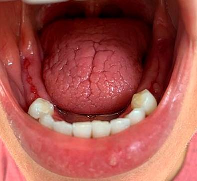
Figure 1 Dorsal tongue with depapilation and deep fissures without ulceration, findings compatible with glossitis resulting from oral xerostomia. Evidence of areas of post-extraction scarring in the lower alveolar ridge mucosa before implant treatment. Common nonspecific oral manifestations associated with inflammatory bowel disease (IBD). Authors’ archives.
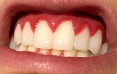
Figure 2 Erythematous gums with enlarged interproximal dental papillae of generalized manifestation compatible with the diagnosis of gingivitis. Common nonspecific oral manifestation associated with IBD. Authors’ archives.
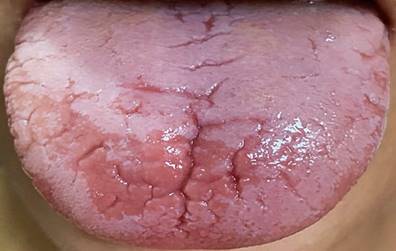
Figure 3 Smooth tongue and glossitis. Common nonspecific oral manifestation associated with IBD. Authors’ archives.
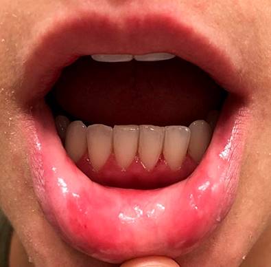

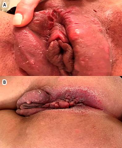
Figure 6 A. Vulvoperineal metastatic Crohn’s involvement. B. Vulvoperineal MCD involvement. Authors’ archives.
A vulvar biopsy showed marked edema of the dermis, dilated lymph nodes with perivascular and interstitial lymphoplasmacytic infiltrate, and noncaseating granulomas, negative for microorganisms. The results are compatible with MCD at this level, without gastrointestinal symptoms and calprotectin levels in the stool in normal ranges. Upper and lower endoscopic studies and small bowel (SB) capsule endoscopy were performed without alterations. Dermatology treated the patient with topical steroids and dentistry with dental implants. She was considered a patient with CD-type IBD with severe MEI. Although she did not have intestinal involvement, we decided to start treatment with anti-tumor necrosis factor (anti-TNF), initially with adalimumab, but she developed paradoxical psoriasis. Subsequently, management with infliximab was started again with the manifestation of severe paradoxical psoriasis, for which it was suspended (Figure 7). Cyclosporine was also used as an immunomodulator, and she presented with intolerable tachycardia.

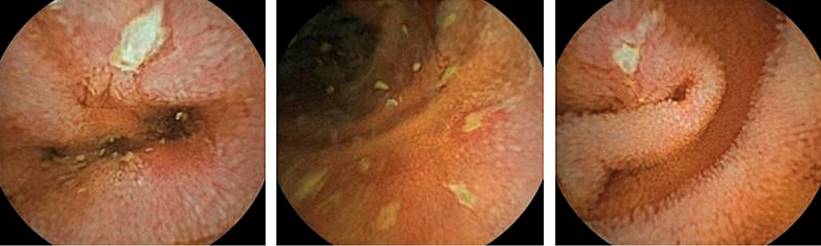
Eighteen months after these symptoms, she exhibited episcleritis of the left eye (Figure 8) and began with colicky abdominal pain and an average of five diarrheal stools daily. High and low endoscopic studies were performed without alterations and a new SI capsule endoscopy revealed CD enteritis involving the duodenum, jejunum, and ileum (Figure 9). Therefore, the patient is considered to have SI CD-type IBD with EIM, vulvoperineal NCD, severe oral involvement, and episcleritis. She is being treated with azathioprine and ustekinumab, with significant clinical improvement.

Discussion
The most frequent EIMs in CD are of the articular type and associated with colonic involvement. At the same time, severe vulvoperineal and oral MCD are rare, and their combination is scarce without previous intestinal involvement2. It has been described that in around 25% of cases of vulvar metastatic CD, its diagnosis may precede digestive CD5, which complicates the situation and may cause a delay in diagnosis with significant physical, mental, and social morbidity. Thus, a multidisciplinary approach is required. In the description in the literature, there is no clear correlation between the activity of metastatic CD, the intestinal clinical picture, and other MEI6. The clinical spectrum ranges from a mild and self-limited lesion to representing the main reason for consultation, so it conditions the treatment4. In the case presented, at the time of onset, the patient was without intestinal activity, and the most relevant clinical data was the cutaneous and mucosal involvement; likewise, articular and ocular manifestations were absent. A high level of suspicion of this GI entity must be maintained even if there are no intestinal manifestations. The refractoriness and severity of this condition require aggressive and comprehensive multidisciplinary management since EIM can become more severe and disabling than intestinal compromise.
MCD is the least frequent cutaneous manifestation of CD. It usually affects young adults. It can manifest as multiple or isolated lesions and affects the genital area and extremities, but it could also appear in any other body part7. The lesions can vary in morphology and occur as ulcers, plaques with or without ulceration, papules, or nodules. According to Barrett et al. (8), four main types of vulvar lesions can be observed: vulvar edema, ulceration, hypertrophic lesions in the form of pseudocondylomas acuminata, and chronic suppuration. The most common clinical manifestation is unilateral vulvar edema associated with chronic vulvar ulceration. It is typically asymptomatic, and the diagnosis is made when vulvar ulcers or hypertrophic lesions are discovered on physical examination. However, symptoms such as vulvar pain, pruritus, vulvar discharge, or dyspareunia have been described, as well as irritative urinary symptoms such as dysuria, among others8. In the case presented, the patient had ulcerated, inflammatory, erythematous, and infiltrated lesions found on physical examination. Given the suspicion of CD based on the clinical picture and histopathology, we proceeded to search for digestive manifestations of CD.
IBD-associated lesions in the oral cavity may precede intestinal manifestations in CD and become important when diagnosing it. Differential diagnoses are multiple and mainly include mycobacterial infection, hidradenitis suppurativa, pyoderma gangrenosum, erythema nodosum, sarcoidosis, and Behçet’s disease8. The differential diagnosis with metastatic CD is difficult in the latter due to the tremendous clinical similarity. Because histology sometimes does not allow them to be differentiated7, the diagnosis must be based on combining the clinical picture with the compatible biopsies. Granulomas in intestinal ulcers are usually employed to diagnose CD, and a positive skin pathergy test shows signs of Behçet’s disease9. The histopathological diagnosis will be based on the finding of chronic or subacute inflammatory infiltrate and epidermal ulceration with aseptic non-caseating granulomas10. This case was diagnostically complex since the skin involvement preceded the intestinal involvement by more than a year, combined with the absence of other suggestive systemic findings, making it necessary to rule out Behçet’s disease first. When HLA-B51 and pathergy test were found to be negative, histopathological studies were conducted on the vulvar lesions, with characteristic findings. Thus, the diagnosis was supported by the chronicity of the lesions, the histological findings, and the exclusion of other granulomatous and infectious diseases.
The therapeutic options are multiple; however, there is no specific treatment regimen since what is known about management is from case reports and a series of few patients whose response to different treatments has been variable and, in many cases, refractory. Most authors have reported a therapeutic response with metronidazole in monotherapy or combination with steroids. Other options include 5-aminosalicylates, antibiotics, and immunosuppressive agents such as azathioprine or cyclosporine8. The introduction of anti-TNF agents in CD has marked a milestone in management; nevertheless, their usefulness has been limited sometimes due to a lack of primary and secondary responses in a considerable proportion of patients11. Ustekinumab and vedolizumab are immunoglobulin G-1 (IgG-1) monoclonal antibodies, which have emerged as a therapeutic alternative for those cases that are refractory or intolerant to conventional treatments or anti-TNF agents and seem to be associated with a rapid and sustained clinical response, a safety profile, inflammatory cascade damping, and low immunogenicity11. In case of failure of pharmacological management, surgical excision can be considered in those patients with severe symptoms or deformities that interfere with the patient’s social and private life12. This clinical case documented skin reactions with the anti-TNF drugs adalimumab and infliximab and intolerable tachycardia due to cyclosporine. Therefore, using ustekinumab was required, with significant and sustained improvement.
Conclusions
MCD poses a diagnostic challenge, especially since it can present without GI manifestations for years, and its clinical and histopathological findings can be seen in other conditions. The possibility of CD must be considered, and where possible, an early benefit in the patient must be sought with a timely diagnosis.

