SciELO - Scientific Electronic Library Online

 
vol.38 issue3Periampullary Mass as an Unusual Manifestation of Coccidioidomycosis. Case Report and Literature ReviewGranulomatous hepatitis, deciphering an elusive spectrum: a clinical case-based approach author indexsubject indexarticles search
Home Pagealphabetic serial listing  

Services on Demand

Journal

Article

Indicators

Related links

  • On index processCited by Google
  • Have no similar articlesSimilars in SciELO
  • On index processSimilars in Google

Share


Revista colombiana de Gastroenterología

Print version ISSN 0120-9957On-line version ISSN 2500-7440

Rev. colomb. Gastroenterol. vol.38 no.3 Bogotá July/Sept. 2023  Epub Jan 19, 2024

https://doi.org/10.22516/25007440.969 

Case report

Crohn’s Disease in a Patient with Tuberculosis: A Diagnostic and Therapeutic Challenge

Santiago Revelo-Moreno1  * 
http://orcid.org/0000-0002-4523-0796

Luz Daniela Gómez-Suta2 
http://orcid.org/0000-0001-9809-3150

Jairo Alfredo Guevara-Guzmán3 
http://orcid.org/0000-0002-4876-0004

1Physician, Internal Medicine specialist, Universidad Tecnológica de Pereira, Colsubsidio SURA Medical Center (PAC). Bogotá, Colombia

2Physician, Internal Medicine resident, Universidad Tecnológica de Pereira. Pereira, Colombia.

3Physician, Internal Medicine, Gastroenterology, and Digestive Endoscopy specialist, Universidad Militar Nueva Granada. Professor of Gastroenterology, Universidad Tecnológica de Pereira, Gastro Elite S. A. S. Pereira, Colombia.


Abstract

Introduction:

Crohn’s disease (CD) is unusual in Colombia. Tuberculosis (TB) occurs more frequently, but intestinal involvement is rare. Differentiating these two entities and treating the cases in which they coexist is a challenge.

Clinical case:

A 28-year-old man with three months of constitutional, respiratory, and GI symptoms was initially diagnosed with pulmonary tuberculosis, and all the clinical manifestations were attributed to this entity. Given the absence of improvement with treatment and the sum of radiological, endoscopic, and pathological findings, CD was confirmed. Treatment was complex due to the coexistence of the two entities, although he finally went into remission with the use of biologicals.

Discussion:

Diagnosing CD requires the sum of clinical and paraclinical findings. A therapeutic test may be necessary to differentiate it from intestinal TB. The treatment of CD in a patient with TB has some limitations; steroids are not contraindicated, and biologicals must be initiated cautiously.

Conclusions:

Differentiating CD from intestinal TB is a diagnostic challenge. Therapeutic management when these two entities coexist requires an interdisciplinary approach.

Keywords: Crohn’s disease; tuberculosis; differential diagnosis

Resumen

Introducción:

la enfermedad de Crohn (EC) es inusual en Colombia. La tuberculosis (TB) se presenta con mayor frecuencia, pero el compromiso intestinal es raro. Diferenciar estas dos entidades y el tratamiento de los casos en los que coexisten es un reto.

Caso clínico:

hombre de 28 años con 3 meses de síntomas constitucionales, respiratorios y gastrointestinales al que en un inicio se le confirmó el diagnóstico de tuberculosis pulmonar y se le atribuyó a esta entidad todas las manifestaciones clínicas. Ante la ausencia de mejoría con el tratamiento y la suma de hallazgos radiológicos, endoscópicos y patológicos, se confirmó EC. El tratamiento fue difícil debido a la coexistencia de las dos entidades, aunque finalmente presentó remisión con el uso de biológicos.

Discusión:

diagnosticar la EC requiere de la sumatoria de hallazgos clínicos y paraclínicos. Para diferenciarla de TB intestinal puede llegar a ser necesaria una prueba terapéutica. El tratamiento de EC en un paciente con TB tiene algunas limitaciones, los esteroides no se contraindican de manera absoluta y el inicio de biológicos debe hacerse con precaución.

Conclusiones:

diferenciar la EC de la TB intestinal es un reto diagnóstico. El enfoque terapéutico cuando coexisten estas dos entidades requiere un abordaje interdisciplinario.

Palabras clave: Enfermedad de Crohn; tuberculosis; diagnóstico diferencial

Introduction

Crohn’s disease (CD) is part of the inflammatory bowel disease, and its manifestation is rare in Colombia: an incidence of 0.74 per 100,000 people per year (1. Meanwhile, tuberculosis (TB) is a more frequent entity, with an incidence of 22.6 to 28.9 cases per 100,000 inhabitants2, but intestinal involvement is uncommon; it only represents 3.5% of extrapulmonary tuberculosis cases3. Intestinal TB mainly affects the terminal ileum and cecum. It may result in the formation of ulcers and fistulas4, and in histopathological studies, granulomas can be found. It is one of the primary differential diagnoses in patients with suspected CD5.

Differentiating these pathologies represents a high level of complexity not only from a diagnostic point of view but also from a therapeutic one since some treatments, such as biologicals, may increase the risk of infections6. We present the case of a patient with pulmonary TB and CD.

Clinical case

A 28-year-old man presented with a 3-month clinical picture of abdominal pain and bloody, non-fatty diarrhea, which woke him up and persisted despite fasting. In the systemic inquiry, he reported occasional cough and involuntary weight loss of 30 kilograms in the last 3 years. Personal and family history was negative. A chest X-ray and CT scan were performed, suggesting TB; this entity was confirmed by bronchoalveolar lavage samples with Ziehl-Neelsen stain, PCR, and positive cultures, with no evidence of drug resistance. Colonoscopy showed colitis and ileitis with histopathology findings of chronic nonspecific ileitis and chronic nonspecific colitis with a focal active component. Due to the clinical context of pulmonary TB and with a high index of suspicion, intestinal TB was diagnosed, so the patient was administered rifampicin, isoniazid, pyrazinamide, and ethambutol.

After 6 months, his respiratory and constitutional symptoms improved, but he was admitted to the hospital due to persistent abdominal pain and diarrhea. Adherence to antituberculous therapy was confirmed, and microbiological studies for TB were negative. The laboratory tests detected fecal calprotectin of 2,060 μg/g (Table 1) and the magnetic resonance enterography (MRE), intestinal sub-occlusion in the ileum (Figure 1). A new ileocolonoscopy identified cecal folds, appendiceal orifice, and ileocecal valve with severe edema covered by ulcers, easy bleeding, and edema involving the proximal ascending colon. In addition, biopsies were taken that reported inflammatory bowel disease with chronic ulceration and granulomas (Figure 2). The diagnosis of CD was made with a Crohn’s disease activity index (CDAI) of 421 points. Controversially, corticosteroids were postponed, starting treatment with azathioprine.

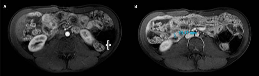
Figure 1 MRE with contrast. A. In the arterial phase: thickening of the ileus (+). B. In the portal phase: thickening of the ileum with a wall of 13 millimeters. Image owned by the authors. 

Table 1 Patient laboratory findings 

Study Result
Blood count Leukocytes: 9.98 x 10³ IU/L, neutrophils: 8 x 10³ IU/L, lymphocytes: 1.2 x 10³ IU/L, hemoglobin: 9.2 g/dL, MCV: 89 fL, RDW: 19.1 %, PLT: 503,000
Albumin 2.82 g/dL
Fecal calprotectin 2,060 μg/g
HIV 1 and 2 antibodies Negative

PLT: absolute platelet count; RDW: red cell distribution width; MCV: mean corpuscular volume; HIV: human immunodeficiency virus. Table owned by the authors.

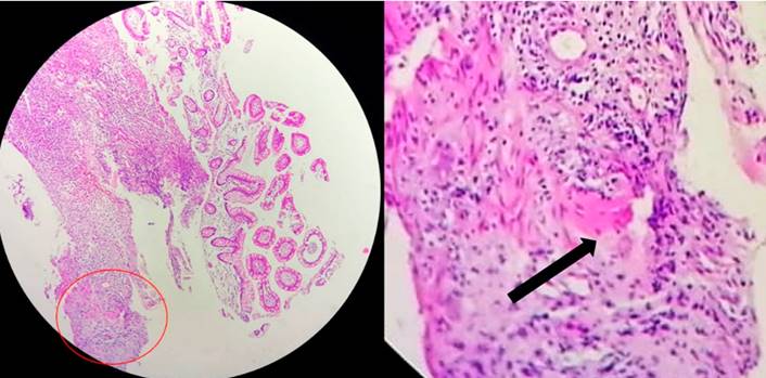
Figure 2 Biopsy: colon mucosa; in the submucosa, there is a non-necrotic, non-caseating granuloma with incipient multinucleated giant cells. Circle: granuloma. Arrow: giant cell. Image owned by the authors. 

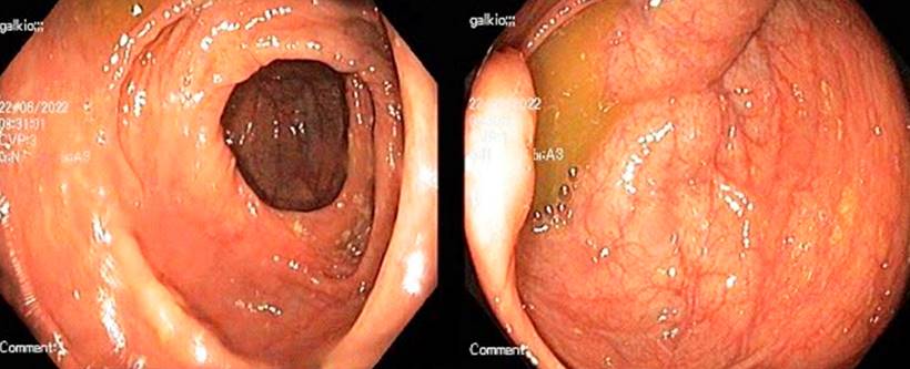
A month later, he consulted for abdominal pain, so a new CT scan was performed, which reported enteritis in the jejunum, pneumoperitoneum, and collections. He was taken to a laparotomy, noting obstruction of the proximal and distal small intestine, granulomas in the middle of the small intestine, perforation of the middle jejunum with a plastron that compromised the distal ileum, and a fistulous tract. An intestinal resection and anastomosis were performed. Once TB treatment was completed, biological therapy was started with adalimumab up to a maintenance dose of 40 mg subcutaneously every 2 weeks. The clinical evolution was favorable; 1 year later, he attended an outpatient gastroenterology check-up on clinical remission and without complications, and a new colonoscopy showed no alterations (Figure 3).

Figure 3 Colonoscopy images of the patient without evidence of disease activity. Image owned by the authors. 

Discussion

CD usually occurs between the second and fourth decades of life, as in the present case7, with a certain predominance in women8. The patient had no risk factors, such as family history or smoking9.

In the case presented, the constitutional and respiratory symptoms led to a rapid diagnosis of TB, which could explain the gastrointestinal findings as an extrapulmonary manifestation. However, given the persistence of symptoms despite having implemented anti-tuberculosis therapy, CD was considered a differential diagnosis.

Although controversial and reserved for very selected cases, when there is confusion between CD and TB in countries where TB is frequent, some authors recommend a therapeutic trial with anti-tuberculosis drugs to corroborate the clinical improvement at 3 months and healthy mucosa at 6 months in almost 100% of patients with TB10, which did not occur in this patient. In addition, the MRE, histopathology, and laboratory findings, such as elevated fecal calprotectin (with approximate sensitivity and specificity of 90% and 80%, respectively)11,12 allowed the diagnosis of CD.

The patient had a moderate CDAI; steroids would be indicated13, but they were not started since the patient was finishing the second phase of treatment for TB. It should be clarified that steroids in doses greater than 15 mg of prednisolone each day confer an odds ratio of 7.7 for infections. However, this is not an absolute contraindication to starting them in cases like this14. Azathioprine is an alternative, although it does not induce remission of an active disease. Another option to consider, since it is a man under 35 years of age, would have been methotrexate to avoid the increased risk of hepatosplenic lymphoma due to thiopurines, but it is not recommended for the induction of remission of the disease13.

The patient had a stenosing and penetrating complication of CD. This event and being under 30 are risk factors for recurrence, which occurs in up to 84% of post-surgical cases15. It was decided to start biological therapy; vedolizumab, an antibody against α4β7 integrin, has shown benefit in CD16 and even good tolerance without an increase in the risk of opportunistic infections compared to placebo17. Even so, no evidence supports it as a first-line medication since it does not seem superior for the induction or maintenance of remission compared to other biologicals18. Thus, and adding to the complications and high risk of the patient, anti-tumor necrosis factor (anti-TNF) was started, which was a difficult decision since infliximab, for example, has shown a relative risk of 18.6 for TB activation19. There is no clarity on when to start anti-TNF in TB. Still, case series show safety in those with an evident clinical response, drug susceptibility results, and who have completed the first two months of the intensive anti-tuberculosis phase19,20.

Patients like this require multidisciplinary management where not only the gastroenterologist but also other specialties such as pulmonology, infectious disease, internal medicine, and coloproctology supervise their treatment.

Conclusion

The diagnosis of CD in a patient with TB is difficult due to the similarity of intestinal involvement between these two entities. The difficulty is also therapeutic since beneficial treatments for CD, such as steroids, can be omitted, and the start of biological therapy can be delayed. Considering the contraindications of these drugs and applying algorithms that allow the two entities to be differentiated is of great clinical use to improve outcomes and avoid complications.

Referencias

1. Juliao F, Damas OM, Arrubla M, Calixto OJ, Camargo JL, Cruz L, et al. The Prevalence of Inflammatory Bowel Disease in Colombia is Increasing: Report on the National Prevalence of IBD and Description of IBD Phenotype. Gastroenterology. 2019;156(6):S-405-S-. https://doi.org/10.1016/S0016-5085(19)37865-5Links ]

2. Cruz Martínez OA. Informe de evento Tuberculosis año 2021. Programa nacional de prevención y control de la tuberculosis [Internet]. Minsalud; 2021 [actualizado noviembre 2021; citado 25 de junio de 2022]. Disponible en: Disponible en: https://www.minsalud.gov.co/sites/rid/Lists/BibliotecaDigital/RIDE/VS/PP/ET/comportamiento-tuberculosis-2020.pdfLinks ]

3. Raviglione MC, Gori A. Tuberculosis. En: Loscalzo J, Fauci A, Kasper D, Hauser S, Longo D, Jameson JL (editores). Harrison’s Principles of Internal Medicine 21.a edición. Nueva York: McGraw-Hill Education; 2022. p. 1357-1381. [ Links ]

4. Eraksoy H. Gastrointestinal and Abdominal Tuberculosis. Gastroenterol Clin North Am.2021;50(2):341-60. https://doi.org/10.1016/j.gtc.2021.02.004Links ]

5. Oñate-Ocaña LF, Pérez-Díaz L. Intestinal Tuberculosis. N Engl J Med. 2022;386(13):e30. https://doi.org/10.1056/NEJMicm2114345Links ]

6. Bonovas S, Fiorino G, Allocca M, Lytras T, Nikolopoulos GK, Peyrin-Biroulet L, et al. Biologic Therapies and Risk of Infection and Malignancy in Patients With Inflammatory Bowel Disease: A Systematic Review and Network Meta-analysis. Clin Gastroenterol Hepatol. 2016;14(10):1385-97.e10. https://doi.org/10.1016/j.cgh.2016.04.039Links ]

7. Johnston RD, Logan RF. What is the peak age for onset of IBD? Inflammatory bowel diseases. 2008;14 Suppl 2:S4-5. https://doi.org/10.1002/ibd.20545Links ]

8. Juliao-Baños F, Puentes F, López R, Saffon MA, Reyes G, Parra V, et al. Characterization of inflammatory bowel disease in Colombia: Results of a national register. Rev Gastroenterol Mex (English). 2021;86(2):153-62. https://doi.org/10.1016/j.rgmx.2020.05.005Links ]

9. Feldman M, Friedman LS, Brandt LJ. Sleisenger and Fordtran’s gastrointestinal and liver disease: pathophysiology, diagnosis, management. 11.a edición. Elsevier Health Sciences; 2020. [ Links ]

10. Kedia S, Das P, Madhusudhan KS, Dattagupta S, Sharma R, Sahni P, et al. Differentiating Crohn’s disease from intestinal tuberculosis. World J Gastroenterol. 2019;25(4):418-32. https://doi.org/10.3748/wjg.v25.i4.418Links ]

11. Waugh N, Cummins E, Royle P, Kandala N, Shyangdan D, Arasaradnam R, et al. Faecal calprotectin testing for differentiating amongst inflammatory and non-inflammatory bowel diseases: systematic review and economic evaluation. Health Technol Assess. 2013;17(55):xv-xix, 1-211. https://doi.org/10.3310/hta17550Links ]

12. Lewis JD. The utility of biomarkers in the diagnosis and therapy of inflammatory bowel disease. Gastroenterology. 2011;140(6):1817-26.e2. https://doi.org/10.1053/j.gastro.2010.11.058Links ]

13. Juliao-Baños F, Ovalle LFP, Regino WO, de Sánchez MTG, Duperly RG, Torres M. Guía de práctica clínica para el tratamiento de la enfermedad de Crohn en población adulta. Rev Colomb Gastroenterol. 2020;35(Supl 2):63-200. https://doi.org/10.22516/25007440.637Links ]

14. Youssef J, Novosad SA, Winthrop KL. Infection Risk and Safety of Corticosteroid Use. Rheum Dis Clin North Am. 2016;42(1):157-76, ix-x. https://doi.org/10.1016/j.rdc.2015.08.004Links ]

15. Lamb CA, Kennedy NA, Raine T, Hendy PA, Smith PJ, Limdi JK, et al. British Society of Gastroenterology consensus guidelines on the management of inflammatory bowel disease in adults. Gut. 2019;68(Suppl 3):s1-s106. https://doi.org/10.1136/gutjnl-2019-318484Links ]

16. Sandborn WJ, Feagan BG, Rutgeerts P, Hanauer S, Colombel JF, Sands BE, et al. Vedolizumab as induction and maintenance therapy for Crohn’s disease. New Eng J Med. 2013;369(8):711-21. https://doi.org/10.1056/NEJMoa1215739Links ]

17. Colombel JF, Sands BE, Rutgeerts P, Sandborn W, Danese S, D’Haens G, et al. The safety of vedolizumab for ulcerative colitis and Crohn’s disease. Gut . 2017;66(5):839-51. https://doi.org/10.1136/gutjnl-2015-311079Links ]

18. Singh S, Fumery M, Sandborn WJ, Murad MH. Systematic review and network meta-analysis: first- and second-line biologic therapies for moderate-severe Crohn’s disease. Aliment Pharmacol Ther. 2018;48(4):394-409. https://doi.org/10.1111/apt.14852Links ]

19. Dobler CC. Biologic Agents and Tuberculosis. Microbiol Spectr. 2016;4(6). https://doi.org/10.1128/microbiolspec.TNMI7-0026-2016Links ]

20. Matsumoto T, Tanaka T, Kawase I. Infliximab for rheumatoid arthritis in a patient with tuberculosis. New Eng J Med. 2006;355(7):740-1. https://doi.org/10.1056/NEJMc053468Links ]

Citation: Revelo-Moreno S, Gómez-Suta LD, Guevara-Guzmán JA. Crohn’s Disease in a Patient with Tuberculosis: A Diagnostic and Therapeutic Challenge. Revista. colomb. Gastroenterol. 2023;38(3):378-382. https://doi.org/10.22516/25007440.969

Received: September 15, 2022; Accepted: December 13, 2022

*Correspondence: Santiago Revelo-Moreno. sanrevmor@gmail.com

Creative Commons License This is an open-access article distributed under the terms of the Creative Commons Attribution License