SciELO - Scientific Electronic Library Online

 
vol.37 número3Nutcracker Syndrome Combined with Wilkie Syndrome: Case ReportRetrorectal Cystic Hamartoma (Tailgut cyst): Case Report and Literature Review índice de autoresíndice de assuntospesquisa de artigos
Home Pagelista alfabética de periódicos  

Serviços Personalizados

Journal

Artigo

Indicadores

Links relacionados

  • Em processo de indexaçãoCitado por Google
  • Não possue artigos similaresSimilares em SciELO
  • Em processo de indexaçãoSimilares em Google

Compartilhar


Revista colombiana de Gastroenterología

versão impressa ISSN 0120-9957versão On-line ISSN 2500-7440

Rev. colomb. Gastroenterol. vol.37 no.3 Bogotá jul./set. 2022  Epub 19-Dez-2022

https://doi.org/10.22516/25007440.798 

Case report

Esophageal and Pulmonary Involvement Caused by Paracoccidioidomycosis in Immunocompromised Patient: Case Report

Néstor Fabián Blanco-Barrera,1 
http://orcid.org/0000-0002-9540-4141

María Alejandra Villamizar-Jiménez,2  * 
http://orcid.org/0000-0002-3247-429X

Diana Valentina Tibaduiza-Upegui,2 
http://orcid.org/0000-0002-3650-0109

Fernando Stiven Ruiz-Julio.2 
http://orcid.org/0000-0002-1298-0355

1Internist, First Year Fellow in Gastroenterology, Universidad Militar Nueva Granada. Bogotá, Colombia.

2Undergraduate Medical Student, Universidad Autónoma de Bucaramanga (UNAB). Bucaramanga, Colombia


Abstract

Paracoccidioidomycosis (PCM) is a fungal infection endemic to South America. It predominantly affects men, depending on their work field: farmers and agriculturists. Paracoccidioidomycosis is caused by the aspiration of the fungus in its micellar form and manifests in three conditions: acute, subacute, and chronic; the latter is more frequent in adults, whose treatment will depend on azoles, amphotericin B, and sulfonamides. This case concerns a 57-year-old Colombian man, a farmer with no pathological history who showed dysphagia for solids that progressed to liquids, sialorrhea, and weight loss for two months. He underwent upper GI endoscopy, and whitish lesions were observed; thus, he was biopsied, displaying yeasts in multiple gemmations compatible with paracoccidioidomycosis. In turn, a chest CT scan showed generalized interstitial parenchymal involvement. Subsequently, he was treated with itraconazole, showing improvement and resolution in his clinical picture. Since the pathology described is endemic in South America and can be disseminated in immunocompromised patients. Given the broad infection spectrum, consideration should be given to patients with risk factors, symptomatology, and findings in extension studies suggesting this disease to provide timely and specific treatment.

Keywords: Paracoccidioidomycosis; blastomycosis; invasive fungal infections

Resumen

La paracoccidioidomicosis es una infección fúngica endémica de América del Sur, que afecta predominantemente a los hombres y, según su campo laboral, granjeros y agricultores. Es ocasionada por la aspiración del hongo en su forma micelar y debuta en tres formas de presentación: aguda, subaguda y crónica; esta última es más frecuente en adultos, cuyo tratamiento dependerá de los azoles, anfotericina B y sulfonamidas. El presente caso trata de un hombre de 57 años, colombiano, agricultor, sin antecedentes patológicos, quien presentaba dos meses de disfagia para sólidos que progresó a líquidos, sialorrea y pérdida de peso, a quien se le realizó endoscopia de vías digestivas altas y se observaron lesiones blanquecinas, por lo cual se realizó una biopsia que evidenció levaduras en múltiple gemación compatibles con paracoccidioidomicosis; a su vez, se observó en una tomografía de tórax compromiso parenquimatoso intersticial generalizado; posteriormente, recibió tratamiento con itraconazol, con el que mostró mejoría y resolución del cuadro clínico. En vista de que América del Sur es endémica de la patología descrita y puede presentarse de forma diseminada en inmunocompetentes, se debe tener en cuenta en aquellos pacientes que poseen factores de riesgo, sintomatología y hallazgos en estudios de extensión sugestivos de dicha enfermedad, dado el gran espectro de presentación de la infección, para así dar tratamiento oportuno y dirigido.

Palabras clave: Paracoccidioidomicosis; blastomicosis; infecciones fúngicas invasoras

Introduction

Paracoccidioidomycosis (PCM) is an infectious fungal disease endemic in South America with greater prevalence in Brazil, Venezuela, Colombia, and Argentina1-3. It is caused by two types of fungi that inhabit the soil in the mycelial form: Paracoccidioides brasiliensis and Paracoccidioides lutzii4,5. Upon inhalation, they enter the pulmonary alveoli, whose body temperature allows their conversion to yeast, which favors their asexual reproduction and generates acute infection of the disease, mainly in children, or chronic infection, primarily in adults6, which sometimes progresses to its secular form and causes pulmonary fibrosis7,8. Since there are few reports in the literature of esophageal involvement caused by PCM, our objective is to bring the case of a farmer who debuts with constitutional syndrome associated with dysphagia, with biopsy findings of lesions evidenced in the upper gastrointestinal endoscopy (UGIE) which were compatible with PCM and confirmed esophageal and pulmonary involvement by chest CT findings.

Case presentation

A 57-year-old male Colombian farmer patient with no pathological history. He presented a clinical picture of two-month evolution, initially characterized by retrosternal foreign-body sensation. Subsequently, progressive dysphagia from solids to liquids, regurgitating 20 minutes after food intake, without nausea and without gagging, accompanied by sialorrhea in the last week of the clinical picture.

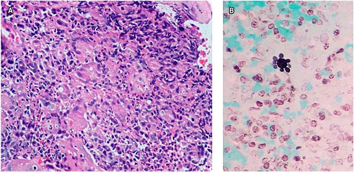
Upon admission, he reported occasional non-productive cough without dyspnea and a 10 kg weight loss in the last month. The patient showed a stable condition, systemic inflammatory response absence, vital signs unaltered, a 17 kg/m² body mass index (BMI), and signs of grade I dehydration. Constitutional syndrome secondary to neoplasia of gastrointestinal origin was initially suspected, so he was taken to UGIE (Figure 1), biopsies were taken, and extension studies such as chest tomography were requested (Figure 2A) showing lesions in the tree-in-bud and esophageal obstruction in the middle and distal third of the esophagus (Figure 2B). Tuberculosis infection was ruled out with 3 serial sputum smear tests and culture-negatives, a negative enzyme-linked immunosorbent assay (ELISA) test for human immunodeficiency virus (HIV), and blood count within normal limits. The histopathological study (Figure 3) of the esophageal biopsy showed a chronic non-necrotizing granulomatous inflammation disease secondary to fungal infection. The Grocott-Gomori’s Methenamine Silver (GMS) stain confirmed extracellular yeasts with narrow-based gemmation and “steering wheels” appearance compatible with PCM. He underwent esophageal balloon dilatation and stent placement and was started on treatment with itraconazole capsules of 100 mg every 24 hours a day for 6 months, improving dysphagia and respiratory symptoms. However, there is no post-treatment imaging follow-up record.
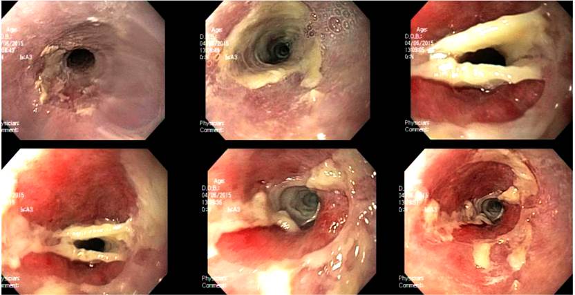
Figure 1 Endoscopic findings, esophageal stricture obstruction, and white endoluminal membranes without ulceration or bleeding. 
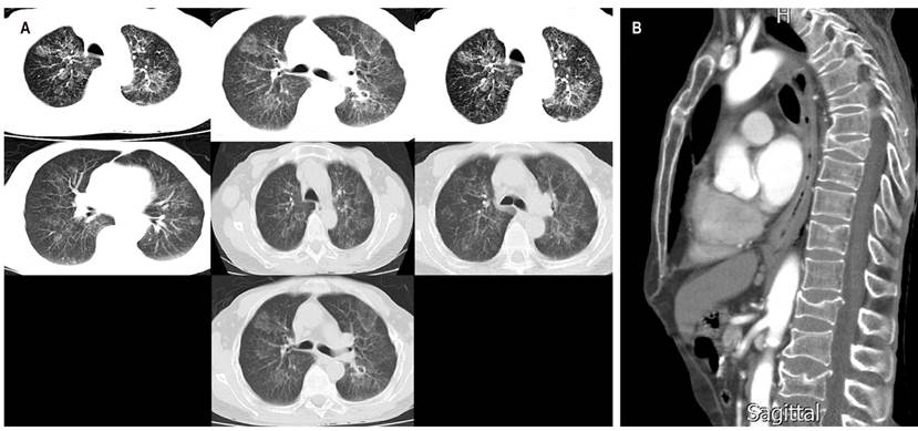
Figure 2 A. High-resolution chest tomography showing generalized interstitial parenchymal involvement with hyperdensities in the tree-in-bud. B. Sagittal chest tomography showing esophageal obstruction in the esophagus in the middle and distal third. 

Figure 3 A. Esophageal biopsy, hematoxylin/eosin 40x. B. Esophageal biopsy, GMS stain, 40x. Microphotographs provided by Dr. Isabel Bolívar, Pathologist, FOSCAL. 

Discussion

Paracoccidioidomycosis is a systemic disease endemic in South America, especially in Santander, Colombia. This disease predominantly affects men between 30-50 years old, but it most often affects farmers, whose primary infection is pulmonary, where the fungus is previously inhaled in mycelial form. The systemic involvement will depend on the host’s immune response. This disease has three clinical presentations: acute, subacute (symptoms occur approximately 45 days after exposure), and chronic (more frequent in adults and characterized by a reactivation of the primary infection that could have been acquired months or even years earlier, and its symptoms depend on the affected organ). Disseminated manifestation, especially esophageal, is rare in immunocompetent individuals. In these patients, the mucous membranes with greater involvement are found in the oral and the laryngeal cavity. However, very few reports exist in the literature9-12. Diagnosis methods range from histological studies and fungal culture to antibody detection6,13-16).

Several risk factors for contracting the infectious agent were considered in this case, including the geographic area where the patient resides or works and gender. Its age group predisposed him to debut with reactivated chronic disease. In addition to the clinical evolution chronicity, he presented constitutional syndrome evidenced by involuntary weight loss; pulmonary involvement, manifested by cough without expectoration and occasional dyspnea, possibly explained by pulmonary parenchymal involvement (Figure 2), and esophageal involvement, characterized by progressive dysphagia, which in turn was confirmed through imaging findings showing endoluminal involvement and endoluminal stenosis (Figures 1 and 2B). The treatment for this disease consists of azoles, especially itraconazole, which has better tolerance, absorption, and fewer adverse effects. This is the treatment that was administered to the patient, showing a subsequent clinical picture improvement. Sulfonamides and amphotericin B, the latter, may be prescribed for severe cases17. Studies have shown that the presence of comorbidities and pulmonary fibrosis is associated with high morbimortality due to exacerbation of the underlying disease and intrinsic complications of the infection, which is why the patient’s prognosis is good since there is no pathological history and there is no evidence of pulmonary fibrosis in the requested lung tests18,19.

Conclusion

In conclusion, it is necessary to consider the geographic area, gender, occupation, and the time of evolution of the signs and symptoms of the patient’s clinical picture must be considered for a PCM diagnostic approach. All of the above must be supported by imaging tests (in our case, chest tomography and UGIE with a histological biopsy study) and antifungal pharmacological management, which consists of azoles. Itraconazole is an excellent option due to its tolerance, absorption, and reduction of adverse drug effects. Sulfonamides and amphotericin B can improve the patient’s clinical evolution. This entity must be considered within the differential diagnoses in people presenting the same symptomatology, whose final prognosis will depend on the degree of pulmonary fibrosis and associated pathological history.

Acknowledgments

To the people who made possible the dissemination of this case report.

Referencias

1. Restrepo A, Gómez BL, Tobón A. Paracoccidioidomycosis: Latin America’s Own Fungal Disorder. Curr Fungal Infect Rep. 2012;6(4):303-11. https://doi.org/10.1007/s12281-012-0114-xLinks ]

2. Sifuentes-Osornio J, Corzo-León DE, Ponce-de-León LA. Epidemiology of Invasive Fungal Infections in Latin America. Curr Fungal Infect Rep. 2012;6(1):23-34. https://doi.org/10.1007/s12281-011-0081-7Links ]

3. Mendes RP, Cavalcante RS, Marques SA, Marques MEA, Venturini J, Sylvestre TF, et al. Paracoccidioidomycosis: Current Perspectives from Brazil. Open Microbiol J. 2017;11:224-282. https://doi.org/10.2174/1874285801711010224Links ]

4. Teixeira MM, Theodoro RC, Nino-Vega G, Bagagli E, Felipe MSS. Paracoccidioides species complex: ecology, phylogeny, sexual reproduction, and virulence. PLoS Pathog. 2014;10(10):e1004397. https://doi.org/10.1371/journal.ppat.1004397Links ]

5. Teixeira M de M, Theodoro RC, Oliveira FFM de, Machado GC, Hahn RC, Bagagli E, et al. Paracoccidioides lutzii sp. nov.: biological and clinical implications. Med Mycol. Oxford University Press; 2014;52(1):19-28. [ Links ]

6. Da Costa MM, Marques da Silva SH. Epidemiology, Clinical, and Therapeutic Aspects of Paracoccidioidomycosis. Curr Trop Med Reports. 2014;1:138-44. https://doi.org/10.1007/s40475-014-0013-zLinks ]

7. Costa AN, Benard G, Albuquerque ALP, Fujita CL, Magri ASK, Salge JM, et al. The lung in paracoccidioidomycosis: new insights into old problems. Clinics (Sao Paulo). 2013;68(4):441-8. https://doi.org/10.6061/clinics/2013(04)02Links ]

8. Bocca AL, Amaral AC, Teixeira MM, Sato PK, Sato P, Shikanai-Yasuda MA, et al. Paracoccidioidomycosis: eco-epidemiology, taxonomy and clinical and therapeutic issues. Future Microbiol. 2013;8(9):1177-91. https://doi.org/10.2217/fmb.13.68Links ]

9. Bicalho RN, Santo MF, de Aguiar MC, Santos VR. Oral paracoccidioidomycosis: a retrospective study of 62 Brazilian patients. Oral Dis. 2001;7(1):56-60. https://doi.org/10.1034/j.1601-0825.2001.70111.xLinks ]

10. Silva CO, Almeida AS, Pereira AA, Sallum AW, Hanemann JA, Tatakis DN. Gingival involvement in oral paracoccidioidomycosis. J Periodontol. 2007;78(7):1229-34. https://doi.org/10.1902/jop.2007.060490Links ]

11. Godoy H, Reichart PA. Oral manifestations of paracoccidioidomycosis. Report of 21 cases from Argentina. Mycoses. 2003;46(9-10):412-7. https://doi.org/10.1046/j.0933-7407.2003.00917.xLinks ]

12. Sant’Anna GD, Mauri M, Arrarte JL, Camargo H Jr. Laryngeal manifestations of paracoccidioidomycosis (South American blastomycosis). Arch Otolaryngol Head Neck Surg. 1999;125(12):1375-8. https://doi.org/10.1001/archotol.125.12.1375Links ]

13. Borges SR, Silva GM, Chambela Mda C, Oliveira Rde V, Costa RL, Wanke B, et al. Itraconazole vs. trimethoprim-sulfamethoxazole: A comparative cohort study of 200 patients with paracoccidioidomycosis. Med Mycol. 2014;52(3):303-10. https://doi.org/10.1093/mmy/myt012Links ]

14. Blotta MH, Mamoni RL, Oliveira SJ, Nouér SA, Papaiordanou PM, Goveia A, et al. Endemic regions of paracoccidioidomycosis in Brazil: a clinical and epidemiologic study of 584 cases in the southeast region. Am J Trop Med Hyg. 1999;61(3):390-4. https://doi.org/10.4269/ajtmh.1999.61.390Links ]

15. Brunaldi MO, Rezende RE, Zucoloto S, Garcia SB, Módena JL, Machado AA. Co-infection with paracoccidioidomycosis and human immunodeficiency virus: report of a case with esophageal involvement. Am J Trop Med Hyg. 2010;82(6):1099-101. https://doi.org/10.4269/ajtmh.2010.09-0751Links ]

16. Moreto TC, Marques ME, de Oliveira ML, Moris DV, de Carvalho LR, Mendes RP. Accuracy of routine diagnostic tests used in paracoccidioidomycosis patients at a university hospital. Trans R Soc Trop Med Hyg. 2011;105(8):473-8. https://doi.org/10.1016/j.trstmh.2011.03.001Links ]

17. Negroni R. Paracoccidiodes brasiliensis (Paracoccidiomycosis) [Internet]. Antimicrobe.org. 2021 [consultado el 12 de octubre de 2021]. Disponible en: Disponible en: http://www.antimicrobe.org/f09.aspLinks ]

18. Shikanai-Yasuda MA, Conceição YM, Kono A, Rivitti E, Campos AF, Campos SV. Neoplasia and paracoccidioidomycosis. Mycopathologia. 2008;165(4-5):303-12. https://doi.org/10.1007/s11046-007-9047-2Links ]

19. Tobón AM, Agudelo CA, Osorio ML, Alvarez DL, Arango M, Cano LE, et al. Residual pulmonary abnormalities in adult patients with chronic paracoccidioidomycosis: prolonged follow-up after itraconazole therapy. Clin Infect Dis. 2003;37(7):898-904. https://doi.org/10.1086/377538Links ]

Citation: Blanco-Barrera NF, Villamizar-Jiménez MA, Tibaduiza-Upegui DV, Ruiz-Julio FS. Esophageal and Pulmonary Involvement Caused by Paracoccidioidomycosis in Immunocompromised Patient: Case Report. Rev Colomb Gastroenterol. 2022;37(3):311-315. https://doi.org/10.22516/25007440.798

Received: June 27, 2021; Accepted: January 14, 2022

*Correspondence: María Alejandra Villamizar-Jiménez. mvillamizar313@unab.edu.co

Conflicts of Interest

The authors have no conflicts of interest to declare

Creative Commons License This is an open-access article distributed under the terms of the Creative Commons Attribution License