


Serviços Personalizados
Journal
Artigo
 texto em
  Inglês (pdf)
 Artigo em XML
 Referências do artigo
 Enviar este artigo por email
Indicadores
-
 Citado por SciELO
 -
 Acessos
Links relacionados
-
 Citado por Google
 -
 Similares em
SciELO
Similares em
SciELO -
 Similares em Google
Compartilhar

 Permalink
Revista MVZ Córdoba
versão impressa ISSN 0122-0268versão On-line ISSN 1909-0544
Rev.MVZ Cordoba vol.24 no.3 Córdoba set./dez. 2019 Epub 08-Jun-2020
https://doi.org/10.21897/rmvz.1431
Case report
First report of avascular necrosis of the femoral head in a goat (Capra aegagrus hircus) in Peru
1 Universidad Científica del Sur, Lima, Perú. Carrera de Medicina Veterinaria y Zootecnia. Panamericana Sur Km 19, Lima 42. Perú.
It is reported the case of a 7-month-old male goat of alpine race, who presented claudication of the right hind limb without definite cause. Clinical and radiographic examinations were performed, suggesting traumatic dislocation or avascular necrosis of the femoral head with consequent degenerative joint disease. It was performed a surgical treatment by an arthroplasty by excision of the head and neck of femur. The histopathological study by staining hematoxylin and eosin described: necrotic bone, lacunae containing necrotic osteocytes, as well as necrotic bone marrow. This information proved the diagnosis of avascular necrosis of the femoral head and neck. In the post-surgical period, the clinical recovery of the patient was considered good, the pain was mitigated and the function of the affected limb was improved. The present case is the first report of this disease in goats in Peru.
Keywords: Aseptic; goat; exeresis; osteoarthritis; osteonecrosis (Source: DeCS)
Se reporta el caso de una cabra macho de raza alpina de 7 meses de edad, que presentaba claudicación del miembro posterior derecho sin causa definida. Se realizaron los exámenes clínicos y radiográficos, sugiriendo luxación traumática o necrosis avascular de cabeza femoral con consecuente enfermedad degenerativa articular. Se realizó tratamiento quirúrgico mediante artroplastia por excisión de cabeza y cuello femoral. El estudio histopatológico mediante la coloración de hematoxilina y eosina describió: hueso necrótico, lagunas conteniendo osteocitos necróticos, además de médula ósea necrótica. Esta información comprobó el diagnóstico de necrosis avascular de la cabeza y cuello femoral. En el post quirúrgico, la recuperación clínica del paciente se consideró buena, mitigando el dolor y mejorando la función del miembro afectado. El presente caso se trata del primer reporte de esta enfermedad en caprinos en el Perú.
Palabras clave: Aséptica; caprino; exéresis; osteoartritis; osteonecrosis (Fuente: DeCS)
INTRODUCTION
The avascular necrosis of the femoral head and neck (AVNFH) is also known as Legg-Calve-Perthes disease or aseptic necrosis of the femoral head. This is a disease related to development and it is described in human being, companion animals and various species used as animal models. 1,2.
In dogs, the AVNFH usually occurs unilaterally with a frequency of up to 80% of cases 1. The causes are not yet well known. However, the pathophysiology is described as a deficit in the blood supply that causes ischemia and necrosis of the femoral head. The deficit of blood supply in this area may be due to several factors: in the case of traumatic lesions in the femur neck, the blood supply and drainage of the head will be compromised by the rupture of vessels at the site of the fracture 3. Experimentally, AVNFH has been reproduced using corticoids 4 and injury products by physical agents such as: temperature, pH, alcoholic solutions and the ligature of the lateral and medial circumflex arteries 5. In the development of the AVNFH, the repetitive forces that are applied on the necrotic bone, at the level of the epiphysis of the femoral head, will cause cracks and fissures in the articular cartilage. This causes the collapse of the subchondral trabeculae, followed by the progressive flattening of the femoral head. Subsequently, the fibrovascular tissue repairs it by beginning at the periphery of the femoral head. Finally, the joint incongruence leads to osteoarthritis of the coxal articulation 6.
The clinical signs commonly shown are unilateral or bilateral claudication of the hind limbs, which progress with the inability to support the weight on the affected limb or both extremities. The palpation of the hip may generate pain and difficulty to perform the movements of abduction, flexion and extension 3. In addition, the affected limb may be shortened, the greater trochanter may become prominent and there may be atrophy of the gluteal and quadriceps muscles. 6
The diagnosis may be complemented by taking radiographs of the hip in ventro-dorsal and medial lateral incidences. The first radiographic indicator is the hip widening. The affected femoral head has a defined vesicle appearance as the interior of the bone begins to be necrotic. The epiphysis looks flattened by the subchondral erosion and if there is sufficient trabecular destruction, the cortical begins to break and collapse 7. At this time, the femoral head may appear fragmented. Finally, the femoral head does not reach its original shape in the healing process, causing the acetabulum to try to accommodate and become arthritic 1.
The treatment is usually aimed at controlling pain and preventing loss of function of the affected limb. In a conservative manner, complete rest and treatment with non-steroidal anti-inflammatories is moderated proposed. The complete replacement of the femoral head and neck is also reported 8. However, the arthroplasty by excision of the head and neck of the femur is the technique most commonly used as a rescue of the affected limb and intended to relieve pain, although pseudoarthrosis and other complications have been reported, which may lead to unpredictable results 9.
PATIENT EVALUATION
Anamnesis. A male goat patient of 7 months of age and 18 kg of weight, with a history of apparent injury due to hip trauma, was presented at the facilities of the Universidad Científica del Sur. The animal had claudication with support of the right posterior limb. The animal is native to Metropolitan Lima and lives in a pasture with other goats of the same age and size.
Findings of the clinical examination. In the clinical examination, the patient was evaluated at station, while walking and at a jog. The patient had a grade 2 claudication of the right hind limb (on a scale of 0 to 5: moderate lameness and clearly visible at the trot). A muscle hypotrophy of the hip and the right hind limb, as well as a notorious prominence of the lateral region on the greater trochanter of the femur were observed at the inspection of the pelvis (Figure 1).
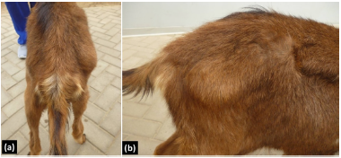
Figure 1 Inspection in posterior (a) and right lateral (b) in a male alpine goat of 7 months of age with claudication of the right hind limb: A prominence is identified in the lateral region on the greater trochanter of the femur
The physical evaluation was carried out with sedation, it was found a difficulty to perform the extension and flexion of the right hip joint and crackling sounds in the coxal articulation were detected. The Ortolani Test was performed with positive results. This information allowed proposing a presumptive diagnosis of traumatic dislocation or avascular necrosis of the femoral head and neck with consequent joint degenerative disease.
Diagnostic help. In order to complement the clinical diagnosis, pelvic radiographs were performed in two incidences (middle lateral and ventro dorsal). The right coxal articulation showed: increased joint space, irregular shape of the femoral head and neck and periarticular osteophytosis (Figure 2).
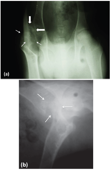
Figure 2 Hip x-ray in ventrodorsal incidence (a) and lateral mid incidence (b) of a 7-month-old male alpine goat. Right coxal articulation: irregularity and porous aspect of the femoral head with areas of decreased density in the epiphysis, compatible with focal bony lysis (thin arrows). The cranial - lateral area of the acetabulum has osteophytes (thick arrows). Flattening and irregularity of the articular surface of the head of the femur that causes subluxation of the joint.
Treatment approach. Based on the findings of the clinical examination and the imaging study, it was decided to perform an arthroplasty by excision of the right femoral head and neck. For pre-anesthetic medication, medetomidine (1 mg/kg), ketamine (2 mg/kg) and butorphanol (0.1 mg/kg) were all used intramuscularly. The cephalic vein was cannulated and induction was performed with Propofol (2 mg/kg) and Fentanyl (0.001 mg/kg). Once the unconsciousness was obtained, the patient was intubated and the anesthetic situation was maintained with isoflurane in 100% oxygen using a non-rebreathing circuit. Assisted ventilation was performed throughout the surgery.
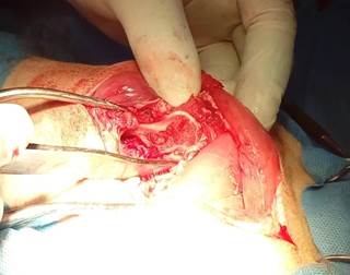
The patient was positioned in the left lateral decubitus position; a vertical incision was made lateral to the right greater trochanter. After dissection of the subcutaneous tissue, retraction of the biceps femoral muscle was caudally performed and the tensor muscle of the fasciae latae cranially. An incision was made in the periosteal insertion of the vastus lateralis muscle and it was ventrally reflected. The joint capsule was identified and incised, which was very thickened, to then perform the section of the round ligament. In the inspection of the femoral head, it presented an irregular and rough surface to the touch; the articular cartilage was thickened and showed ulcerations and numerous subchondral cracks
In order to perform the ostectomy, an assistant turned the limb outwards until the patella is directed upwards; this maneuver produced the dislocation of the joint. The exeresis was performed with an osteotome and an impactor. The line of ostectomy belonged to a straight line between the junction of the femoral neck and the third trochanter (Figure 3).
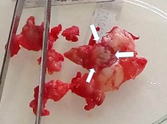
Figure 3 Surgical piece after the femoral head and neck exeresis. The tissue is multifragmented with irregular fibrous tissue, a region of collapsed and necrotic articular cartilage (white arrows), as well as subchondral cracks (black arrows) is evident in the femoral head.
To perform the ostectomy, an assistant turned the limb out until the patella was directed upwards; this maneuver caused dislocation of the joint. The exeresis was performed with an osteotome and an impactor. The ostectomy line corresponded to a straight line between the junction of the femoral neck and the third trochanter (Figure 4).
Soft tissue closure was performed with 1-0 absorbable suture material and skin suture with non-absorbable 1-0 material. The pain handling was performed with ketoprofen at a dose of 1 mg/kg intramuscularly every 24 hours and antibiotic therapy with cephalotin at a dose of 30 mg/kg intramuscularly every 12 hours. The patient went on to recovery in a cage, starting his rehabilitation at 24 hours with controlled walks and promoting the early use of the limb. The recovery was considered good. The full support of the operated limb and no signs of pain at 30 days of post-surgery were achieved.
Histopathological diagnosis. The femoral head and neck surgical piece was fixed in 10% formaldehyde and sent for histological-pathological study with haematoxylin and eosin staining in the Histopathology Laboratory of the Universidad Científica del Sur. Histopathologically, a wide variety of trabecular bone necrosis was observed with limited inflammatory changes. The diagnosis corresponded to avascular necrosis of the femoral head and neck (Figures 5 and 6).
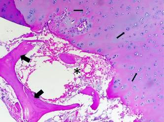
Figure 5 Section of necrotic bone, the articular cartilage is thickened and abnormal chondrocyte dispositions are observed (thin arrows). Area of subchondral bone necrosis, there are empty trabecular bone gaps containing necrotic osteocytes (thick arrows). The hematopoietic tissues show depression and you can see atrophic adipose tissue (asterisk) (Hematoxylin and eosin, 20x).
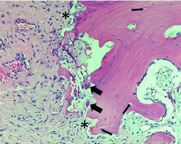
Figure 6 The bone marrow has been replaced by fibrotic marrow in an eosinophilic matrix, this tissue as a whole is partially connected to the necrotic bone (asterisks). Some multinucleated giant cells can be seen in the periphery of the matrix (large arrows). There are some empty gaps in the trabecular bone containing necrotic osteocytes (thin arrows). (Hematoxylin and eosin, 40x).
DISCUSSION
Goats and other minor ruminants stand out for their anatomy and vasculature similar to humans; therefore, they have been used as experimental models for the knowledge and management of AVNFH 10,2,11. The femur of these animals was also used as a reference to know aspects of the evolution and regeneration of AVNFH 12. However, no reports were found in the literature consulted of experimental and natural cases of NACF in goats in Peru; so we consider the present case as a first report.
The AVNFH is not well described in small ruminants. Thus, it is believed that it could have a hereditary component and affect more young animals, as it is recognized in other species 1. A probable explanation of the few reports of this disease may be a consequence of the characteristics of goat breeding (in herds or extensive breeding). It is common for animals affected with chronic lameness and that are unproductive being discarded or may finally have cachexia and dead without a definite cause. In that sense, it is also possible that the AVNFH is underdiagnosed in goats and is confused with diseases such as caprine arthritis encephalitis and other conditions that lead to degenerative joint disease 13.
In this case, the simple radiography was the tool used to approximate the diagnosis of AVNFH, which showed irregular bone density and degenerative changes typical of osteonecrosis of the head and neck of the femur. Radiologically, signs of osteonecrosis were observed, with a decrease in the radio-density of the femoral head and abnormal contours of the same 1. These findings agree with the findings available in experimental studies in goats and sheep 12.
The histological findings also belonged to the expected findings in a necrotic bone affected by AVNFH 11,14. These changes were reported as empty osteocyte gaps or with necrotic osteocytes. This necrotic bone was surrounded by fibrotic bone marrow, which was disorganized and with abundant collagen fibers. An eosinophilic matrix and some giant multinucleated cells adjacent to the bone trabeculae were also identified in the marrow.
In this report, the surgery was performed in order to control the pain in the affected coxal articulation, by limiting the unstable bony contact between the femoral head and the acetabulum. This intervention generated the formation of a fibrous false joint, thus improving the comfort and function of the affected limb 15,16. It is important to highlight postoperative care in this case, in which the use of the limb was promoted as soon as possible after surgery. With this aim, daily walks were made using a harness conditioned to the patient, as well as the application of analgesics to ensure comfort and adequate mobility. The purpose of this measure was based on preventing the fibrous tissue that will form during the postoperative period will be restrictive and limiting the range of motion of the operated coxal articulation 9.
On the other hand, it could be considered that age and weight were additional factors that contributed to the recovery of the patient, since in dogs and cats, for example, it has been indicated that at greater age and greater weight, the capacity of compensate for mechanical disadvantages in the coxofemoral joint after surgery 8.
No controlled studies were found that would have evaluated the surgical results of complete excision of the femoral head and neck in goats or other minor ruminants. Therefore, the present case is considered as a first report in Peru of NACF in goats.
Acknowledgements
To the Doctors of Veterinary Medicine and Animal Husbandry (MVZ) Astrid Morillo, Brenda Benavides and Claudia Maguiña of the surgery and orthopedics unit at UCSUR who contributed in the development of this report.
REFERENCES
1. Jankovits DA, Liska WD, Kalis RH. Treatment of avascular necrosis of the femoral head in small dogs with micro total hip replacement. Vet Surg. 2012; 41(1):143-147. https://doi.org/10.1111/j.1532-950x.2011.00925.x [ Links ]
2. Boss JH, Misselevich I. Osteonecrosis of the Femoral Head of Laboratory Animals: The Lessons Learned from a Comparative Study of Osteonecrosis in Man and Experimental Animals. Vet Pathol. 2003; 40:345-354. https://doi.org/10.1354/vp.40-4-345 [ Links ]
3. Cardoso CB, Rahal SC, Mamprim MJ, Salvador H, Melchert A, Figueroa J, et al. Avascular Necrosis of the Femoral Head in Dogs - Retrospective Study. Acta Scientiae Veterinariae. 2018; 46:1537. https://doi.org/10.22456/1679-9216.81845 [ Links ]
4. Xie XH, Wang XL, Yang HL, Zhao DW, Qin L. Steroid-associated osteonecrosis: Epidemiology, pathophysiology, animal model, prevention, and potential treatments (an overview). J Orthop Translat. 2015; 3(2):58-70. https://doi.org/10.1016/j.jot.2014.12.002 [ Links ]
5. Yang S, Wu X, Mei R, Yang C, Li J, Xu W, Ye S. Biomaterial-loaded allograft threaded cage for the treatment of femoral head osteonecrosis in a goat model. Biotechnol Bioeng. 2008; 100(3):560-56. https://doi.org/10.1002/bit.21792 [ Links ]
6. DeCamp CE, Johnston SA, Déjardin LM, Schaefer SL. The hip joint. In: Piermattei D.L. (Ed). Brinker, Piermattei, and Flo’s handbook of small animal orthopedics and fracture repair. 5th edn. St. Louis: Elsevier; 2016. https://doi.org/10.1016/b978-1-4377-2364-9.00025-2 [ Links ]
7. Scherzer C, Windhagen H, Nellesen J, Crostack HA, Rohn K, Witte F, Thorey F, et al. Comparative structural analysis of the canine femoral head in Legg-Calvé-Perthes disease. Vet Radiol Ultrasound. 2009; 50(4):404-411. https://doi.org/10.1111/j.1740-8261.2009.01557.x [ Links ]
8. Fitzpatrick, N, Pratola, L, Yeadon, R. Total hip replacement after failed femoral head and neck excision in two dogs and two cats. Vet Surg . 2011; 41:136-142. https://doi.org/10.1111/j.1532-950x.2011.00940.x [ Links ]
9. Harper T. Femoral Head and Neck Excision. Veterinary Clinics of North America: Small Animal Practice. 2017; 47:885-897. https://doi.org/10.1016/j.cvsm.2017.03.002 [ Links ]
10. Zhu ZH, Gao YS, Luo SH, Zeng BF, Zhang CQ. An animal model of femoral head osteonecrosis induced by a single injection of absolute alcohol: An experimental study. Medical Science Monitor: International Medical Journal of Experimental and Clinical Research. 2011; 17(4):97-102. https://doi.org/10.12659/msm.881708 [ Links ]
11. Crawford C, Allen B, Powers D. Growth Profiles and Articular Cartilage Characterization in a Goat Model of Legg-Calvé-Perthes Disease. Journal of lnvestigative Surgery. 1995; 8:391-408. https://doi.org/10.3109/08941939509031606 [ Links ]
12. Tang TT, Lu B, Yue B, Xie XH, Xie YZ, Dai KR, et al. Treatment of osteonecrosis of the femoral head with hBMP-2-gene-modified tissue engineered bone in goats. The Journal of Bone and Joint Surgery. 2007; 89(1):127-132. https://doi.org/10.1302/0301-620x.89b1.18350 [ Links ]
13. Harwood D, Mueller K. Goat Medicine and Surgery. CRC Press. 2018. https://www.crcpress.com/Goat-Medicine-and-Surgery/Harwood-Mueller/p/book/9781498748636#googlePreviewContainer [ Links ]
14. Kobayashi R, Kurotaki T, Yamada N, Kumabe S, Doi T, Wako Y, Tsuchitani M. Spontaneous and bilateral necrosis of the femoral head in a young experimental beagle dog. Toxicol Pathol. 2015; 28:121-124. https://doi.org/10.1293/tox.2014-0060 [ Links ]
15. Winders CLB, Vaughn WL, Birdwhistell KE, Holsworth IG, Franklin SP. Accuracy of femoral head and neck excision via a craniolateral approach or a ventral approach. Veterinary and Comparative Orthopaedics and Traumatology. 2018; 31(2):102-107. https://doi.org/10.3415/vcot-17-07-0099 [ Links ]
16. Off W, Matis U. Excision arthroplasty of the hip joint in dogs and cats. Clinical, radiographic, and gait analysis findings from the Department of Surgery, Veterinary Faculty of the Ludwig-Maximilians-University of Munich, Germany. Vet Comp Orthop Traumatol. 2010; 23(5):297-305. https://www.orthovet.org/files/vcot_2010-23-5_13649_0.pdf [ Links ]
How to cite (Vancouver) Salinas CE, Celi MI. First report of avascular necrosis of the femoral head in a goat (Capra aegagrus hircus) in Peru. Rev MVZ Cordoba. 2019; 24(3):7372-7377. DOI: https://doi.org/10.21897/rmvz.1352
Received: August 2018; Accepted: May 2019; Published: September 2019
 Este es un artículo publicado en acceso abierto bajo una licencia Creative Commons
