




 English (pdf)
 Article in xml format
 Article references

 Automatic translation
 Send this article by e-mail
 Cited by SciELO
 Cited by Google
 Similars in
SciELO
 Similars in Google

 Permalink

INTRODUCTION
The history of herbal medicine began centuries ago through empirical knowledge in which, through observation of the behavior of animals in relation to the ingestion of this or that vegetable, it was possible to understand the useful or harmful properties of medicinal plants; they are part of human evolution and were the first therapeutic resources used by generations [1- 3]. Ancient civilizations have their own historical references about medicinal plants and, long before any form of writing appeared, man already used plants and, among these, some as food and others as medicine [4, 5].
The Hamamelis virginiana L. (Hamamelidaceae) is a small tree or deciduous and branched shrub native to the United States and Canada bearing the name, Virginiana which refers to the place in the United States where it was found, it was also introduced in England and later throughout Europe [6, 7]. Due to the presence of tannins, proteins precipitate from the surface cells of the mucosa and tissues, forming a protective coating, reducing the formation of secretions and protecting against infections; in addition, it has anti-inflammatory and antioxidant action and is associated with the presence of flavonoids [8, 9].
The Maytenus ilicifolia Mart. ex Reissek (Celastraceae) has been known for many years by the Indians, it has this name for the appearance of its leaves, which have thorns on the edges, being originally from Brazil, found in the regions of Minas Gerais to Rio Grande do Sul, being more found in the south of Parana [10, 11].
It has a toning action that is obtained due to the reintegration of the stomach effects that it promotes, it has a potent anti-gastric ulcer effect due to the action of the tannins present in its structure that increases the pH and volume of the gastric content, also promoting a healing effect on the ulcer [12, 13].
The Casearia sylvestris Sw. (Salicaceae), widely used for years by the natives as an antidote for snakebite, is a Brazilian plant found in almost all territory, and the parts used for therapeutic purposes are the leaves and the stem bark [14]. It has cytoprotective activity that acts on all levels of ulcerations, thus contributing mainly to the healing of gastrointestinal ulcers [15, 16].
In skin wounds, there are stimuli for the migration and proliferation of cells (fibroblasts, epithelial cells, and keratinocytes) from their margins, the basal cells close to the wound region, when they lose interaction with adjacent cells, are activated, acquire mitotic properties and proliferate towards the center of the lesion [17].
Even when the wound space is filled with granulation tissue, the margins move towards each other due to the differentiation of some fibroblasts from the wound margins to myofibroblasts, therefore, to fibroblasts with contractile capacity [18].
Among the growth factors, those that seem to have more prominence in wound healing are the fibroblast growth factors (FGF), with the basic reaction FGF (FGFβ) exerting a greater number of physiological reactions [19, 20]. The main sources of these cytokines are macrophages, lymphocytes, and platelets [21, 22].
This study aimed to determine the action of the tinctures of Hamamelis virginiana, Maytenus ilicifolia and Casearia sylvestris on the growth of Escherichia coli cultures, in order to simulate, in this type of model, the potential regenerative activity of connective and epithelial cells, especially the fibroblast, by means of the disk-diffusion and spectrophotometric.
MATERIAL AND METHODS
Plant Samples
Samples of Hamamelis virginiana L. (Hamamelidaceae), Maytenus ilicifolia Mart. ex Reissek (Celastraceae) and Casearia sylvestris Sw. (Salicaceae) were obtained through the acquisition from the company Ely Martins.
All characterization and identification were performed in the pharmacognosy laboratory of the Pontifical Catholic University of Campinas (PUC-CAMPINAS).
Agar Diffusion Method
First, dilutions were made in sterile water of the glycolic tinctures of Hamamelis virginiana, Maytenus ilicifolia and Casearia sylvestris in the following dilutions: 100%, 50%, 25%, 12.5%, 6.25%, 3.12%, 1.56%, 0.8% and 0.4%, according to Table 1.
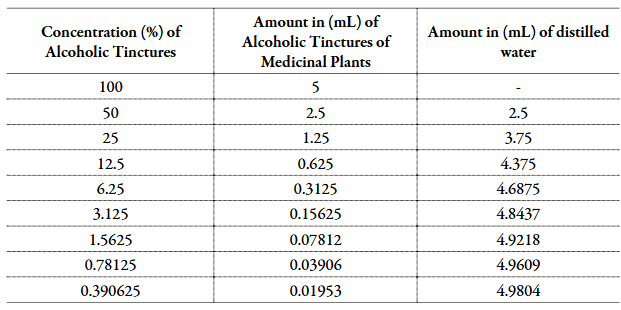
The application of the diffusion method is limited to fast-growing microorganisms, whether aerobic or facultative anaerobes. The microorganism used was Escherichia coli, which takes the form of a bacillus and belongs to the Enterobacteriaceae family. They are facultative aerobic and anaerobic. Its natural habitat is the intestinal lumen of humans and other warm-blooded animals. It has multiple flagella arranged around the cell.
Three well-isolated colonies of the same morphological type as the agar plate were selected.
The surface of each colony was touched with a loop, and the microorganisms transferred to a tube containing 4-5 mL of a suitable culture medium, such as tryptic soy broth. It was incubated in broth at 35 °C until reaching or exceeding the turbidity of a standard 0.5 MacFarland solution for 6 hours.
After 6 hours, the growing culture was adjusted to turbidity with sterile saline compatible with that of the 0.5 MacFarland standard solution.
This resulted in a suspension containing approximately 1 to 2 × 10⁸ CFU/mL of Escherichia coli ATCC® 25922. It was necessary to perform a reading with a spectrophotometer at this stage so that the turbidity adjustment is within the MacFarland scale standards 0.5.
On each plate add 20 mL of the molten culture medium. Promote smooth homogenization with figure-eight movements. Allow to solidify in a homogeneous and smooth layer.
Under ideal conditions, a sterile cotton swab was dipped into the suspension for 15 minutes after adjusting the turbidity of the inoculum suspension. The swab was rotated several times and pressed firmly against the tube wall above the liquid level to remove excess inoculum on the swab.
The dry surface of the TSA plate was inoculated by rubbing the swab across the entire sterile surface, rubbing twice more, rotating approximately 60° each time, and finally swabbing the edge of the agar plate, ensuring uniform distribution of the inoculum.
Recommended inoculation conditions are 35-37 °C temperature for bacteria for 24 hours to 48 hours. As a variation of the method, the petri dishes were incubated for 14 h at 37 °C, and at the end of this time, the inhibition halos were measured, in millimeters, with the aid of a caliper ruler.
For the application of the tinctures of Hamamelis virginiana, Maytenus ilicifolia and Casearia sylvestris, in the perforated disks of the plate deposited on the solidified culture medium containing the bacterial inoculum, an automatic pipette was used, applying 100 µg/mL of the tinctures (and their dilutions in decreasing concentrations) in each hole. The technique was performed in a sterilized medium close to a Bunsen burner. At least three plates were prepared for each standard concentration.
Technique for spectrophotometric determination of bacterial growth in broth
To prove the results obtained by the disk-diffusion technique, the broth dilution method was also performed. This method considers the relationship between the proportion of growth of the challenged microorganism in the liquid medium and the concentration of the substance tested. The evaluation is compared against a biological reference standard, in this work the inert culture medium was used. During the conduction of the method, 10.36 g was dissolved in distilled water, completing the volume to 280 mL according to the manufacturer's instructions, with volumes of 5 mL in test tubes sterilized in an autoclave at 121 °C/15 minutes. For the preparation of the microorganism on the MacFarland 0,5 scale, three colonies, well isolated, of the same morphological type of the agar plate were selected.
The surface of each colony was touched with a loop, and the microorganisms transferred to a tube containing 4-5 mL of a suitable culture medium, such as tryptic soy broth. It was incubated in broth at 35 °C until reaching or exceeding the turbidity of a standard 0,5 MacFarland solution for 6 hours. After 6 hours, the growing culture was adjusted to turbidity with sterile saline compatible with that of the 0,5 MacFarland standard solution. This resulted in a suspension containing approximately 1 to 2 × 10⁸ CFU/mL of Escherichia coli ATCC® 25922. It was necessary to perform a reading with a spectrophotometer at this stage so that the turbidity adjustment is within the MacFarland scale standards 0.5.
After 24 hours, the test tubes were inoculated, where 54 test tubes were used, the test was performed in triplicate, to obtain more accurate results. To the previously prepared BHI broth, 100 µg/mL were added with an automatic and sterile pipette of the alcoholic tincture of the plant in each test tube, previously diluted in decreasing concentrations. This step was repeated for the three alcoholic tinctures under study.
Under ideal conditions, 100 µg/mL was added with an automatic and sterile pipette of the microorganism previously prepared within the 0.5 MacFarland scale standards in all tubes, except for the blank sample, while for the negative control, culture medium is used with the solvent used to dissolve the sample and the microbial suspension.
The technique was performed in a sterilized medium close to the Bunsen burner, after inoculation of the bacteria in the test tubes, they were properly closed with a lid to avoid contamination. The test tubes were incubated at 35-37 °C for approximately 24 hours. At the end of this time, it is understood that by proportion the turbidity density was caused by microbial growth.
Quantitative analysis was performed using the spectrophotometric technique, at a wavelength of 625 nm. The reading was performed in all test tubes, containing the alcoholic tinctures in their different concentrations, being used for calibration the sample containing the culture medium and the microorganism studied. The method provides quantitative results and is not influenced by the growth rate of microorganisms.
RESULTS
Disk diffusion method in TSA
The plates were satisfactorily seeded, and the inoculum was correct and showed uniformly circular inhibition halos and a confluent growth mat. The diameters of the halos were measured including the diameter of the disc, calculated the diameter of the halos with a ruler, leaning against the back of the inverted petri dish with reflected light. Figure 1 shows the diameter of the inhibition halos for each dilution of the studied tinctures.
As shown in Figure 1, it was observed that in the holes in the plate where the tinctures in low concentration were placed, there was an increase in the proliferation of Escherichia coli colonies, which indicates that the tinctures in high concentrations have an inhibitory action.

Figure 1 Mean diameter of inhibition zones (mm) of Escherichia coli growth under the addition of tinctures of Hamamelis virginiana, Maytenus ilicifolia and Casearia sylvestris in decreasing concentrations. The analysis of variance (ANOVA with repetition of measures) showed a significant difference (p<0.05) between the groups, and the Tukey test indicated a significant difference (p<0.05) between the Maytenus ilicifolia and Casearia sylvestris groups and between Maytenus ilicifolia and Hamamelis virginiana.
Technique for Spectrophotometric Determination of Bacterial Growth in Broth
The use of this technique aimed to confirm the previous results, mainly in relation to the observation that at low concentrations there was a bacterial growth. In fact, this was verified according to the results presented in Figure 2. In the tested dilutions (less than 1.6%), there was an absorbance (turbidity), as seen in Figure 3, greater in the tubes containing low concentrations of the tinctures than in those in that there was only the culture of bacteria (control). It is also possible to observe that the tincture of which Hamamelis virginiana proved to be less efficient in this parameter.

Figure 2 Diluted BHI absorbance after growth of Escherichia coli under the addition of tinctures of Hamamelis virginiana, Maytenus ilicifolia and Casearia sylvestris in decreasing concentrations. The analysis of variance (ANOVA with repetition of measures) showed a significant difference (p<0.05) between the groups, and the Tukey test indicated a significant difference (p<0.05) between the Hamamelis virginiana group and the Maytenus ilicifolia groups (p<0.05) and Casearia sylvestris (p<0.001).

DISCUSSION
The method used to observe bacterial growth was initially disk-diffusion in agar, where using different dilutions of tincture of the three species studied, it was observed that at higher concentrations the tinctures caused bacterial inhibition, however at lower concentrations bacterial growth was stimulated. The agar diffusion method is normally used to evaluate the antimicrobial activity at different concentrations with a focus on determining the minimum inhibitory concentration (MIC), however this study differs in its objective, as it was prioritized the concentration that instead of inhibiting stimulates bacterial growth.
Antiseptic activity for Maytenus ilicifolia, being an indication of the inhibitory potential of microorganisms when used in medium and high dilutions [23]. Casearia sylvestris has a well-defined inhibitory activity for fungi such as Saccharomyces cerevisiae and Aspergillus niger, in addition to being an important antiseptic [24- 26]. In the case of Hamamelis virginiana, there are no reports of its use as an antiseptic or antimicrobial activity, however this was verified in the present work, although with a more discreet efficiency in relation to the other two tested tinctures.
As a confirmatory method of the first procedure, and aiming at better precision, the spectrophotometric technique was used to determine bacterial growth in broth, confirming and detailing the results presented in disk-diffusion, mainly regarding the stimulation of bacterial growth, which was observed in an imprecise way in the first technique. In the tested dilutions (less than 1.6%), there was a higher absorbance (turbidity) in the tubes containing low concentrations of the tinctures than in those in which there was only the bacterial culture (control). It is also possible to observe that the tincture of which Hamamelis virginiana proved to be less efficient in this parameter.
For bacterial proliferation it is necessary: (A) increase in ribosomal activity as these correspond to the site of protein synthesis [27]. Protein synthesis involves a complex cycle in which various ribosomal components play specific roles [27]; the ribosome can be interpreted as a dynamic structure, which interacts with many other cellular proteins [27]; (B) production of glycocalyx as the bacteria adhere to each other and to the substrate through protein-protein interactions present on the surface of the pathogen and host cells [28]. Furthermore, as the outer polysaccharide layers are likely to be associated with significant amounts of water, it is believed that the glycocalyx may confer some resistance to desiccation [29].
In the above perspective, there is a similarity between bacterial growth and fibroblast proliferation in a regenerative process, since these cells undergo phenotypic changes from migrating and replicating immature cells at the beginning of the process to cells actively engaged in protein synthesis, that is, its cytoplasm becomes voluminous and presents a rough endoplasmic reticulum with abundant ribosomes. As a result, they begin to secrete large amounts of collagen. It gradually replaces proteoglycans and fibronectin until it becomes the main component of the scar being formed [30].
In addition, with regard to the glycocalyx in epithelial cells, it represents an integral part of the cell membrane, which plays an important role in different forms of cellular interaction, such as, for example, cell adhesion processes, lymphocyte circulation and other signaling processes recognition, since glycocalyx frequently intervenes in the formation of receptors on the cell surface [31, 32].
The results obtained with the tincture of Hamamelis virginiana are in agreement with the scientific literature since isolated fractions of the tinctures of this plant stimulate and increase the proliferation of keratinocytes in epidermal cell culture [33, 34].
Similarly, the results obtained for the tincture of Maytenus ilicifolia corroborate reports in the literature that this plant accelerates the healing of peptic ulcers, and in comparative models with cimetidine, who ingested the Maytenus ilicifolia tinctures had their lesions healed earlier [35- 37]. For Casearia sylvestris, whose hypothesis below led to the development of this experimental design, there are reports that its active components stimulate the proliferation of fibroblasts, intensifying the healing process, and these data are indirectly in agreement with the results obtained in the present study for the tincture of this species [38, 39].
CONCLUSION
According to the results obtained in this study, it is concluded that the tinctures of Hamamelis virginiana, Maytenus ilicifolia and Casearia sylvestris at high and medium concentrations (above 1.6%) inhibit the development of Escherichia coli in culture, but at low concentrations (from 0.4 to 1.6%) induce bacterial proliferation, especially in the case of Maytenus ilicifolia and Casearia sylvestris tinctures.
The induction of bacterial growth, in the experimental design used, is a strong indication that these tinctures, if used in regenerating epithelial tissue, stimulate the production of extracellular matrix by the fibroblast, collaborating in the healing process, as well as inducing the production of glycocalyx in the epithelial cells facilitating adhesion and migration of these for recovery of the injured region.
Evidently, the present work is a simulation, but its results corroborate the existing scientific literature on the subject, which brings relevance to the results.
Finally, it is evaluated that the methodology used allowed the proposed objectives to be met, highlighting as positive points the ease of method development, the low cost and contribution to the rationalization in the use of animals. As an unfavorable point, the results are not directly applicable to animals, requiring an in vivo test to ensure efficacy and safety.

