
SciELO - Scientific Electronic Library Online

 
vol.52 issue2Factors related to adherence to treatment in patients with chronic noncommunicable diseases in three Colombian citiesDrugs solubility prediction in mono-solvents at various temperatures using a minimum number of experimental data points author indexsubject indexarticles search
Home Pagealphabetic serial listing  

Services on Demand

Journal

Article

Indicators

Related links

  • On index processCited by Google
  • Have no similar articlesSimilars in SciELO
  • On index processSimilars in Google

Share


Revista Colombiana de Ciencias Químico - Farmacéuticas

Print version ISSN 0034-7418On-line version ISSN 1909-6356

Rev. colomb. cienc. quim. farm. vol.52 no.2 Bogotá May/Aug. 2023  Epub May 01, 2023

https://doi.org/10.15446/rcciquifa.v52n2.104480 

Scientific Research Articles

Biofilm formation on toothbrushes by mutans group streptococci and Candida spp. isolated from oral cavity of students of the State University of Goiás, Brazil

Formação de biofilme em escovas dentais por estreptococos do grupo mutans e Candida spp. isolados da cavidade oral de alunos da Universidade Estadual de Goiás, Brasil

Formación de biopelículas en cepillos de dientes por estreptococos del grupo mutans y Candida spp. aislados de cavidad oral de estudiantes de la Universidad Estatal de Goiás, Brasil

1Postgraduate Program in Sciences Applied to Health Products. Bioassay Laboratory, Central Campus of the State University of Goiás


SUMMARY

Introduction:

Tooth brushing is an important measure in promoting oral health. However, the brush suffers the deposition and proliferation of microorganisms in its bristles during its use. In addition, a correlation has been demonstrated between the microbial load found on toothbrushes and the dental status of patients.

Aim:

To evaluate the biofilm formation on toothbrushes by mutans group streptococci and Candida spp. isolated from the oral cavity of students at the State University of Goiás.

Methodology:

The methodology employed in this study allowed the collection, isolation, and presumptive morphological identification of oral microorganisms, and allowed the evaluation of biofilm formation by both microorganisms in sterile toothbrushes.

Results:

All microbial isolates were able to form biofilms under the tested conditions, with microbial cell counts adhered to the biofilm ranging from 6.75 to 11.00 log10 cfu·mL-1 for mutans group streptococci, and 4.94 to 5.79 log10 cfu·mL-1 for Candida spp. Mutans group streptococci and Candida spp. were isolated from 92.31% of brushes used in the collection of samples from the oral cavity of the volunteers.

Conclusion:

All microorganisms isolated were able to form biofilm on toothbrushes, which corroborates the importance of care with decontamination of toothbrushes in promoting oral health.

Keywords Microbial biofilm; mouth; tooth decay; oral candidiasis; students

RESUMO

Introdução:

a escovação dentária é uma medida importante na promoção da saúde bucal. No entanto, a escova sofre a deposição e proliferação de micro-organismos em suas cerdas durante o seu uso. Além disso, foi demonstrada uma correlação entre a carga microbiana encontrada nas escovas de dente e o estado dentário dos pacientes.

Objetivo:

avaliar a formação de biofilme em escovas dentais por estreptococos do grupo mutans e Candida spp. isolados da cavidade oral de estudantes da Universidade Estadual de Goiás.

Metodologia:

a metodologia empregada neste estudo permitiu a coleta, o isolamento e a identificação morfológica presuntiva de micro-organismos bucais, bem como a avaliação da formação de biofilme por ambos os micro-organismos em escovas dentais estéreis.

Resultados:

que todos os isolados microbianos foram capazes de formar biofilmes nas condições testadas, com contagens de células microbianas aderidas ao biofilme variando de 6,75 a 11,00 log10 ufc·mL-1 para estreptococos do grupo mutans e 4,94 a 5,79 log10 ufc·mL-1 para Candida spp. Foram isolados estreptococos do grupo mutans e Candida spp. em 92.31% das escovas usadas na coleta de amostras da cavidade bucal dos voluntários.

Conclusão:

todos os micro-organismos isolados foram capazes de formar biofilme nas escovas, o que reforça a importância dos cuidados com a descontaminação das escovas dentais na promoção da saúde bucal.

Palavras-chave Biofilme microbiano; boca; cárie dentária; candidíase bucal; estudantes

RESUMEN

Introducción:

el cepillado de dientes es una medida importante en la promoción de la salud bucal. Sin embargo, el cepillo sufre la deposición y proliferación de microorganismos en sus cerdas durante su uso. Además, se demostró una correlación entre la carga microbiana encontrada en los cepillos de dientes y el estado dental de los pacientes.

Objetivo:

evaluar la formación de biofilm en cepillos de dientes por estreptococos del grupo mutans y Candida spp. aislados de la cavidad oral de estudiantes de la Universidad Estatal de Goiás.

Metodología:

la metodología utilizada en este estudio permitió la recolección, el aislamiento y la identificación morfológica presuntiva de los microorganismos orales, así como la evaluación de la formación de biofilms por los microorganismos en cepillos dentales estériles.

Resultados:

todos los aislados microbianos pudieron formar biofilms en las condiciones probadas, con recuentos de células microbianas adheridas a la biopelícula entre 6,75 - 11,00 log10 ufc·mL-1 para los estreptococos del grupo mutans y entre 4,94 - 5,79 log10 ufc·mL-1 para las Candida spp. Los estreptococos del grupo mutans y Candida spp. fueron aislados en el 92,31% de los cepillos utilizados para la toma de muestras de la cavidad bucal de los voluntarios.

Conclusión:

todos los microorganismos aislados fueron capaces de formar biofilms en los cepillos de dientes, lo que refuerza la importancia del cuidado de la descontaminación de los cepillos de dientes en la promoción de la salud bucal.

Palabras clave Biopelícula microbiana; boca; caries dental; candidiasis oral; estudiantes

Introduction

Oral microbiota refers to the term used to determine the group of more than 700 species of microorganisms that symbiotically inhabit the oral cavity and that may be beneficial or harmful to the human host. Current evidence demonstrates that the microbiota plays a crucial role in the associations between oral and general systemic health [1].

Streptococci of the mutans group constitute about 70% of the cultivable microorganisms in the oral cavity. They are Gram-positive, ovoid bacteria, organized in pairs or chains that ferment dietary sugars and have acidogenic and adherence capacity. In addition, they initiate and have a strong role in the production, increase and maturation of biofilms [2].

Previously, Streptococcus mutans were thought to be the only cause of dental caries [3]. However, it has recently been shown that Candida albicans also plays a critical role in the development of root caries, causing significant microbial dysbiosis and increased abundance of acidogenic/aciduric S. mutans, which leads to the formation of a more cariogenic polymicrobial biofilm [4].

The microbial biofilm is the polymicrobial grouping involved in a viscous matrix composed of water, exopolysaccharides, proteins, nucleic acids and substances absorbed by the attached microorganisms that bind to the film acquired on the surface of the tooth [5]. This film is formed within 15 minutes after cleaning and polishing the dental enamel and is considered the first step for the fixation of S. mutans and the formation of dental biofilm [6].

Adequate control of the biofilm in the oral cavity results in a lower risk for the development of oral and systemic diseases arising from the oral microbiota [7], and the toothbrush is an important instrument in the promotion of oral health.

Apposite tooth brushing provides partial removal of oral biofilm with reduction of microorganisms related to the etiology of dental caries [8].

However, the brush itself can become an environment for deposition and proliferation of microorganisms in its bristles [9]. In addition, a correlation has been demonstrated between the microbial load found on toothbrushes and the dental status of patients [10].

In this context, the objective of this work was to evaluate, under laboratory conditions, the formation of biofilm on toothbrushes by streptococci of the mutans group and Candida spp. isolated from the oral cavity of students at the State University of Goiás in Brazil.

Material and methods

Obtaining saliva samples

This study was submitted to the Research Ethics Committee of the State University of Goiás (UEG) in accordance with resolutions 196/96 and 466/12 for research involving human beings of the Ministry of Health and the National Health Council through the Brazil Platform with approval number 3.604.978. Consultation and free, prior and informed consent (FPIC) was signed and a copy delivered to the participant before any procedure in this research.

Saliva samples were collected with stimulation by tooth brushing, from 13 students at the Central Campus of the State University of Goiás in the city of Anápolis. Volunteers were included according to the inclusion criteria: being of legal age, agreeing to participate in the research by signing the necessary documentation, having good systemic health, not having eaten in the period between the last brushing and collection, not having brushed their teeth before collection and not having any missing tooth in the oral cavity, except for third molars.

In the act of collecting the material, information was collected and then the volunteers were instructed on brushing with the modified Bass technique [11]. An illustrated insert was presented in order to facilitate brushing and standardize sample collection by the participants, and the brushing time was timed. Toothpaste or dental floss was not used and the tongue was not brushed.

Saliva samples were collected with new, disposable brushes with a size 30 rounded head, 20 tufts, 840 bristles and 14 cm long soft bristles that were previously sterilized in an autoclave at 125 °C for 15 minutes.

Volunteers were taken to the bathroom and requested that, as soon as brushing was completed, the brushes were immediately returned to be placed in Falcon tubes containing 15 mL of 0.9% sodium chloride sterile saline solution for transport, and subsequent processing in the Microbiology Laboratory.

Microbiological processing

The samples were submitted to sonication to detach the microorganisms adhered to the brushes in an ultrasonic bath at 40 kHz (Ultronique Q 5.9/40A, Indaiatuba, São Paulo) for 5 minutes at room temperature [12]. Then the brushes were discarded and the supernatants were diluted and plated up to 10-8 for the isolation of the collected microorganisms.

Microbial samples were subcultured onto mitis salivarius agar (MSB) (Himedia, Mumbai, India) supplemented with 20% sucrose PA (Neon, São Paulo, Brazil), 0.2 IU bacitracin (Dr. Ehrenstorfer, Augsburg, Germany) and 1% potassium tellurite (Dinâmica, Indaiatuba, Brazil) for the isolation of mutans group streptococci and on Sabouraud dextrose chloramphenicol agar (SDA) (KASVI, São José dos Pinhais, Brazil) for isolation of Candida spp. [13]. The plates were incubated at 35.5 °C in microaerophilia for 72 hours (Figure 1).

After incubation, growth readings were performed in the culture media with a colony counter (MA-6000, Marconi, Piracicaba, Brazil) for the presumptive identification of the microorganisms recovered from each brush.

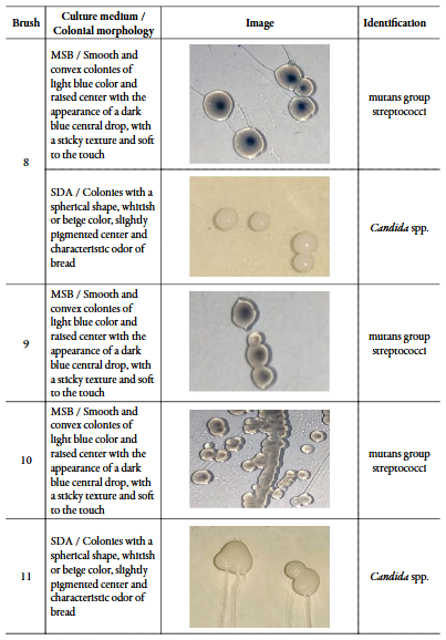
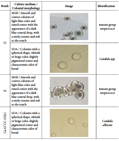
The presumptive identification of the isolated microorganisms was carried out based on the analysis of colonial aspects such as morphology, color, texture and odor after growth in the respective selective culture media. The enriched MSB medium is highly selective for S. mutans, S. sobrinus, S. salivarius and S. mitis. Smooth and convex colonies of light blue color and raised center with the appearance of a dark blue central drop, with a sticky texture and soft to the touch were identified as mutans group streptococci in this study [14], [15].
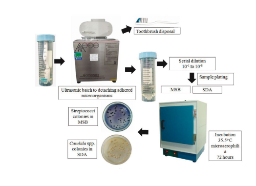
Figure 1 Microbiological processing of samples. 

For the isolation and identification of Candida spp., the criteria used were growth in SDA with chloramphenicol, with formation of whitish, spherical colonies, with a slightly darker center and odor of the plates similar to bread fermentation [16].

After identification, the isolated microorganisms were frozen in aliquots for further studies of biofilm formation [17].

Biofilm formation on brushes

Aliquots of the isolated microorganisms were subcultured in the respective culture media and after incubation at 35.5 °C in microaerophilia for 72 hours, and then the purity of the cultures was checked. Subsequently, three to five typical colonies were dissolved in 0.9% sterile saline solution (0.9% SSS) at approximately 1.5 × 10⁸ cfu·mL-1 with the 0.5 McFarland scale. Then, 200 µL of the suspension was transferred to Falcons tubes with 19,800 µL of Brain Heart Infusion Broth (BHI) (Himedia, Mumbai, India) adjusting and the initial inoculum for the biofilm formation assay at 1.5 × 10⁵ cfu·mL-1.

After preparation of the microbial inocula, sterile toothbrushes were aseptically placed in Falcon tubes and the samples were incubated. Then, the brushes were removed with sterile forceps and then rinsed in a Falcon tube with 20 mL of 0.9% SSS. After this procedure, the brushes were transferred to other tubes with 20 mL of 0.9% SSS and sonicated in an ultrasonic bath at 40 kHz (Ultronique Q 5.9/40A, Indaiatuba, São Paulo) for 5 minutes at room temperature [12] for detachment of attached colonies and subsequent counting in MSB and SDA media, for growth of mutans streptococci and Candida spp., respectively (Figure 2).
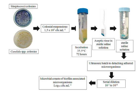
Figure 2 Biofilm formation assay on sterile toothbrushes. 

All microorganisms were evaluated in three independent replicates. The results of the counts were transformed into log10 and the means and standard deviations were calculated. Sterility controls and the Candida albicans ATCC 10231 were included in the assay.

Results

Isolation and presumptive identification

Of the total of 13 brushes processed, microorganisms were isolated in 12 (92.31%), in 1 (7.69%) brush were not isolated. A toothbrush that showed microbial growth was removed from the study due to the waiving of a volunteer from participating in the investigation.

A total of 15 types of colonies were obtained in the 11 brushes with microbial growth. Of these, 10 colonies were suggestive of mutans group streptococci and 5 colonies of Candida spp. In 6 (54.54%) brushes were isolated only streptococci of the mutans group, in 4 (36.36%) brushes were isolated both microorganisms and in 1 (9.10%) brush only Candida spp. was found.

Table 1 presents the morphological description and presumptive identification of the isolated microorganisms.
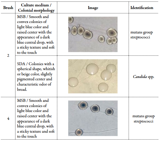
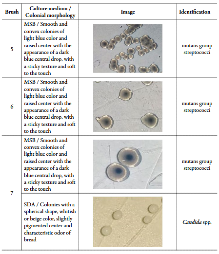
Table 1 Presumptive identification based on colonial morphology of the isolated microorganisms. 

Biofilm formation on brushes

All microbial isolates were able to form biofilms under the tested conditions, with microbial cell counts adhered to the biofilm ranging from 6.75 to 11.00 log10 cfu·mL-1 for mutans streptococci, and 4.94 to 5.79 log10 cfu·mL-1 for Candida spp.

The isolates MS7 and MS14 were the major biofilm formers, with counts of 11.00 ± 2.51 and 10.28 ± 1.70 log10 cfu·mL-1, respectively. The other streptococci remained with more homogeneous counts. The isolated yeast counts were more uniform, as shown in Figures 3 and 4.
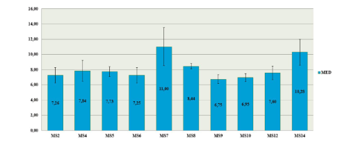
Figure 3 Mutans group streptococci cell count (log10 cfu·mL-1) recovered from the microbial films adhered in toothbrushes. MS: mutans group streptococci; cfu: colony forming unit. 
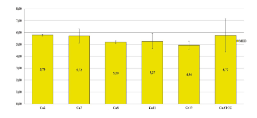
Figure 4 Candida spp. cell count (log10 cfu·mL-1) recovered from the microbial films adhered in toothbrushes. Ca: Candida spp.; cfu: colony forming unit; ATCC: American Type Culture Collection 

Discussion

The oral cavity is colonized by several types of microorganisms, with the establishment of a transient microbiota, generally controlled by microbial antagonism with the resident microbiota and by routinely employed hygiene [1]. In the present study, brushes were used to isolate mutans group streptococci and Candida spp. of the oral cavity of volunteers. Subsequently, we verified the ability of the isolates to form biofilms on sterile brushes. In the first stage of the study, microorganisms were isolated in 92.31% of the brushes used to obtain the microbial samples. Of the 15 microorganisms isolated, 10 were mutans group streptococci and 5 were Candida spp.

Tooth brushing is essential for oral hygiene, however the use of a toothbrush requires care such as proper cleaning and frequent replacement to minimize contamination, because during brushing, part of the oral microbiota can be transferred to the brush and the toothbrush, and from then on, the microbial load tends to increase and diversify with each use [18], [19].

The mutans group streptococci are present in 90% of the world population, while C. albicans are isolated in 30-50% [20]. S. mutans are related to the installation and progression of dental caries due to their ability to primarily adhere to the smooth surface of the tooth and allow the subsequent aggregation of other acidogenic microorganisms, with increased sucrose metabolism, lactic acid release and maturation of polymicrobial biofilm [21].

Yeasts of the Candida genus are part of the oral microbiota in situations of normality of the host, but they can behave as pathogens when local and systemic factors are triggered [22]. In addition, C. albicans has the ability to colonize the oral mucosa and denture surfaces, and is reported as a frequent cause of oropharyngeal infections and has cariogenic potential, being found in 10% of decayed teeth without S. mutans[23].

By itself, C. albicans has no cariogenic capacity, but in the presence of sucrose, aggregation with S. mutans occurs, increasing the amount of aciduric acid-tolerant microorganisms and the production of extracellular matrix. Therefore, there is a relationship between the levels of caries, the amount of microorganisms and their ability to form biofilms. In this way, the mixed biofilm of these microorganisms can intensify the carious processes [24], [25]. Furthermore, Candida is an opportunistic fungus, and oral pathological manifestations are directly related to the immune status of the host [26] since the occurrence of microbial biofilm in this anatomical site is related to the oral health of the patient.

The data presented in figures 3 and 4 indicate that all the isolates were able to form biofilms on the brushes, with high counts of microorganisms associated with the biofilms. These results are similar to the results obtained by other authors, although using different substrates and methodologies. In a study [27] on S. mutans biofilm formation on bovine tooth enamel, the counts obtained in the control group ranged from 5.00 to 10.00 log10 cfu·mL-1. The results found in the viability controls of studies that evaluated the inhibition of biofilm formation by S. mutans were 6.5 and 10.00 log10 cfu·mL-1, respectively [28], [29]. Regarding the cell counts associated with Candida biofilm, other authors found counts ranging from 4.02 to 9.50 Log10 cfu·mL-1 cells [30]-[33]. Biofilms constitute a form of existence of microorganisms encapsulated in an extracellular matrix that holds cells together and forms a three-dimensional structure resistant to extrinsic adversities [34]. In the oral cavity, the biofilm is adhered to the surfaces of the mouth in general and tends to establish itself and increase if there is no chemical or mechanical interference [35]. Thus, microbial control methods are used to reduce microbial biofilm and maintain oral health, the use of mouthwashes and tooth brushing are common methods used for this purpose.

Conclusion

In summary, in this work we evaluated the biofilm formation on toothbrushes by mutans group streptococci and Candida spp. isolated from the mouths of students at the State University of Goiás. The sample collection methodology proved to be effective, as microorganisms were not recovered in just one brush collected. In the second part of the study, it was detected that all isolates were able to form biofilms on the brushes, and that the isolates showed considerable counts of microorganisms associated with the biofilm, with emphasis on the counts of streptococci of the mutans group. In agreement with other authors [36], [37], the results obtained in this study highlight the importance of taking care with the decontamination of toothbrushes in the promotion of oral health.

Acknowledgements

We thank our laboratory colleagues Kamila Cecílio, Osvaldo Gomes, Pabline Gasparotti, Amanda Correia and valéria Oliveira for their contributions at various stages of the work, and to the volunteers who kindly participated in the sample collection stage. We also thank the Coordination for the Improvement of Higher Education Personnel (CAPES).

References

[1] C. Barboza-solís, L.A. Acuña-amador, The oral microbiota: A literature review for updating professionals in dentistry. Part I, Odovtos - International Journal of Dental Sciences, 22(3), 59-68 (2020). Doi: https://doi.org/10.15517/ijds.2020.39178. [ Links ]

[2] A. Schubert, R. Bürgers, F. Baum, O. Kurbad, T. Wassmann, Influence of the manufacturing method on the adhesion of Candida albicans and Streptococcus mutans to oral splint resins, Polymers, 13(15340), 1534 (2021). Doi: https://doi.org/10.3390/polym13101534. [ Links ]

[3] N. Philip, B. Suneja, L. Walsh, Beyond Streptococcus mutans: Clinical implications of the evolving dental caries aetiological paradigms and its associated microbiome, British Dental Journal, 224, 219-225 (2018). Doi: https://doi.org/10.1038/sj.bdj.2018.81. [ Links ]

[4] Q. Du, B. Ren, J. He, X. Peng, Q. Guo, L. Zheng, J. Li, H. Dai, X. Vivian, L. Zhang, Z.X. Xu, Candida albicans promotes tooth decay by inducing oral microbial dysbiosis, The ISME Journal, 15, 894-908 (2021). Doi: https://doi. org/10.1038/s41396-020-00823-8. [ Links ]

[5] T. Gong, X. He, J. Chen, B. Tang, T. Zheng, M. Jing, Y. Lin, Y. Pan, Q. Ma, Y. Li, X. Zhou, Transcriptional profiling reveals the importance of RcrR in the regulation of multiple sugar transportation and biofilm formation in Streptococcus mutans, mSystems. 6(4), e00788-21 (2021). Doi: https://doi.org/10.1128/mSystems.00788-21. [ Links ]

[6] J.B. Faria, M.B. Santiago, C.B. Silva, V.R. Geraldo-Martins, R.D. Nogueira, Development of Streptococcus mutans biofilm in the presence of human colostrum and 3′- sialyllactose, The Journal of Maternal-Fetal & Neonatal Medicine, 35(4), 630- 635 (2020). Doi: https://doi.org/10.1080/14767058.2020.1730321. [ Links ]

[7] M.B. da Silva, K.S. Andrade, J.K.M. Oliveira, M.F. Farias, K.T. Fragoso, A.M.R. Cardoso, Condição bucal e doenças respiratórias em pacientes internados em Unidade de Terapia Intensiva. Archives of Health Investigation, 2021, 10(1), 147- 152 (2021). Doi: https://doi.org/10.21270/archi.v10i1.4935. [ Links ]

[8] E. Greenfield, A.L.F. Marangoni, Avaliação comparativa entre escovas dentais manuais e ultrassônicas para remoção do biofilme, Revista Odonto, 27(54), 19-24 (2019). Doi: https://doi.org/10.15603/2176-1000/odonto.v27n54p19-24. [ Links ]

[9] A. Ansari, H. Torabzadeh, Z.S. Nabavi, P.S. Hassani, Comparing the effect of dry and wet brushing on dental plaque removal in children, Journal of Indian Society of Pedodontics and Preventive Dentistry, 37(1), 292-296 (2019). Doi: https://doi.org/10.4103/JISPPD.JISPPD_71_19. [ Links ]

[10] M. Bargrizan, R. Fekrazad, N. Goudarzi, N. Goudarzi, Effects of antibacterial photodynamic therapy on salivary mutans streptococci in 5- to 6-year-olds with severe early childhood caries, Lasers in Medical Science, 34(3), 433-440 (2018). Doi: https://doi.org/10.1007/s10103-018-2650-2. [ Links ]

[11] J. Lindhe, N.P. Lang, T. Karring, Tratado de Periodontia Clínica e Implantologia Oral, 5st ed., Guanabara Koogan, Rio de Janeiro, 2010. [ Links ]

[12] R.A.C. Chaves, D.M.L. Ribeiro, J.E. Zaia, E.G. Alves, M.G.M. Souza, C.H.G. Martins, S.F. Mestriner, Avaliação de soluções antibacterianas na descontaminação de escovas dentárias de pré-escolares, Revista de Odontologia da UNESP, 36(1), 29-33 (2007). [ Links ]

[13] A.L.S. Cota, R.G. Alvim, Effect of storage temperature on Streptococcus mutans viability, Revista de Odontologia da UNESP, 47(2), 74-78 (2018). Doi: http://dx.doi.org/10.1590/1807-2577.08317. [ Links ]

[14] M.E. Saravia, L.A.B. Silva, R.A.B. Silva, N.M. Cudmani, S. Tineo, N.E. Hillen, M.P. Lucisano, A.M. Queiroz, C.-G. Emilson, P. Nelson-Filho, Morphological identification of Streptococcus mutans and Streptococcus sobrinus in SB-20M culture medium has efficiency comparable to proteomic identification by the MALDI-TOF mass spectrometry technique, Archives of Oral Biology, 110, 104595 (2020). Doi: https://doi.org/10.1016/j.archoralbio.2019.104595. [ Links ]

[15] X. Zhou, Y. Li, Atlas of oral microbiology: from healthy microflora to disease, 2nd ed., Elsevier, Rio de Janeiro, 2020. [ Links ]

[16] S. Sridhar, S. Suprabha, R. Shenoy, E. Suman, A. Rao, Association of Streptococcus mutans, Candida albicans and oral health practices with activity status of caries lesions among 5-year-old children with early childhood caries, Oral Health & Preventive Dentistry, 18(1), 911-919 (2020). Doi: https://doi.org/10.3290/j.ohpd.a45411. [ Links ]

[17] J.O. Silva, P.P. Costa, S.H.C. Reche, Manutenção de leveduras por congelamento a -20°C, Revista Brasileira de Análises Clínicas, 40(1), 73-74 (2020). [ Links ]

[18] R.P. Mantovani, A. Sandri, M. Boaretti, A. Grilli, S. Volpi, P. Melotti, G. Burlacchini, M.M. Lleò, C. Signoretto, Toothbrushes may convey bacteria to the cystic fibrosis lower airways. Journal of Oral Microbiology, 11(1), 1647036 (2019). Doi: https://doi.org/10.1080/20002297.2019.1647036. [ Links ]

[19] A.P. Watanabe, Toothbrush contamination by Candida spp. and efficacy of mouthrinse spray for their disinfection, Mycopathologia, 169, 133-138 (2010). Doi: https://doi.org/10.1007/s11046-009-9239-z. [ Links ]

[20] P.S. Dabas, An approach to etiology, diagnosis and management of different types of candidiasis, Journal of Yeast and Fungical Research, 4(6), 63-67 (2013). Doi: https://doi.org/10.5897/JYFR2013.0113. [ Links ]

[21] A. Kumar, A. Alam, M. Rani, N.Z. Ehtesham, S.E. Hasnain, Biofilms: Survival and defense strategy for pathogens, International Journal of Medical Microbiology, 307(8), 481-489 (2017). Doi: https://doi.org/10.1016/j.ijmm.2017.09.016. [ Links ]

[22] D.W. Williams, R.P.C. Jordan, X.-Q. Wei, C.T. Alves, M.P. Wise, M.J. Wilson, M.A.O. Lewis, Interactions of Candida albicans with host epithelial surfaces, Journal of Oral Microbiology, 5, 22434 (2013). Doi: https://doi.org/10.3402/jom.v5i0.22434. [ Links ]

[23] D.F.A. Pereira, C.J. Seneviratne, C.Y. Koga-Ito, L.P. Samaranayake, Is the oral fungal pathogen Candida albicans a cariogen? Oral Diseases, 24(4), 518-526 (2018). Doi: https://doi.org/10.1111/odi.12691. [ Links ]

[24] H. Koo, D.R. Andes, D.J. Krysan, Candida-streptococcal interactions in biofilm associated oral diseases, PLoS Pathogens, 14(12), e1007342 (2018). Doi: https://doi.org/10.1371/journal.ppat.1007342. [ Links ]

[25] S.A. Sounah, A.A. Madfa, Correlation between dental caries experience and the level of Streptococcus mutans and Lactobacillus in saliva and carious teeth in a Yemeni adult population, BMC Research Notes, 13, 112 (2020). Doi: https://doi.org/10.1186/s13104-020-04960-3. [ Links ]

[26] A. Sharma, Oral candidiasis: An opportunistic infection: A review, International Journal of Applied Dental Sciences, 5(1), 23-27 (2019). [ Links ]

[27] C.M. de Souza, M.T. Garcia, P.P. de Barros, L.L.C. Pedroso, R.A.C. Ward, J.F. Strixino, V.M.M. Melo, J.C. Junqueira, Chitosan enhances the Antimicrobial Photodynamic Inactivation mediated by Photoditazine® against Streptococcus mutans, Photodiagnosis and Photodynamic Therapy, 32, 102001 (2020). Doi: https://doi.org/10.1016/j.pdpdt.2020.102001. [ Links ]

[28] L.M. Tokubo, P.L. Rosalen, J.C.O. Sardi, I.A. Freires, M. Fujimaki, J.E. Umeda, P.M. Barbosa, G.O. Tecchio, N. Hioka, C.F. de Freitas, R.S.S. Terada, Antimicrobial effect of the photodynamic therapy using erythrosine/methylene blue combination on Streptococcus mutans biofilm, Photodiagnosis and Photodynamic Therapy, 23, 94-98 (2018). Doi: https://doi.org/10.1016/j.pdpdt.2018.05.004. [ Links ]

[29] H. Jafri, G. Benerjee, M.S.A. Khan, H.H. Abulreesh, A.S. Althubiani, Synergistic interaction of eugenol and antimicrobial drugs in eradication of single and mixed biofilms of Candida albicans and Streptococcus mutans, AMB Express, 10(1), 185 (2020). Doi: https://doi.org/10.1186/s13568-020-01123-2. [ Links ]

[30] F. Khan, D. Oh, P. Chandika, D.-M. Jo, N.I. Barmunarachchi, W.-K. Jung, Y.-M. Kim, Inhibitory activities of phloroglucinol-chitosan nanoparticles on mono- and dual-species biofilms of Candida albicans and bacteria, Colloids and Surfaces B: Biointerfaces, 211, 112307 (2022). Doi: https://doi.org/10.1016/j.colsurfb.2021.112307. [ Links ]

[31] F.C.T. Coimbra, M.M. Rocha, V.C. Oliveira, A.P. Macedo, V.O. Pagnano, C.H. Silva-Lovato, H.d.F.O. Paranhos, Antimicrobial activity of effervescent denture tablets on multispecies biofilms, Gerodontology, 38(1), 87-94 (2021). Doi: https://doi.org/10.1111/ger.12500. [ Links ]

[32] F.C. da Silva, P.L.F. Rodrigues, T.S.D. Araújo, M.S. Santos, J.M. de Oliveira, L.P. Rosa, G.P.O. Santos, B.P. de Araújo, V.S. Bagnato, Fluorescence spectroscopy of Candida albicans biofilms in bone cavities treated with photodynamic therapy using blue LED (450 nm) and curcumin, Photodiagnosis and Photodynamic Therapy, 26, 366-370 (2019). Doi: https://doi.org/10.1016/j.pdpdt.2019.05.002. [ Links ]

[33] A.L.R. Palma, L. de Paula-Ramos, N. Domingues, G.N. Back-Brito, L.D. de Oliveira, C.A. Pereira, A.O.C. Jorge, Biofilms of Candida albicans and Streptococcus sanguinis and their susceptibility to antimicrobial effects of photodynamic inactivation, Photodiagnosis and Photodynamic Therapy, 24, 95-101 (2018). Doi: https://doi.org/10.1016/j.pdpdt.2018.07.003. [ Links ]

[34] R. Li, X. Yuan, J. Wei, X. Zhang, G. Cheng, Z.A. Wang, Y. Du, Synthesis and evaluation of a chitosan oligosaccharide-streptomycin conjugate against Pseudomonas aeruginosa biofilms, Marine drugs, 17(1), 43 (2019). Doi: https://doi.org/10.3390/md17010043. [ Links ]

[35] M.L.F.V. de Menezes, Y.V.G. de MAcedo, N.M.P. Ferraz, K.F. MAtos, R.O. Pereira, N.M. Fontes, M.I.H.M. Batista, M.R. Paulino, A importância do controle do biofilme dentário: uma revisão da literatura, Revista Eletrônica Acervo Saúde, 55(1), e3698 (2020). Doi: https://doi.org/10.25248/reas.e3698.2020. [ Links ]

[36] E.C.F. Queiros, M.A.N. Passos, Aspectos de contaminação e descontaminação das cerdas de escovas dentais, Revista Ciências e Odontologia, 3(1), 1-5 (2019). [ Links ]

[37] A. Unahalekhaka, P. Butpan, R. Wongsaen, P. Phunpae, K. Preechasuth, Contamination of antimicrobial-resistant bacteria on toothbrushes used with mechanically ventilated patients: A cross sectional study, Intensive and Critical Care Nursing, 68, 103120 (2022). Doi: https://doi.org/10.1016/j.iccn.2021.103120. [ Links ]

Conflict of interestAll authors report that they do not have any conflicts of interest.

How to cite this articleR.M. Rocha, P.L.F. Naves, Biofilm formation on toothbrushes by mutans group strep-tococci and Candida spp. isolated from oral cavity of students of the State University of Goiás, Brazil, Rev. Colomb. Cienc. Quim. Farm., 52(2), 889-907 (2023). https://doi.org/10.15446/rcciquifa.v52n2.104480

Received: August 27, 2022; Revised: March 30, 2023; Accepted: May 07, 2023

Creative Commons License All the contents of this journal, except where otherwise noted, is licensed under a Creative Commons Attribution License