


 text in
  English (pdf)
 Article in xml format
 Article references
 Send this article by e-mail
 Cited by SciELO
  Cited by Google
  Similars in
SciELO
Similars in
SciELO  Similars in Google
 Permalink
Introduction
Osteoporosis is a systemic skeletal disease characterized by low bone mineral density (BMD) and bone architecture deterioration, resulting in reduced bone elasticity and resistance and, consequently, a higher risk of fractures 1. The main clinical manifestation of osteoporosis is a fragility fracture, and approximately 80% of all fractures are related to osteoporosis 2. Since fractures are associated with hospitalization, comorbidities, complications and disability, they make activities of daily living difficult, negatively affect the quality of life and cause a greater financial burden. Only a third of patients with hip fractures return to their previous functional level, and a third of these patients must be institutionalized in nursing homes; this justifies the importance of detecting people at risk of fractures and providing appropriate therapeutic options 3.
The clinical practice guidelines recommend that postmenopausal women with a high risk of fractures be treated with bisphosphonates, in order to reduce the risk of fractures, vertebral and non-vertebral injuries, and hip fractures 4. However, case reports of unusual fragility fractures in the subtrochanteric region and femoral diaphysis, in patients treated with bisphosphonates, are a cause for concern 5,6.
Published case reports and epidemiological studies examining the relationship between the use of bisphosphonates and these fractures have been on the rise since 2007 7. Bisphosphonates are not the only medications related to these fractures; there are also reports of unusual fractures with the use of denosumab 8,9 odanacatib 10 and romosozumab 11. These medications use different mechanisms to inhibit bone resorption and recover the balance between bone formation and resorption.
The concern regarding the relationship between bisphosphonates and atypical hip fractures gained importance with the Food and Drug Administration (FDA) warning in 2010 12, with a subsequent reduction in the prescription of bisphosphonates in the United States and United Kingdom 13.
The age-adjusted incidence of atypical fractures is estimated to be 1.8 per 100,000 person-years in patients who have been taking bisphosphonates for less than two years, increasing to 113 per 100,000 person-years in people with more than eight years of treatment (14). On the other hand, there is data from European Union countries reporting underutilization of osteoporosis medications 15.
It has been hypothesized that reduced bone resorption in bisphosphonate users causes bone turnover suppression with an accumulation of microfissures and homogeneously mineralized bone, making the bones more fragile and allowing spontaneous fractures, which are especially frequent in the femur 16. It is unlikely that these atypical fractures occur due to high-energy mechanisms. In fact, although a fall from a standing height is generally reported as the mechanism of injury, it is not unusual for patients to report that they felt the fracture occur before falling; 34 to 70% of patients feel pain in the groin or thigh before the injury. Therefore, patients should be asked about pain in the opposite extremity, because they may be experiencing similar prodromal symptoms on the uninjured side 17.
We present this case in order to describe the diagnostic and therapeutic process of an unusual pathology associated with the use of medications commonly prescribed to treat osteoporosis, with defined clinical and radiological criteria, but surgical and pharmacological treatment that is still under debate.
Case presentation
We present the case of a married mestiza patient in her seventies who was previously independent in her activities of daily living (ADLs) and had no cognitive disorders. She presented with a three-month history of pain in the proximal third of her right thigh, with no history of trauma, which had an intensity of 7-8 on the visual analogue scale (VAS) and was exacerbated by exercise and mitigated by analgesics and rest.
This symptom inhibited her walking, forced her to use a single point cane, and led to an emergency room visit at the beginning of 2023, in which no abnormalities were found on x-ray, and hydrotherapy was prescribed. Later, during a hydrotherapy session, with no additional weight or resistance, she felt an intense pain (10/10 on the VAS) in the hip area of her right leg, radiating to the ipsilateral groin, associated with difficulty walking, pain with movement and limb deformity, and was therefore referred to the emergency room.
Her personal medical history included menopause at age 48 (not treated), migraines, chronic gastritis, dyslipidemia, constipation for the last two years and osteoporosis. The latter was treated with 5 mg of zoledronic acid every year from 2017 to 2021, followed by 60 mg of subcutaneous denosumab, with the first dose in January 2023. In addition, she was taking 20 mg per day of omeprazole and 600 mg/200 IU of calcium + vitamin D per day.
Her vital signs on admission were: blood pressure 112/72 mmHg in the supine position, heart rate 84 beats per minute, respiratory rate 16 breaths per minute, and temperature 36.4 degrees Celsius; pulse oximetry was 95% on room air, weight was 55 kg, and height was 157 cm, with a body mass index (BMI) of 22.31 kg/m2. On physical exam, she had an external rotation deformity of the right lower limb, with limited movement due to pain and no sensitivity or vascular abnormalities. The opposite limb was within normal limits.
An x-ray of the pelvis and both femurs showed a displaced and angulated oblique fracture of the proximal third of the femur, minimally comminuted (as seen in Figure 1A), and V-shaped periosteal thickening, shown in Figures 1B and 1C.
Blood chemistries showed vitamin D insufficiency (Table 1). Seven days after admission she underwent fracture repair with a cephalomedullary nail, and eight days after this procedure she underwent prophylactic contralateral cephalomedullary nailing (images available in Figure 1D), and was discharged that same day with a prescription for physical rehabilitation and Teriparatide.
Discussion
The diagnostic criteria for atypical fractures were defined in 2010 18 and reaffirmed in 2014 in the second report of the American Society for Bone and Mineral Research working group. These included the location in the femoral diaphysis, absent or minimal trauma, the lineal nature of the fracture, minimally comminuted fractures, and V-shaped periosteal and endosteal thickening 19. Our patient's symptoms occurred three months before the atypical fracture, with a minimally comminuted fracture point in the proximal third of the right femur, no prior trauma, and periosteal and endosteal thickening seen on imaging (Figures 1B and 1C), which enabled a clinical and imaging diagnosis of the condition in this case. Although atypical fractures due to the use of bisphosphonates and other pharmacological treatments used to reduce the risk of osteoporotic fractures are rare, the likelihood of occurrence is related to antiresorptive treatment lasting more than four or five years 14. Our patient had received the treatment for five years, with a year-long pause and treatment resumption in 2023.
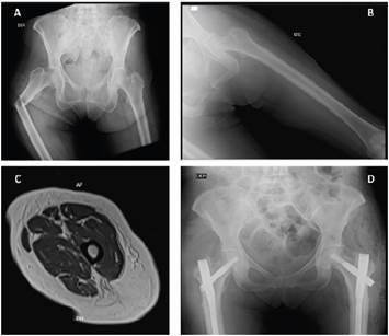
Figure 1 A: a pelvic x-ray showing an oblique, displaced, angulated fracture of the proximal third of the femur. B: an x-ray of the left femur shows V-shaped periosteal thickening. C: nuclear magnetic resonance imaging of the femur showing regular, circumferential, hypointense cortical thickening of up to 8 mm in the diaphysis, proximal metaphysis and distal metaphysis, in all sequences, without expansile lesions or cortical destruction. D: a pelvic x-ray showing osteosynthesis material in both femurs.
Atypical fractures are a clinical challenge associated with disability, functional limitations, immobility and all the complications potentially related to fractures, with the aggravating factor of more consolidation failure compared to other types of fractures. Coupled with this, patients over the age of 55 have a higher likelihood of having other chronic health conditions that can add complexity to treatment and rehabilitation.
Surgical management of bisphosphonate-related atypical fractures should be approached with caution and careful planning. In addition, treatment of the contralateral side mainly depends on the patient's symptoms or lack of symptoms and the femoral x-ray (17). Our patient had radiographic signs in the contralateral femur that suggested microfractures (Figures 1B and 1C). Therefore, although the decision to perform surgery to correct the fracture point was made quickly, there was no consensus at first on whether to perform surgery on the contralateral femur. This discussion involved evidence that was still under construction and intense debate, which therefore required a multidisciplinary team of geriatricians, orthopedic surgeons, and rehabilitation professionals to determine that the insertion of a cephalomedullary nail in the contralateral femur was the best strategy to prevent a future fracture (Figure 1C).
There is no guideline at present to indicate the best pharmacological approach after an atypical fracture, but the evidence suggests that the risk of causing new atypical fractures should be weighed against the risk of fragility fractures. Teriparatide, a parathyroid hormone analog, has been suggested as a safe option for treating osteoporosis in these patients, especially because it can also have a beneficial effect on curing the atypical fracture itself (20). Thus, in light of this patient's very high risk of fracture, 20 micrograms/day of Teriperatide, 1,200 mg/day of calcium and 2,000 IU/day of vitamin D were prescribed. However, what treatment to use once this treatment's two-year period is up, or what to do in the event of treatment failure, has still not been resolved.
Conclusion
The study of the causes of these potential complications of bisphosphonate treatment requires prospective, doubleblind randomized trials which would ideally include incident variables like plasma vitamin D and B12 levels, ferremia, albuminemia, thyroid hormones and natremia, among others.
Table 1 The patient's blood chemistry.
| Laboratory test | Result | Normal range |
|---|---|---|
| Chloride | 103 | 98-107 mmol/L |
| Potasium | 3.8 | 3.5-5.1 mmol/L |
| Sodium | 135 | 137-145 mmol/L |
| Total calcium | 10.00 | 8.4 - 10.2 mg/dL |
| Serum phosphorus | 2.6 | 2.5 - 4.5 mg/dL |
| Blood urea nitrogen | 11 | 7-17 mg/dL |
| Serum creatinine | 0.6 | 0.52-1.04 mg/dL |
| Leukocytes | 9.97 | 3.98 - 10.04 x 10^3/uL |
| Neutrophils | 7.16 | 1.56 - 6.13 x 10^3/uL |
| Lymphocytes | 2.23 | 1.18 - 3.74 x 10^3/uL |
| Hematocrit | 38.9 | 34.1 - 44.9 % |
| Hemoglobin | 12.9 | 11.2 - 15.7 g/dL |
| Mean corpuscular volume | 87.9 | 79.4 - 94.8 fL |
| Platelets | 395 | 182 - 369 x10^3/uL |
| Lactate dehydrogenase | 204 | 120 - 246 U/L |
| TSH | 3.02 | 0.4001 - 4.049 mUI/L |
| Albumin | 4.50 | 3.5 - 5 g/dL |
| Intact parathyroid hormone | 55.18 | 13.6 - 85.8 pg/ml |
| Total 25-hydroxy vitamin D | 22 | Sufficiency: > 30 ng/ml |
| Erythrocyte sedimentation rate (ESR) | 19 | 1-20 mm/hour |
| C-reactive protein | 7.69 | 0-9.99 mg/L |
| TSH= thyroid-stimulating hormone | ||
Fragility fracture prophylaxis should not itself become a fracture risk factor. To avoid this, patients who have started treatment with these medications should have appropriate monitoring and follow up, including careful history taking for symptoms that would indicate the existence of risk, to allow the clinician to suspect the risk and order confirmation tests to evaluate whether to discontinue the related medication and facilitate joint assessment with orthogeriatrics to determine the pertinence of prophylactic interventions like the one described in this case.
There are many limitations and challenges in dealing with these patients, but as was clearly seen in this case, these barriers can be overcome and are an opportunity to improve the institutional care protocols and the articulation of care to achieve the treatment goals, patient satisfaction and the interdisciplinary quality inherent in human health care. The future question of this patient's long-term level of functioning remains to be answered.

