SciELO - Scientific Electronic Library Online

 
vol.49 issue2An atypical hip fracture associated with the use of bisphosphonatesHandgrip strength and long COVID A test for evaluating capacity author indexsubject indexarticles search
Home Pagealphabetic serial listing  

Services on Demand

Journal

Article

Indicators

Related links

  • On index processCited by Google
  • Have no similar articlesSimilars in SciELO
  • On index processSimilars in Google

Share


Acta Medica Colombiana

Print version ISSN 0120-2448

Acta Med Colomb vol.49 no.2 Bogotá Apr./June 2024  Epub May 24, 2024

https://doi.org/10.36104/amc.2024.3123 

INTERNAL MEDICINE IMAGES

Pyomyositis and necrotizing fasciitis

LUIS GERARDO DOMÍNGUEZ-GASCAa  * 

LUIS GERARDO DOMÍNGUEZ-CARRILLOb 

aOrtopedista. Cirugía articular, División de Cirugía del Hospital ángeles León

bEspecialista en Medicina de Rehabilitación. Catedrático de la Facultad de Medicina de León, Universidad de Guanajuato. Guanajuato (México).


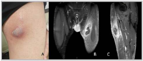
Figure 1 A: a photograph of the anterior internal aspect of the left thigh showing an abscess in the vertex of Scarpa's triangle. B: magnetic resonance imaging, coronal section, C: sagittal section showing an abscess and area of pyomyositis in the left thigh..  

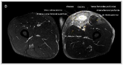
Pyomyositis 1 is an acute bacterial infection of the skeletal muscle with abscess formation. The incidence is 33-40%, mainly in young adult males. Staphylococcus aureus is responsible for 90% of the cases 2. It occurs due to dissemination from a distal infection or direct extension from an adjacent process. It has three phases: a) invasive: one to two weeks; b) purulent: the next 1021 days (90% of the patients consult at this stage); and c) systemic inflammatory response 3. A culture of the secretion and diagnostic ultrasound are required 4; magnetic resonance imaging is the test of choice 5. It is initially treated with antibiotics and requires surgical drainage in the second and third stages. Broad spectrum coverage is used in patients with comorbidities or immunosuppression, including Gram negative and anaerobic coverage. The reported mortality ranges from 2 to 20%, and is highest in patients with comorbidities.

Figure 2  Axial section on magnetic resonance imaging of the upper third of the left thigh showing an abscess, pyomyositis and necrotizing fascitis; compare with the opposite side.  

References

1. Pérez YC, Rodríguez PY, Moreno ES, Aviles RM. Absceso de músculo iliopsoas y piomiositis extensa en un adolescente inmunocompetente. Rev Enferm Infec Ped 2013;26: 270-273. Doi eip131. [ Links ]

2. Ntusi N, Khaki A. Primaiy multifocal pyomyositis due to Staphylococcus aureus, Q J Med 2011;104:163-165. doi.org/10.1093/qjmed/hcq075Links ]

3. Moreno RA, Baraia EJ, Gutierrez MA, Ferrero BO. Primary pyomyositis: a new case in an immunocompetent patient, Galicia Clin 2011;72:131-132. doi: 10.1007/s10067-010-1569-1. [ Links ]

4. Chou H, Teo H, Dubey H, Peh W. Tropical pyomyositis and necrotizing fasciitis. Semin Musculoskelet Radiol 2011;15:489-505. doi: 10.1055/s-0031-1293495. [ Links ]

5. Soler R, Rodríguez E, Aguilera C, Fernández R. Magnetic resonance imaging of pyomyositis in 43 cases. Eur J Radiol 2000;35:59-62. doi: 10.1016/s0720-048x(99)00108-4. [ Links ]

Received: February 05, 2024; Accepted: March 20, 2024

*Correspondencia: Dr. Luis Gerardo Domínguez-Carrillo. Guanajuato (México) E-Mail: lgdominguez@hotmail.com

Creative Commons License This is an open-access article distributed under the terms of the Creative Commons Attribution License