


Services on Demand
Journal
Article
 text in
  English (pdf)
 Article in xml format
 Article references
 Send this article by e-mail
Indicators
-
 Cited by SciELO
 -
 Access statistics
Related links
-
 Cited by Google
 -
 Similars in
SciELO
Similars in
SciELO -
 Similars in Google
Share

 Permalink
Colombian Journal of Anestesiology
Print version ISSN 0120-3347
Rev. colomb. anestesiol. vol.40 no.2 Bogotá Apr./June 2012
https://doi.org/10.1016/S0120-3347(12)70032-1
http://dx.doi.org/10.1016/S0120-3347(12)70032-1
Case report
Acute Airway Obstruction During an Interventional Radiology Procedure in a Patient With a Mediastinal Mass.
Case Report
Obstrucción aguda de la vía aérea en paciente con masa mediastinal
durante procedimiento de radiología intervencionista.
Reporte de caso
John Bautistaa*, Oscar Suárezb, Plutarco García-Herrerosc, Francisco Valero-Bernald
a Anesthesiology and Resuscitation Resident, Universidad Nacional de Colombia, Bogotá, Colombia "Corresponding author: calle 155 # 9 45 apto 429, torre 1, Bogotá, Colombia. E-mail: johnbautistas@yahoo.com (J. Bautista).
bAnesthesiology and Resuscitation Resident, Fundación Universitaria San Martín, Bogotá, Colombia
cPulmonology Specialist, Universidad Nacional de Colombia, Bogotá, Colombia
dAnesthesiology Specialist, Associate Professor, Universidad Nacional de Colombia, Bogotá, Colombia
ARTICLE INFO
Article history: Received: July 1, 2011 Accepted: February 10, 2012
ABSTRACT
Mediastinal masses are relatively frequent in patients coming to specialized oncology services. It is important to recognize the effects and influences of these lesions on the anesthetic management of the patient, considering that associated complications are usually devastating, and even fatal. We present the case of an adult patient with a large anterior mediastinal mass compressing the airway, who required endobronchial stenting before treatment of the tumor with radiotherapy. It was decided to perform the procedure under sedation in the operating room. After the onset of sedation, the patient developed acute airway obstruction leading to catastrophic consequences. There are descriptions in the literature of how anterior mediastinal masses affect the airway with different degrees of severity depending on factors such as the size and location of the tumor, the degree of invasion of the tracheobronchial lumen, etc. There are clinical and paraclinical elements that help predict airway-associated complications, and that need to be assessed carefully as part of the preanesthetic evaluation. The challenge for anesthesiologists in the face of patients with this type of tumor is to understand the mechanical and physiological effects of these lesions, to recognize predicting factors of intra-operative and post-operative complications, and to initiate rapid and appropriate management when difficulties arise during the anesthetic procedure.
Keywords: Airway management Intraoperative complications Mediastinum Mediastinal neoplasms.
© 2011 Sociedad Colombiana de Anestesiología y Reanimación. Published by Elsevier.
All rights reserved.
RESUMEN
Las masas mediastinales son lesiones relativamente frecuentes en pacientes en centros de atención especializada en oncología. Es necesario conocer los efectos y las influencias de estas masas en el manejo anestésico del paciente, ya que las complicaciones derivadas suelen tener repercusiones devastadoras, incluso fatales. Presentamos el caso de un paciente adulto con una gran masa mediastinal anterior, con compresión sobre la vía aérea, que requería colocación de stents endobronquiales con el fin de llevarlo posteriormente a tratamiento del tumor con radioterapia. Se decidió realizar el procedimiento bajo sedación en salas de cirugía. Iniciada la sedación, el paciente presentó obstrucción aguda de la vía aérea, la cual desencadenó consecuencias catastróficas. La literatura describe que las masas mediastinales anteriores ejercen efectos en la vía aérea, con un grado variable de severidad que depende de factores como el tamaño del tumor, la localización, el grado de invasión a la luz traqueobronquial, etc. Existen elementos clínicos y paraclínicos que ayudan a predecir complicaciones relacionadas con la vía aérea y hay que evaluar minuciosamente durante la valoración preanestésica.
El reto para el anestesiólogo frente a los pacientes con este tipo de tumores consiste en entender los efectos mecánicos y fisiológicos de tales lesiones, reconocer oportunamente los factores predictores de complicaciones intraoperatorias y postoperatorias e instaurar un manejo rápido y apropiado en caso de que se presenten dificultades durante el procedimiento anestésico.
Palabras clave: Manejo de la vía aérea Complicaciones intraoperatorias Mediastino Neoplasias del mediastino.
© 2011 Sociedad Colombiana de Anestesiología y Reanimación. Publicado por Elsevier.
Todos los derechos reservados.
Clinical case
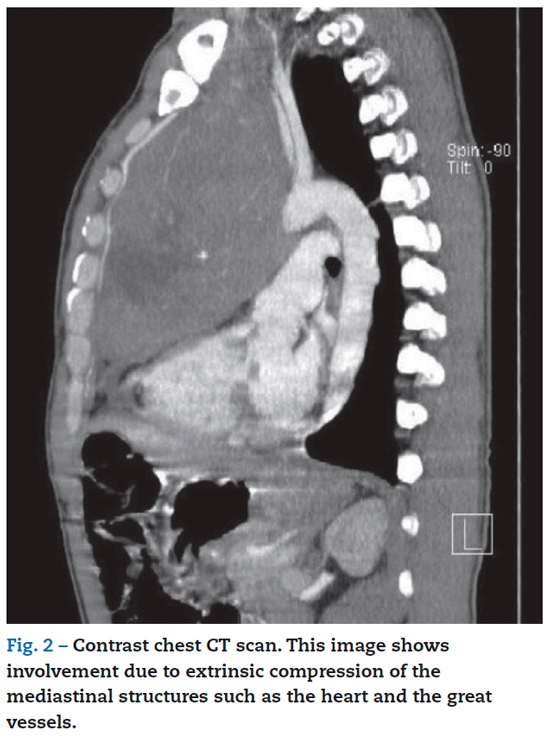
Thirty-five year-old male patient with an anterior mediastinal mass diagnosed as a neuroendocrine carcinoma by histology. The patient responded poorly to initial management with chemotherapy and went on to develop a superior vena cava syndrome and tracheobronchial obstruction. He underwent uncomplicated caval stenting and was then scheduled for endobronchial stenting before proceeding with radiotherapy (figs. 1 and 2).
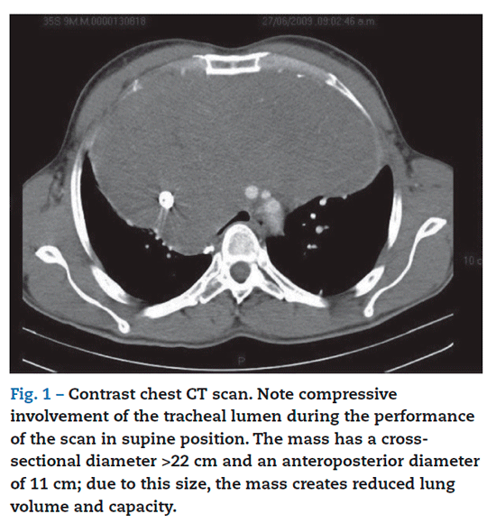
The patient did not have a prior medical or surgical history. He reported heavy smoking and, in the review by systems, he reported difficulty breathing while lying on his back and explained that he slept with several pillows or, occasionally, even in a sitting position. He was scheduled for placement of endobronchial stents by the pulmonology and interventional radiology services. During the preanesthetic assessment, the patient was classified as ASA-PS 4, with no predictors of difficult intubation. Based on the findings of the chest CT scan and the flow-volume curve, he was considered as having a high risk of intraoperative complications associated with airway compromise due to compression (fig. 3).
It was decided to perform the procedure under sedation and local endotracheobronchial anesthesia with fiber optic bronchoscopy, and the use of fluoroscopy for stenting. Basic monitoring was set up using continuous electrocardiography, non-invasive blood pressure monitoring, pulse oxymetry and capnography. The patient was placed in anti-Trendelemburg position. Total collapse of the right main bronchus and
partial left bronchial obstruction was reported during fiber optic bronchoscopy. It was necessary to place the patient in a supine position in order to proceed to stenting because of reasons associated with the use of the fluoroscopy
equipment. After a few seconds in that position, the patient developed significant arterial desaturation of oxygen and bradycardia, requiring the interruption of the procedure and returning to the anti-Trendelemburg position. However, the signs of respiratory distress, desaturation and bradycardia
persisted, requiring the use of positive pressure ventilation and pharmacological treatment with intravenous atropine. In view of impeding ventilatory failure, intravenous induction without muscle relaxant was given, using orotracheal intubation with a ring-design tube. During mechanical ventilation, peak pressures in the airway reached 50 cmH2O but no adequate effective ventilation was achieved. There was marked hemodynamic deterioration and the cardiac rhythm changed progressively from sinus rhythm to bradycardia and then asystole. Resuscitation maneuvers were initiated, the orotracheal tube was repositioned by introducing it through the fiber optic bronchoscope down to the left main bronchus beyond the partial obstruction reported initially. Ventilation improved, followed by improved output rhythm and oxygen saturation. The patient was again placed in the anti-Trendelemburg position, improving ventilation further. This, together with the recovery of spontaneous ventilation, improved the hemodynamic performance. The patient was taken to the intensive care unit, where he died twelve days later from sepsis originating in the lung.
Discussion
Multiple reports have been published for over 30 years of cases of anterior mediastinal masses causing serious complications, with occasional fatal outcomes. Complications are associated mainly with hemodynamic changes and airway obstruction, and they may occur at the time of induction, during extubation, or even days after the procedure.1,2 Many mediastinal tumors may remain asymptomatic and may be identified only as an incidental finding in imaging studies. Manifestations appear when there is a mass effect on the neighboring structures, creating the characteristics signs and symptoms of nervous, cardiovascular o respiratory involvement.3
These patients require thorough preoperative assessment, because inaccurate diagnosis and inadequate planning of
the anesthetic technique may increase the incidence of complications.1 Certain determining factors of intraoperative complications have been considered traditionally, although they have been revised recently.2
The predictors of complications have included the degree of compression of the airway,4 mass volume,5 functional pulmonary tests such as spirometry,6 and other variables such as the clinical spectrum of the patient's signs and symptoms.1 Spirometry in particular is not useful for predicting anesthesia-related complications.6 Hnatiuk et al6 observed that despite some abnormal spirometric results, anesthesia was not modified when compared with patients with normal results. Moreover, this test did not show a good correlation with the degree of airway obstruction. For this reason, imaging studies are now considered more adequate for assessing the compromise created by the lesion, when used together with the clinical history of associated signs and symptoms such as orthopnea, stridor, dyspnea, etc.2,7 In a retrospective study of 98 patients with risk factors for intraoperative and postoperative complications, Bechard et al2 found a significant association with the mixed restrictive-obstructive pattern and the reduced peak expiratory flow in the flow-volume curve.
Patients with anterior mediastinal masses must be taken to preoperative chest CT in order to determine the site, severity and degree of airway obstruction. Traditionally, an obstruction of the tracheal lumen greater than 50% has been considered to render a procedure under general anesthesia unsafe, because of the greater risk of complications.7 However, no association has been found between this predictor and the development of complications, at least during the intraoperative period.2
Another valuable component of the preoperative assessment, perhaps the most important, is the patient's clinical status, where the greater the severity of the symptoms, the greater the risk of complications. Asymptomatic patients may lie in decubitus without showing symptoms. Patients with mild symptoms may develop coughing or an oppressive sensation while in decubitus. Patients with moderate symptoms sometimes cannot tolerate the supine position, and patients with severe symptoms usually cannot tolerate decubitus.7 Other manifestations associated with the tumor mass include dyspnea, orthopnea, syncope, superior vena cava syndrome, chest pain, etc.; these suggest additional cardiovascular involvement.2,8 When severe, clinical manifestations may be a reliable predictor of complications, as shown by the work by Bechard et al.
Anesthetic management of these patients starts with a careful anesthetic plan including not only preventive measures but also prompt and effective intervention strategies should complications arise. Apnea must be considered during anesthetic induction, and consideration must be given to the fact that deep anesthesia in patients with very large masses may result in the inability to provide ventilation due to the weight affecting an airway where the integrity of the supporting cartilages has been impaired. Not using muscle relaxants has become an option because of the loss of the mechanics of spontaneous ventilation resulting from the loss of muscle tone and active inspiratory forces that lead to the inability to ventilate.8 If neuromuscular
blockade is required, short half-life muscle relaxants like succinylcholine should be preferred.7 It is important to place the patient on a surgical table that allows for rapid changes in positioning, because in the event of difficult ventilation or severe hemodynamic alterations, the first thing that needs to be done is to reposition the patient in a semi-sitting position. This maneuver reduces gravity-influenced compression of the airway and the cardiovascular structures. An additional useful strategy is to advance the tracheal tube until the end passes beyond any airway obstruction caused by the mass. In the event of persistent obstruction due to mass compression, the use of the rigid bronchoscope has been described as a rescue option in order to achieve adequate ventilation.1
The use of cardiopulmonary bypass has been described in order to prevent or manage complications associated with airway collapse.9 However, this option is not widely available and would take several minutes to set up in case it is required; moreover, and there is no way to guarantee that the patient will not develop sequelae as a result of transient hypoxia.
Conclusions
To date, there are no reliable predictors of intraoperative complications associated with the inability to ventilate the patient, although there are some related that are associated with less severe intraoperative and postoperative complications. Some patients may present severe signs of clinical compromise as a result of the tumor, and they should be considered as high risk for complications.
It is important to consider the usefulness of special airway management techniques such as intubation with the fiber optic bronchoscope with the patient awake. This allows direct evaluation not only of the effect of the mass on the caliber of the airway, but also of the impact of the supine position on ventilation mechanics. Likewise, rigid bronchoscopy may be a rescue strategy in patients with immediate intraoperative complications associated with ventilation.
Funding
Authors' own resources.
Conflict of interests
None declared.
REFERENCES
1. Erdôs G, Tzanova I. Perioperative anaesthetic management of mediastinal mass in adults. Eur J Anaesthesiol. 2009;26:627-32.
2. Bechard P, Letourneau L, Laçasse Y, Cote D, Bussières J. Perioperative cardiorespiratory complications in adults with mediastinal mass. Incidence and Risk Factors. Anesthesiology. 2004;100:826-34.
3. Guzmán F, Morales D, Guerrero Y. Evaluación, diagnóstico y tratamiento quirúrgico de las neoplasias del mediastino. Rev Venez Oncol. 2006;18:19-27.
4. Shamberger RC, Holzman RS, Griscom NT, Tarbell NJ, Weinstein HJ. CT quantitation of tracheal cross-sectional area as a guide to the surgical and anesthetic management of children with anterior mediastinal masses. J Pediatr Surg. 1991;26:138-42.
5. Turoff RD, Gomez GA, Berjian R, Park JJ, Priore RL, Lawrence DD, et al. Postoperative respiratory complications in patients with Hodgkin's disease: Relationship to the size of the mediastinal tumor. Eur J Cancer Clin Oncol. 1985;21:1043-6.
6. Hnatiuk OW, Corcoran PC, Sierra A. Spirometry in surgery for anterior mediastinal masses. Chest. 2001;120:1152-6.
7. Slinger P, Karsli C. Management of the patient with a large anterior mediastinal mass: recurring myths. Curr Opin Anaesthesiol. 2007;20:1-3.
8. Gothard JW. Anesthetic considerations for patients with anterior mediastinal masses. Anesthesiol Clin. 2008;26:305-14.
9. Takeda S, Shinichiro M, Omori K, Okumura M, Matsuda H. Surgical rescue for life-threatening hypoxemia caused by a mediastinal tumor. Ann Thorac Surg. 1999;68:2324-3.
1. Erdös G, Tzanova I. Perioperative anaesthetic management of mediastinal mass in adults. Eur J Anaesthesiol. 2009;26:627-32. [ Links ]
2. Bechard P, Letourneau L, Lacasse Y, Cote D, Bussières J. Perioperative cardiorespiratory complications in adults with mediastinal mass. Incidence and Risk Factors. Anesthesiology. 2004;100:826-34. [ Links ]
3. Guzmán F, Morales D, Guerrero Y. Evaluación, diagnóstico y tratamiento quirúrgico de las neoplasias del mediastino. Rev Venez Oncol. 2006;18:19-27. [ Links ]
4. Shamberger RC, Holzman RS, Griscom NT, Tarbell NJ, Weinstein HJ. CT quantitation of tracheal cross-sectional area as a guide to the surgical and anesthetic management of children with anterior mediastinal masses. J Pediatr Surg. 1991;26:138-42. [ Links ]
5. Turoff RD, Gomez GA, Berjian R, Park JJ, Priore RL, Lawrence DD, et al. Postoperative respiratory complications in patients with Hodgkins disease: Relationship to the size of the mediastinal tumor. Eur J Cancer Clin Oncol. 1985;21: 1043-6. [ Links ]
6. Hnatiuk OW, Corcoran PC, Sierra A. Spirometry in surgery for anterior mediastinal masses. Chest. 2001;120:1152-6. [ Links ]
7. Slinger P, Karsli C. Management of the patient with a large anterior mediastinal mass: recurring myths. Curr Opin Anaesthesiol. 2007;20:1-3. [ Links ]
8. Gothard JW. Anesthetic considerations for patients with anterior mediastinal masses. Anesthesiol Clin. 2008;26:305-14. [ Links ]
9. Takeda S, Shinichiro M, Omori K, Okumura M, Matsuda H. Surgical rescue for life-threatening hypoxemia caused by a mediastinal tumor. Ann Thorac Surg. 1999;68:2324-3. [ Links ]
