


 text in
  English (pdf)
 Article in xml format
 Article references
 Send this article by e-mail
 Cited by SciELO
  Cited by Google
  Similars in
SciELO
Similars in
SciELO  Similars in Google
 Permalink
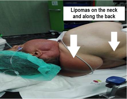
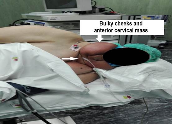
The accompanying images exhibit a large round, subcutaneous fat mass protruding from the posterior cervical area and also several other masses along the back, in a patient with Madelung disease (Image 1). From an anterior view, it is also possible to see big bulky cheeks and another fat mass on the anterior neck (Image 2).
Source: Authors.
Image 1 Large round, subcutaneous fat mass protruding from the posterior cervical area and also several other masses along the back, in a patient with Madelung disease.
Source: Authors.
Image 2 Big bulky cheeks and another fat mass on the anterior neck, in a patient with Madelung disease.
Madelung's disease, also known as multiple symmetric lipomatosis or Launois-Bensaude syndrome is a rare lipid metabolic disorder characterized by diffuse, progressive growth of encapsulated subcutaneous adipose tissue, in the neck, shoulder and other areas, as can be seen in the image. The exact cause of Madelung's disease has not yet been fully understood, but there's one theory that argues that a defect in the adrenergic-stimulated fat breakdown (lipolysis) process could result in improper fat deposits. Madelung's disease affects more males than females (ratio of 15-30:1) and is usually diagnose between 30-70 years of age. For unclear reasons, this disorder appears to be more prevalent in Mediterranean and European population as compared to others regions in the world. 1
As described, the characteristics of Madelung's disease imposes a careful anaesthetic approach because it makes airway management very challenging and peripheral or spinal block difficult, depending on where the masses are located.
The accompanying images show the patient on the operating table after preoperative physical examination of a man suffering from Madelung's disease who required sedation for a colonoscopy (taking into account the patient disease, it was decided that the best local to do the procedure was the operating room). The images reveal a significant cervical mass around the neck forming a typical "Madelung's collar", the posterior cervical lipoma makes cervical mobility nearly impossible and the presence of the anterior cervical lipomas makes it impossible to evaluate thyromental distance, tracheal orientation and also difficult access to front of the neck, if needed. The bulky cheeks predict difficult mask ventilation.
Although it is a procedure where a "common" sedation can be performed, all the predictors of a difficult airway that the patient presents make it also a reliable option to opt to secure the airway with fiberoptic endotracheal intubation or awake videolaryngoscopy before the procedure because in the case an oversedation occur during the procedure, ventilation and secure of the airway would then be time sensible. 2,3
Nevertheless, in this case a sedation was carried out with caution to avoid oversedation and potential hypoventilation and hypoxia.
For these patients it is imperative to perform a regional anaesthesia whenever possible, besides, there should be a plan A, B and C for the management of the airway and carefully discussion with all the members of the team in order to prevent a "can't intubate, can't ventilate" situation.

