




 English (pdf)
 Article in xml format
 Article references

 Automatic translation
 Send this article by e-mail
 Cited by SciELO
 Cited by Google
 Similars in
SciELO
 Similars in Google

 Permalink

INTRODUCTION
Disorders in oral functions such as mastication, breathing and swallowing and their physiological manifestations such as incompetent lips can determine the occurrence or worsening of occlusal disorders. These can also be seen in the facial appearance of patients. 1-3. Muscle management of disorders in oral functions emphasizes the need for non-invasive methods to qualify and quantify these disorders. Non-invasive methods may help give objective information about the muscle activity and muscle strength in patients with normal swallowing and atypical swallowing (AS) both with incompetent lips (IL) and with competent lips (CL). Surface electrode electromyography (sEMG), myoscan analysis and dynamometry 1 are some measurements of choice for these cases because of their low cost and easy implementation.
Swallowing is an automatic motor action, in which respiratory muscles and gastrointestinal tract intervene. The purpose of swallowing is the transport of food bolus and the cleansing of the respiratory tract. Swallowing involves up to 30 muscles and 6 encephalic pairs 4-6.
Atypical swallowing is defined as the anterior or lateral pressure of the tongue against the dental arches, or placed in an anterior or lateral position during swallowing. Atypical swallowing can be diagnosed by visual observation of perioral muscles, and using Payne's technique 7. Incompetent lips are diagnosed by clinical observation of lip position at rest. These diagnoses do not allow for quantitative control to guarantee greater accuracy in the assessment of muscle activity and muscle strength involved in the function. Electromyography, myoscan analysis, and dynamometry could be the solution to this problem.
Techniques to record the muscle activity via EMG are well defined and tested. Potential errors in methodology such as electrode placement have been well described 8,9.
Clinical use of surface electromyography has been proposed in the fields of diagnosis, treatment and control of oral disorders at the muscular and functional levels 10-13 . This is based on the concept that multiple diseases or dysfunctional conditions can be differentiated by means of surface electromyography (sEMG) records of muscle activity14-18, abnormal occlusal positions 19-21, functional hypoactivity and hyperactivity 22,23, muscle spasms 24-25, fatigue 26-27 and muscle imbalance 28. Surface electromyography has been suggested as a useful means to record changes in muscle function prior and posterior to therapeutic interventions as an evidence of treatment success 10,12. For more than a decade, surface electromyography (sEMG) was considered as an alternative to previous studies in order to assess muscle activity associated with swallowing 29; particularly, sEMG studies showed promising results in both adults 30,31 and children 5 in this knowledge area.
In the 1970s, myoscanners and dynamometers were described as measurement instruments of orofacial muscle activity. These devices were designed by Dr. Richrad Shepard and Dr. Daniel Garliner, who have a reference scale of values for the U.S. children and adult population 32.
The purpose of this study was to characterize the values of muscle activity using sEMG and muscle strength using myoscan and dynamometry analyses in patients with atypical swallowing and competent lips or incompetent lips versus a control group.
MATERIALS AND METHODS
This analytical case-control study used the Statcalc calculator in the Epi Info 6 software for the calculation of the sample. Sample was calculated considering an 80% power (1-Beta), a ratio of 1:1 and an expected prevalence of factor in the non-exposed group of 30% (presence of an abnormal sEMG in normal patients), an OR of 2.5 (72% of probability to show an abnormal electromyography in the exposed group) and a prevalence of factor in the exposed group of 51.72 %. The sample would include 88 patients in the exposed group and 88 in the control group. Finally, 90 school students (51 girls and 39 boys) aged between 7 and 13 (10.55 ± 1.21) in the control group and 88 school students (55 girls and 33 boys) aged between 7 and 13 (10.07 ± 1.23) diagnosed with atypical swallowing were examined.
School students were selected from three high schools in the city of Manizales (Caldas, Colombia). This study requested an authorization from the Ethics Committee of the Autonoma University of Manizales (Minutes No. 12 of February 2, 2011) and met all the requirements of the 8430 rule, governing health research applied to patients. Parents or authorized guardians of students received the necessary information about the experience in which they would participate and they previously signed the informed consent. Similarly, students were requested their authorization to participate in the research project.
Diagnosis of atypical swallowing was performed with the traditional protocol, using a Payne black light, model UVL-56, Black Ray lamp, long wave UV (nm), 115 volts, 60 Hz, 0.16 Amps, UVP, Upland.CA91786, USA. This was used to know the specific location of the tongue during swallowing.During the examination, the patient was sitting with the back straight, head against the wall, and feet on the floor. The patient should have pulled the tongue forward to place a 10% fluorescein solution in the anterior part, apex and lateral sides. Patients were asked to swallow in order to observe, then, the place where the solution was recorded intra-orally using the black light.
SEMG PROCEDURE
SEMG records were performed with the elec-trophysiograph and Cadwell Sierra Wave software. Acquisition parameters were: sampling frequency of 76.8 kHz, the bandpass filter 10 to 10k Hz and gain 200uV. For acquisition and storage of records, the EMG protocol was selected. 302139-200 stainless steel disk electrodes (Cadwell) of a 10 mm diameter were used.To perform the SEMG record, school students were sitting against the back of a chair with the back straight, arms relaxed, feet on the floor, eyes open and staring at a point in front of them. Prior to the positioning of the electrodes, their faces were cleaned with a piece of cotton with 95% ethanol, Cadwell conductivity gel model 202153-000 was used in order to reduce the artifact occurrence due to the low conductance. The ground electrode, common to all electrode pairs, was placed 2 cm above the nasion.This study assessed the muscle activity of the right and left masseter muscles and left orbicularis oris and mentalis muscles; these were assessed unilaterally because of their symmetric function; these muscles work predominantly in the first phase of swallowing. A bipolar configuration was selected for masseter, orbicularis oris and mentalis muscles. These configurations are sensitive to changes in the strength exerted by a muscle 33. Electrodes were placed as follows: Left masseter: subjects were asked to close teeth with maximal force. Masseter muscle was then examined. An active electrode was placed on the motor point, reference electrode was placed 1 cm under the ear lobe. Left orbicularis oris: Active electrode was placed 1 cm above the upper lip vermilion and a reference electrode was placed on the zygomatic bone. Finally, Mentalis muscle: active electrode was placed on the chin, 1 cm from the midline and a reference electrode was placed 2 cm above the distal direction.
Every muscle was assessed independently. Maximal voluntary contraction (MVC) of the orbicularis oris muscle (10 seconds of pursed lips) was determined. Nine swallows of 25 ml of water (three by each muscle) were requested. Subjects were requested to keep the liquid in the mouth until staying at rest. Subjects were instructed to "swallow as usual" to promote the normal way of doing it by each subject. Elapsed time during the SEMG process did not exceed 10 minutes.
This study examined the amplitude of muscle activity while swallowing and the MVC of the orbicularis oris muscle. Amplitude corresponds to the peak to peak voltage of the interference pattern of the muscle activity at the time of swallowing.
MYOSCAN PROCEDURE
The myoscan used was the Pounds Myos-canner, Neilco Technology Inc., D-926119T8 Hoxt, 1volt=F.s., pt 2500. The physiological force of muscles was estimated by means of a plug. This allowed the measurement of the lip compression strength, strength of tongue projection and contraction of masseter muscles. To do the examination, patients were asked to sit comfortably with their head against a wall and feet on the floor. First, the right masseter muscle contraction was assessed. Patients were asked to close their teeth tightly. The examiner touched the muscle with the tips of the index and middle fingers; the same action was repeated three times in order to determine its location. Then, the test began by calibrating the myoscan in order for its needle to be exactly 0.0 pounds. A plate was set on the masseter muscle and patients were asked again to have a maximal occlusion. The same procedure was made on the left masseter (Reference values: the result was from 0.4 to 0.6 pounds for students aged between 4 and 10 years old and the result was from 0.6 to 0.8 pounds for students older than 10 years old).
The measurement of the compression strength of the orbicularis oris muscle began with the myoscan calibration set at 0.0 pounds. Patients were asked to close the teeth, stretch lips forward and press the plate strongly. (Reference values: the result was from 0.2 to 0.4 pounds for students aged between 4 and 10 years old and the result was from 0.4 to 0.6 pounds for students older than 10 years old). Finally, the tongue tensile strength was assessed. Patients were instructed to protrude the tongue through the hole in the plate held by the examiner. (Reference values: the result was from 0.6 to 0.8 pounds for students aged between 4 and 10 years old and the result was from 0.8 to 1.2 pounds for students older than 10 years old). All measurements of the myoscan procedure were repeated three times and the average was written for each of them.
DYNAMOMETRY PROCEDURE
The dynamometer used was Chatillon Inc. (New York, NY, USA), model CHA LN-10. Results were measured in pounds of force and were taken with a dynamometer with a hook attached to a six-inch cord, ending in a button of 1 inch in diameter. The button was placed in front of the incisors and the back of the lips. The researcher pulled the dynamometer until the button was expelled and then the result was read. The normal range for lip force, listed by Garliner, is of 3 to 5 pounds 7. To do the examination, patients were sitting, head against the wall, arms at their sides, and feet on the floor.
STATISTICAL ANALYSIS
Statistical analysis was performed with the spss statistical package (spss® ibm® v.20). Comparisons were made with the Mann-Whitney U test because most variables did not meet the normality tests (Kolmogorov-Smir-nov) nor homogeneity of variance (Levene's test). These variables were analyzed under a 95% confidence interval.
RESULTS
The amplitude of the SEMG record of orbicularis oris muscle when swallowing was signicantly higher in the IL group than in the CL group and the control group (Table 2). This difference among groups was only significant for the right masseter. The amplitude of the maximal voluntary contraction of the orbicularis oris muscle was significantly lower for patients with atypical swallowing than for the control group. In the mentalis muscle, when swallowing, the amplitude of the SEMG showed no significant differences among the groups (Table 1).
Table 1 Amplitudes of the EMG record and strength of the muscles studied in patients with AS versus controls
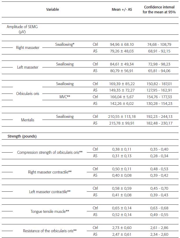
Table 1. Amplitude of sEMG waveforms and strength measurements of studied muscles. Comparison of measurements of patients with atypical swallowing and the control group. (Ctrl) Control group (n =90). (AS) Atypical swallowing group (n =88). (a) Calculation made with Mann-Withney U test for two independent samples. (*) p<0.05. (**) p<0.01.
The student group with as was divided into patients with competent lips (CL) and incompetent lips (IL). The CL group showed an amplitude of the SEMG record of the right masseter muscle significantly higher than that of the control group. The amplitude of the SEMG record of orbicularis oris muscle when swallowing was significantly in the IL group than in the CL group and the control group (Table 2).
Table 2 Amplitudes of the EMG record and strength of the muscles studied in patients with SA and CL and IL versus controls
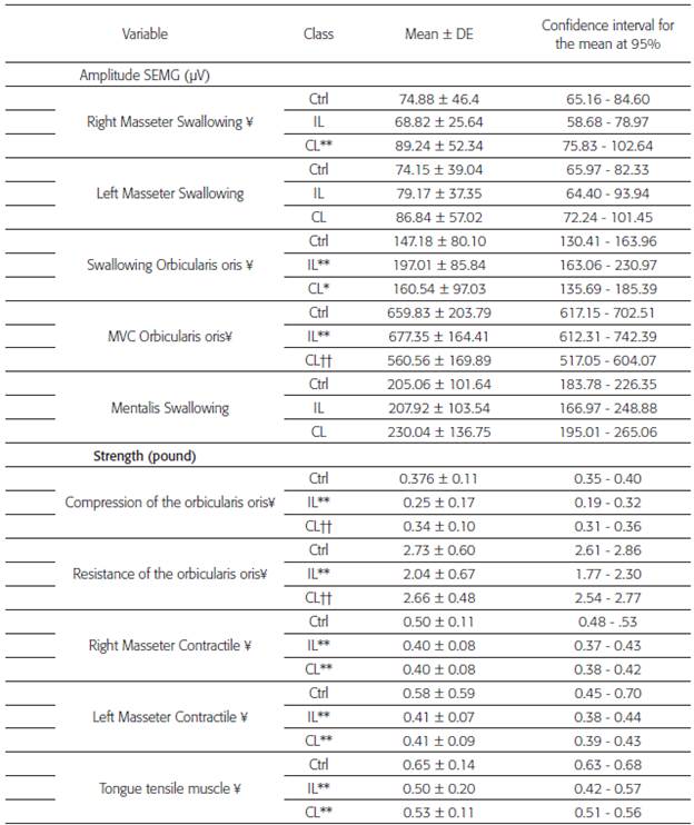
Table 2. Amplitude of the sEMG waveforms and measurements of strength of the muscles studied. Comparison of measurements of patients with atypical swallowing and the control group. Ctrl: Control group (n =90). IL: Group of atypical swallowing with incompetent lips (n =27). CL: Group of atypical swallowing with competent lips (n =61).
(¥) x2 of the Kruskal-Wallis test with p<0.05.
(1) Calculation made with Mann-Withney U test for the two independent samples.
(*) Differences in respect to the control group p<0.05.
(**) Differences in respect to the control group p<0.01.
(††) Differences among the AS-CL and AS-IL groups p<0.01.
In maximal contraction of the orbicularis oris, the amplitude of the SEMG record was significantly lower in the CL group than in the IL group and the control group.
Contractile forces of the masseter muscles, the compression strength of the orbicularis oris muscle and tongue tensile strength were measured by means of the myoscan analysis and the resistance force of the orbicularis oris muscle via dynamometry. Patients diagnosed with atypical swallowing showed a decrease in the compression strength and resistance strength of the orbicularis oris, contractile force of masseter muscles, and tongue tensile muscle (Table 1).
When analyzing the strength of these muscles in the cl and IL groups, the findings were as follows. The control group showed higher contractile forces of the masseter muscles, tongue tensile muscle, and resistance of the orbicularis oris compared to both IL and cl groups (p <0.05). There were no differences between these two groups. The IL group showed a compression force and resistance of the orbicularis oris lower than the control group and cl group (Table 2).
DISCUSSION
In this research, school students with atypical swallowing (AS) showed greater electromyographic activity than patients in the control group. The contraction of mentalis and orbicularis oris muscles during atypical swallowing has been considered a clinical sign of abnormality. Stormer et al. 34 reported that patients with as, during saliva or water swallowing, may have an increased electromyographic activity of perioral muscles, contrary to the findings of this research work. They also found a decreased activity of the masticatory muscles, which was similar to the findings of this research work.
Patients with as and IL showed higher electrical amplitude than LC and controls in the orbicularis oris. These patients with as would show higher amplitude in the waveforms of these muscles due to a harder effort when swallowing. The more motor units activated to produce the same force, the higher the reduction of the electromechanical efficiency. When more effort is exerted on these muscles, there is higher amplitude recorded in the activity. Thus, the increase in amplitude would be caused by a higher amount of recruited fibers, not necessarily by higher strength, which is similar to Archer's records.
In this work, the average contractile force of the tongue, registered with the myoscan, was lower in the AS group. The role of the tongue in swallowing is well known, however, as Lear Moores 36, the tongue produces four times more pressure during swallowing than the perioral musculature. In normal swallowing, most of this pressure is exerted on the palate, but not on the teeth, unlike to what happens in atypical swallowing, where these forces are directly applied on the teeth, causing protrusion of the incisors and an increase in the horizontal dimension. Rogers 37 studied swallowing patterns and concluded that the majority of the children who presented tongue pressure in swallowing had overjet with 'short' lips, and the tongue moved itself freely through the anterior teeth without touching the lips. In his study, Stormer 34 found a lower SEMG activity in the masticatory muscles of patients with AS than in controls. This author stated these patients seem to have perioral muscle activity without the intervention of the temporalis muscle and higher activity in the orbicularis oris and mentalis muscles; these findings are different from the ones in this research work.
Electromyographic studies done by Ahlgren 38 and Tosello et al. 39 have shown that the orbicularis oris muscle has a greater activity in patients class II/I with incompetent lips during swallowing than in patients with competent lips. Findings in these studies show that patients with increased lower facial third proportions and/or maxillomandibular anteroposterior discrepancies are more susceptible to an increased perioral muscle activity and incompetent lips. Gustafsson and Ahlglen 8 found greater activity of the orbicularis oris (upper and lower) in the resting posture of the lips, during swallowing. They state that perioral contraction during atypical swallowing is a common characteristic for individuals with incompetent lips, independent of tongue function.
The pressures exerted by the lips and cheeks in the buccal stage of swallowing are associated mainly with labial sealing and with the expulsion of liquids from the buccal vestibule rather than with the labial musculature contraction in the elevation of the larynx. In this first stage, larger potentials are generated by the orbicularis oris 40.
Lowe et al.41 claim that patients with IL have a reduced activity of the orbicularis oris muscle, which is opposite to the findings in this study. Patients with IL have difficulties to chew with labial contact; this explains the higher activity of the perioral muscle, which would be reflected on the results of this research. Pallú et al. also observed a great activity of the orbicularis oris in patients with IL 42. This would mean that these patients should make a greater effort to obtain good closure during swallowing.
In the study of Dutra et al.43 patients with incompetent lips showed masseter muscle hyperactivity during swallowing, suggesting a visceral type of swallowing. In this study, patients with AS and incompetent lips showed no masseter muscle hyperactivity, but there was a muscle hyperactivity of the orbicularis oris. Balata 44 found that patients with incompetent lips showed a higher amplitude and a lower coefficient of variation of muscle activity, which are consistent with the records available for this study. During closure of the lips, it has been found that patients with incompetent lips show a muscle activity of the upper orbicularis oris and mentalis muscles greater than subjects with competent lips, who show, in most cases, a slight increase in mentalis muscle activity 8.
CONCLUSIONS
Amplitude of the SEMG record of left and right masseter, orbicularis oris and mentalis muscles in school students diagnosed with as was lower than that of the control group; that amplitude was statistically significant in the orbicularis oris.
Patients with AS diagnosis showed a decrease in the compression and resistance forces of the orbicularis oris, contractile of masseter muscles and tongue tensile strength. The difference with respect to the control group was statistically significant.
The IL group recorded a higher activity in the orbicularis oris than LC and control groups; MVC of the orbicularis oris was statistically significant.
Recommendation
The use of the support materials described in this study (EMG, myoscan and dynamometry) gives the possibility to go beyond the subjective clinical criteria of the clinical diagnosis and to achieve more objective parameters to attain a reliable diagnosis.
Disclosure
The authors report no conflict of interests related to materials and methods used in this study or the findings reported in this project.

