




 text in
  English (pdf)
 Article in xml format
 Article references
 Automatic translation
 Send this article by e-mail
 Cited by SciELO
  Cited by Google
  Similars in
SciELO
Similars in
SciELO  Similars in Google
 Permalink

Introduction
Hydatidosis is a zoonotic infection caused by parasites of the Echinococcus genus, most commonly E. granulosus and E. multilocularis, which are responsible for its two primary forms: hepatic echinococcosis (HE) and alveolar echinococcosis (AE), respectively1. In endemic regions for E. granulosus, such as South America, Eastern Europe, the Middle East, Russia, and China, incidence rates can reach up to 50 cases per 100,000 people per year, with prevalence ranging from 20% to 95%, varying in high-risk areas such as those where animals are slaughtered for human consumption2. Regarding mortality, untreated or inadequately treated HE has a rate of 2% to 4%, while AE can reach up to 90% within 10 years of diagnosis1.
Imaging and serology are used for diagnosis, with ultrasound being the first-line choice as it allows for lesion characterization and staging3,4. Treatment involves an interdisciplinary approach-medical, surgical, or minimally invasive-including the administration of anthelmintics such as albendazole. Depending on the patient’s symptoms and cyst characteristics, procedures like hepatectomy, cystopericystectomy, or capsulectomy may be performed4,5.
Case Report
A 66-year-old female patient with a history of type 2 diabetes mellitus and hypertension, residing in an urban area, presented to the emergency department with a one-month history of intermittent right hypochondrium pain and a palpable mass associated with erythema, edema, localized warmth, and unquantified febrile episodes. Physical examination revealed no abdominal distension or clinical signs of peritoneal irritation; however, hepatomegaly of 15 cm below the costal margin was noted. Initial blood biochemistry showed no significant abnormalities.
A full abdominal ultrasound revealed a hypoechoic area measuring 75 x 60 x 41 mm (95 mL in volume) in segment VI of the liver and another well-defined, heterogeneous subhepatic expansile lesion measuring 90 x 73 x 58 mm. Possible etiologies included a liquefying liver abscess, a neoproliferative expansile lesion, or a hydatid cyst. An abdominal computed tomography (CT) scan was subsequently performed, which confirmed an intraparenchymal collection in hepatic segment VI, an underlying intraperitoneal collection, and an intramuscular collection in the right flank (Figure 1). Tumor markers were assessed due to suspicion of a neoplastic lesion but showed no abnormalities. Consultations with Infectious Diseases and Hepatopancreatobiliary Surgery led to a diagnosis of hepatic hydatidosis, and empirical therapy with albendazole (400 mg every 12 hours for 30 days) was initiated, with outpatient follow-up scheduled.
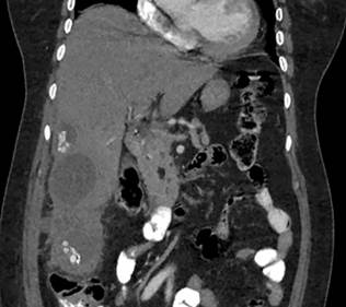
Image property of the authors.
Figure 1 Coronal section of an abdominal CT scan showing hepatic and peritoneal involvement.
The patient experienced a poor clinical course due to persistent symptoms. Upon reevaluation by the treating team, spontaneous rupture of the hydatid cyst into the peritoneal cavity was suspected. Follow-up blood biochemistry studies revealed neutrophilic leukocytosis and elevated C-reactive protein (CRP). Subsequent serology for Echinococcus returned positive (enzyme-linked immunosorbent assay [ELISA] = 1/800). An abdominal magnetic resonance imaging (MRI) scan was performed, clearly demonstrating extensive hepatic involvement as well as abdominal wall invasion (Figure 2).
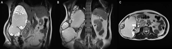
Image property of the authors.
Figure 2 Follow-up abdominal MRI. A. Coronal view showing extensive hepatic involvement with multiple hydatid cysts. B and C. Coronal and axial views confirming hydatid cyst infiltration into the abdominal wall.
The patient underwent image-guided percutaneous drainage, yielding approximately 200 mL of frank purulent fluid. Gram staining revealed gram-positive cocci, though subsequent culture was negative. The case was discussed in a multidisciplinary medical meeting, and surgical management was decided. During the procedure, a large mass composed of multiple mixed-component (solid and liquid) cysts was found, involving hepatic segments V, VI, and part of VII, with spontaneous drainage into the abdominal wall. The cysts fully infiltrated the transverse abdominal muscle and 80% of the right internal oblique. A hepatic resection of the hydatid cysts and concurrent abdominal wall cyst excision were performed. Postoperatively, the patient recovered well and was discharged after 17 days to complete outpatient antiparasitic therapy. Histopathology confirmed the presence of a germinal membrane, a proliferative capsule, and protoscolices of E. granulosus, along with significant pericystic inflammatory reaction and marked infiltration of mononuclear and giant cells in the muscle tissue.
Discussion
Hepatic hydatidosis, or echinococcosis, is a zoonotic infection with hepatic and alveolar presentations, caused primarily by Echinococcus granulosus and E. multilocularis1. Other species (E. vogeli and E. oligarthra) are associated with polycystic and rare unicystic forms, respectively6. It is a public health concern, particularly in developing countries, and the World Health Organization (WHO) has classified it among the 17 neglected tropical diseases targeted for control or elimination by 20501,3.
Endemic regions report incidence rates of up to 50 cases per 100,000 people/year, with prevalence ranging from 20% to 95% in high-risk areas. Although surgical cases are reasonably documented, they represent only a fraction of total infections. Low socioeconomic status, plant-based subsistence, regional climate, and livestock rearing with poor sanitary controls are established risk factors2. Humans are incidental hosts and do not participate in the parasite’s life cycle, which requires a definitive host (typically canines) for adult development and an intermediate host (e.g., pigs, sheep, or goats) for the larval stage. Humans ingest infectious eggs excreted by definitive hosts before larval development begins (Figure 3)3,6,7. Human-to-human transmission has not been reported3.
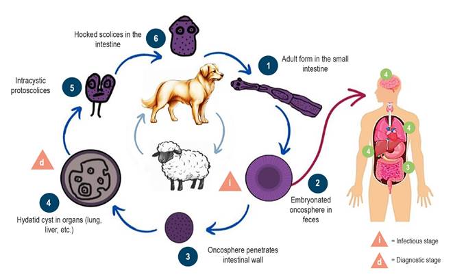
Figure 3 Life cycle of Echinococcus granulosus. Prepared by the authors based on information from the Centers for Disease Control and Prevention (CDC), Atlanta.
The organs most frequently affected by echinococcosis are the liver (50-70% of cases) due to intestinal venous drainage, followed by the lungs (20-30%), and less commonly the spleen, heart, kidneys, bones, and central nervous system (CNS)2,3. Symptoms vary depending on the affected organ and are often absent in early stages. Hepatic symptoms typically emerge when the cyst(s) occupy more than 70% of the organ’s volume or exceed 10 cm in diameter, compressing or destroying biliary ducts or vascular structures. In this case, the hepatic lesion measured 75 × 60 × 41 mm, while the extrahepatic lesion reached 90 × 93 × 58 mm.
Clinical manifestations of hepatic hydatidosis often include abdominal discomfort/pain, decreased appetite, nausea/vomiting, and jaundice in cases of biliary obstruction. Physical exam may reveal a palpable upper abdominal mass, hepatomegaly (the most common sign), or abdominal distension. Cyst rupture can cause symptoms ranging from fever, urticaria, and eosinophilia to anaphylactic shock and death1. This patient’s presentation aligned with the most frequently reported manifestations, including physical exam findings, though eosinophilia was absent on blood biochemistry.
Due to slow growth (approximately 1-5 cm/year)8, most cases are incidentally detected on imaging during evaluation for nonspecific symptoms or unrelated conditions3,5. For symptomatic patients or incidental findings suggestive of the disease, diagnosis relies on combined imaging and serology. A detailed clinical history should identify risk factors (e.g., residence in/travel to endemic areas, contact with definitive hosts) alongside thorough physical examination3.
The WHO Informal Working Group on Echinococcosis (WHO-IWGE) classifies cysts based on ultrasonographic findings and corresponding treatments (Tables 1 and 2)4. Liver ultrasound is the preferred diagnostic modality, with 95% sensitivity9 for lesion identification, characterization, and staging, as well as post-treatment follow-up in endemic areas. It is also valuable for surveillance and screening5 and is the most sensitive tool for detecting membranes, septations, and hydatid sand2. Here, ultrasound identified hepatic lesions and suggested the diagnosis, classifiable as CE1 based on sonographic features.
Table 1 WHO-IWGE Classification of Hydatid Cysts
| Stage | Ultrasonographic Appearance (WHO-IWGE) |
|---|---|
| CL | Unilocular anechoic cyst, no internal echoes/septations |
| CE1 | Cyst with fine internal echoes (hydatid sand): active cyst |
| CE2 | Multiseptated cyst with "honeycomb" appearance (unilocular primary cyst): active cyst |
| CE3 | Unilocular cyst with detached membranes ("water lily sign," CE3a) or daughter vesicles with hypo-/hyperechoic images (CE3b): transitional cyst |
| CE4 | Mixed hypo-/hyperechoic content, no daughter vesicles ("ball of wool" appearance): degenerative cyst |
| CE5 | Partially/fully calcified wall: inactive cyst |
Table prepared by the authors.
Table 2 Hydatid Cyst Therapy Protocol
| Stage | Size | First-Line Treatment | Alternative Therapy |
|---|---|---|---|
| Contraindication/invasive treatment refused | ABZ (6 months) | ||
| CE1, CE3a | Small | ABZ alone (6 months) | PAIR + ABZ (1 month) |
| Medium | Surgery + ABZ (1-6 months) | PAIR + ABZ (1 month) | |
| Large | Surgery + ABZ (1-6 months) | MoCaT + ABZ (1 month) | |
| CE2, CE3b | Small | ABZ alone (6 months) | MoCaT + ABZ (1 month) |
| Medium | Surgery + ABZ (1-6 months) | MoCaT + ABZ (1 month) | |
| Large | Surgery + ABZ (1-6 months) | MoCaT + ABZ (1 month) | |
| CE4, CE5 | Any | "Watch and wait" | "Watch and wait" |
| Complicated cysts (any stage) | Any | Surgery (± interventional endoscopy for biliary rupture) + ABZ (6 months) | Surgery (rupture) / percutaneous drainage (infection) + ABZ (1 month) |
ABZ: albendazole; MoCaT: modified catheterization; PAIR: puncture, aspiration, injection, reaspiration. Table prepared by the authors.
CT is typically the complementary imaging study of choice, providing more accurate assessment of cyst location, size, and hepatic parenchymal integrity. It is particularly useful for small cysts or when extrahepatic, vascular, or adjacent structure involvement (e.g., biliary tract) is suspected, with 95% sensitivity9. Additionally, it enables detailed surgical planning for complex or atypical cases like this one, where it clearly delineated the extensive abdominal wall involvement and guided surgical strategy. MRI is more valuable for CNS involvement2,5.
Serologic testing for Echinococcus serves as another diagnostic tool, especially when imaging findings are suggestive but not pathognomonic. However, results require careful interpretation since specific antibodies may remain positive for years even after radical surgery, not necessarily indicating active infection. In this case, the patient’s 1/800 titer suggested active infection. Several factors influence interpretation: WHO-IWGE stage (discussed earlier), size (>15 cm vs. <15 cm), hepatic/extrahepatic location, cyst multiplicity, and complications. One study found hepatic CE1 and CE4-5 cysts were seronegative in 30-58% and 50-87% of cases, respectively, whereas CE2/CE3 cysts had lower negativity rates (5-20%). Thus, diagnostic challenges arise with small CE1 cysts and inactive CE4-5 cysts exhibiting nonspecific imaging.
A positive correlation exists between active cyst size and serologic positivity, attributed to increased antigenic mass from cyst wall compromise (whether due to immune exposure or therapy)10. Fine-needle aspiration or biopsy may aid diagnosis when results remain inconclusive5. Other laboratory findings are nonspecific but may include leukopenia, thrombocytopenia, or liver profile abnormalities. Neutrophilia or eosinophilia may suggest secondary infection or cyst rupture-consistent with this patient’s neutrophilia, supporting the suspected rupture during reevaluation for persistent symptoms8.
Treatment options include medical, surgical, or minimally invasive approaches11. Medical management involves anthelmintics like albendazole (10-15 mg/kg/12h for 3-6 months) or mebendazole (if albendazole-intolerant). Adding praziquantel (25 mg/kg/24h) to benzimidazoles may enhance efficacy. After one year of medical therapy alone, ~30% of cysts resolve, 30-50% shrink, and 20-40% remain unchanged. Antiparasitics are also used adjunctively: preoperatively to reduce cyst viability and postoperatively to prevent intraoperative contamination and recurrence4,5,8.
As previously described, the patient was initially treated with albendazole upon diagnosis and continued postoperatively to complete eradication therapy. For symptomatic, complicated, large, or active cysts, surgery remains the primary treatment. Surgical approaches include hepatectomy, pericystectomy, and capsulectomy, with technique selection depending on surgeon preference, patient characteristics, preoperative imaging planning, and intraoperative findings. Margin assessment and lymph node dissection have not proven beneficial. All procedures require peritoneal cavity protection-isolating the cyst to prevent content spillage and shielding the surgical field with hypertonic saline (typically 20%)-soaked gauze or anthelmintics. Laparoscopic intervention is preferred for anterior liver surface cysts (<3 cysts) without biliary involvement4,8. Cyst aspiration/injection carries complication rates of 1.4-13.7%, high therapeutic failure risk, and potential peritoneal dissemination9.
Follow-up with imaging and serology is recommended every six months for the first two years, then annually based on clinical judgment and patient status2.

