




 text in
  English (pdf)
 Article in xml format
 Article references
 Automatic translation
 Send this article by e-mail
 Cited by SciELO
  Cited by Google
  Similars in
SciELO
Similars in
SciELO  Similars in Google
 Permalink

Introduction
Fibrovascular polyps are benign submucosal and intraluminal tumors composed of fibrous tissue, adipocytes, and vascular structures, and are covered by normal squamous epithelium. They are extremely rare tumors, accounting for less than 2% of all benign esophageal tumors, and their most common symptoms include dysphagia, weight loss, odynophagia, gastrointestinal bleeding, and respiratory symptoms. They are most frequently located in the proximal third of the esophagus, are usually slow-growing, and are characterized by reaching large dimensions: polyps up to 25 cm in length have been described in the literature1.
We present the case of a patient with a large fibrovascular polyp located in the distal third of the esophagus.
Case Report
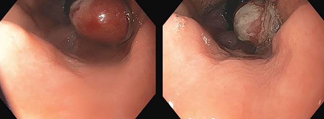
A 54-year-old male patient presented to the outpatient clinic with symptoms of progressive dysphagia and weight loss. He denied a history of gastroesophageal reflux, hematemesis, or melena, a family history of neoplasms, or other relevant pathologies. The physical examination revealed no abnormalities, with documented absence of lymphadenopathy and positive abdominal findings. Given the diagnosis of dysphagia, an upper gastrointestinal endoscopy was requested, which revealed a polyp at 38 centimeters from the dental arches (Figure 1).
Images property of the authors.
Figure 1 Polyp located 38 cm from the dental arches, adjacent to the Z-line.
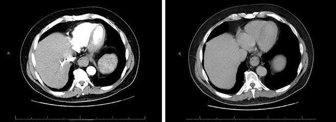
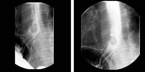
A thoracoabdominal computed tomography (CT) scan was requested (Figure 2), which ruled out probable neoplastic pathology or lesions suggestive of metastasis. Consequently, an esophagogram was ordered, which reported a 25 mm esophageal polyp in the distal esophagus, with normal esophageal transit, no evidence of stenosis, dysmotility, or other significant alterations (Figure 3).
Images property of the authors.
Figure 2 Computed tomography scan describing the esophageal lesion as a polyp and ruling out lesions suggestive of metastasis.
Images property of the authors.
Figure 3 The polyp is observed in the distal third of the esophagus.
Given the endoscopic finding, an endoscopic ultrasonography was performed, which reported findings consistent with a fibrovascular polyp of the same size. No other masses or lymphadenopathy were observed in the upper digestive tract. The patient underwent endoscopic polypectomy with a hot snare, and the diagnosis was confirmed histologically; the outcome was adequate, with no recurrence of the mass.
Discussion
Fibrovascular polyps are rare submucosal tumors that originate predominantly in the cervical part of the esophagus at the pharyngoesophageal junction. Although their true incidence is unknown, they are estimated to account for less than 2% of benign esophageal tumors2. They are observed more frequently in middle-aged and elderly men3. Due to an initial lack of symptoms, they can reach a considerable size, with the largest reported size being 25 cm4.
Fibrovascular polyps are often completely asymptomatic and only become clinically significant when they have reached a considerable size; 62% of patients may present with dysphagia, 38% with regurgitation of the mass, and 25% with a persistent lump sensation in the throat5. Other less frequent symptoms include retrosternal discomfort, odynophagia, dyspnea, cough, and weight loss. It is important to mention that serious complications have been reported, such as gastrointestinal bleeding from ulceration of the polyp and even death from asphyxiation6,7. The presence of dysphagia and weight loss in the presented patient is consistent with the typical clinical presentation of a fibrovascular polyp described in the literature, despite the absence of other common symptoms.
The diagnosis of an esophageal fibrovascular polyp is a complex process that requires a combination of techniques and a multidisciplinary approach, as these polyps can be confused with other conditions, such as achalasia or mediastinal tumors causing extrinsic esophageal compression, underscoring the importance of an accurate diagnosis8.
The diagnostic process should begin with a detailed medical history and a thorough physical examination. These initial steps are crucial for identifying the aforementioned characteristic symptoms, which may point towards the presence of an esophageal polyp. More specific studies should then be performed, with the barium swallow or esophagogram being the diagnostic technique of choice9. This test typically reveals an intraluminal filling defect in the cervical esophagus, which may extend distally. The characteristic image is that of a “sausage-shaped” mass originating in the upper esophagus and potentially reaching the lower esophagus.
Upper gastrointestinal endoscopy plays a fundamental role in diagnosis, allowing visualization of a mobile intraluminal mass covered by normal mucosa, and a careful examination of the upper esophageal sphincter may reveal the stalk of the mass. This technique allows for biopsy sampling, identification of ulcerated areas, and, in some cases, polyp resection. However, endoscopic diagnosis can be challenging because the squamous epithelium covering the polyp may be indistinguishable from the normal esophageal mucosa10.
Endoscopic ultrasound complements conventional endoscopy by providing valuable information about the internal structure of the polyp. This technique is especially useful for delineating the polyp’s stalk, assessing its histological composition and vascularization, and reporting on possible tumor infiltration and the presence of lymphadenopathy. Endoscopic ultrasound also allows determination of the submucosal origin of the polyp, which is crucial for the differential diagnosis, as was performed in our patient11.
CT and magnetic resonance imaging (MRI) are valuable complementary tools in the diagnostic process. These advanced imaging techniques provide detailed information about the extent and composition of the polyp. Neck and chest MRI, in particular, can be decisive for treatment planning, as it accurately demonstrates the origin of the stalk and the composition of the polyp11. Furthermore, these techniques are useful for ruling out the possibility of mediastinal involvement and for preoperative evaluation.
It is important to note that endoscopic biopsies may be inconclusive due to the submucosal nature of the lesion. Therefore, the definitive diagnosis is established by histopathological examination of the resected specimen. This analysis reveals the presence of fibrovascular tissue covered by normal squamous epithelium, confirming the benign nature of the polyp11, as occurred in our patient.
The treatment for fibrovascular polyps is surgical or endoscopic resection, depending fundamentally on the site of stalk insertion, the size of the lesion, its mobility, and the number of feeding vessels. Those with predominant fatty composition are good candidates for endoscopic resection, whereas if they are highly vascularized, the risk of bleeding increases, and surgical excision may be preferable, since hemostasis is achieved more safely with open surgical techniques12. Surgical resection is preferred due to the potential risk of respiratory compromise and hemorrhage, in addition to serving to exclude cancer and avoid the small risk of malignant degeneration13.
For polyps larger than 8 cm in length or those with a thick stalk, rich vascularization, and originating in the upper third of the esophagus, surgical resection via cervical esophagotomy is preferred as the primary intervention. Total esophagectomy is reserved for more severe cases. Thoracotomy is considered for more caudal or larger lesions. Generally, polyps less than 2 cm in diameter with thin stalks can be removed by endoscopic ligation of the stalk and electrocautery, as was performed in the patient in this case report13,14.
The procedure allowed for complete excision of the esophageal fibrovascular polyp. Following the intervention, the patient showed a favorable recovery. During postoperative follow-up, no recurrence of the tumor mass was detected, and the patient did not present complications such as bleeding, perforation, or surgical site infection.
Conclusions
Fibrovascular polyps are an unusual pathology that should be considered as a differential diagnosis in adult patients presenting with dysphagia. Diagnosis is made by upper gastrointestinal endoscopy and confirmed histologically. Complementary examinations such as computed tomography and endoscopic ultrasonography should be performed. Endoscopic resection is the best therapeutic option.

