


 text in
  English (pdf)
 Article in xml format
 Article references
 Send this article by e-mail
 Cited by SciELO
  Cited by Google
  Similars in
SciELO
Similars in
SciELO  Similars in Google
 Permalink
Introduction
Pyloric syndrome, or gastric outlet obstruction (GOO), is a condition characterized by the inability of the stomach to empty its contents, secondary to motility disorders or, more commonly, mechanical causes1,2. The latter arise from intrinsic or extrinsic obstruction at the level of the distal stomach, pylorus, or duodenum. These conditions cause acute or chronic symptoms such as abdominal pain, nausea, vomiting, and postprandial fullness4. Prior to the introduction of proton pump inhibitors, peptic ulcer disease was the most common cause of gastric outlet obstruction, although its incidence varied across different regions of the world. Currently, this condition is strongly associated with malignant disease and accounts for approximately 50% to 80% of cases. It predominantly affects men, with a male-to-female ratio of 3:15-7.
The onset of symptoms varies primarily according to the underlying etiology, with acute causes such as pancreatitis, gallstones, peptic ulcer disease, and volvulus more commonly producing pain, abdominal distension, and post-prandial fullness. Malignant causes, in addition to abdominal pain and food intolerance, are also associated with weight loss and chronic malnutrition3,8,9.
Diagnostic suspicion is established through appropriate clinical evaluation and a detailed history of symptoms and prior conditions. Imaging studies may be required depending on clinical suspicion, such as computed tomography, which can provide information regarding intrinsic or extrinsic pathology. However, confirmation is generally achieved through endoscopy, which allows objective evaluation of the etiology and enables additional diagnostic procedures, such as biopsy, according to endoscopic findings10,11.
The management of GOO depends on the underlying etiology. In both benign and malignant conditions, different therapeutic alternatives may be required to achieve adequate relief of the obstruction. These range from conservative approaches focused on local control of inflammation to surgical management (gastroenteric bypass) and endoscopic interventions such as dilation, stent placement, and diversion procedures, including natural orifice transluminal endoscopic surgery (NOTES) and endoscopic ultrasound-guided gastroenterostomy (EUS-GE)10,12,13. The latter has emerged as a safe alternative, as it avoids surgical morbidity and reduces the risk of recurrent obstruction, stent migration, or tumor ingrowth14.
The aim of this article is to present the experience of a high-complexity referral center in Colombia with EUS-GE, as well as to describe technical aspects of the procedure and patient outcomes.
Patient and methods
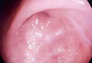
The case of a 62-year-old female patient with a medical history of arterial hypertension, type 2 diabetes mellitus, chronic peptic ulcer disease, and hypertrophic pyloric stenosis is presented. She had previously undergone endoscopic stent placement on two occasions, the most recent two months prior. She presented to the emergency department with a 24-hour history of burning epigastric pain associated with multiple episodes of emesis and intolerance to oral intake. Over the previous two months, she reported an approximate weight loss of 8 kg, as well as early satiety and nausea following food intake during the previous two weeks. Given the patient’s symptoms and medical history, upper gastrointestinal endoscopy was performed. Imaging demonstrated punctate benign pyloric stenosis (Figure 1) with complete luminal occlusion and no evidence of malignant tissue.
Considering the patient’s history of two prior dilations and the findings on upper gastrointestinal endoscopy, a structured clinical evaluation was performed. This evaluation revealed a high comorbidity burden and high nutritional risk. The patient was deemed unsuitable for surgical gastrojejunostomy due to the high risk of complications. Therefore, endoscopic ultrasound-guided gastrojejunostomy was selected using a lumen-apposing metal stent (LAMS).
Technical procedure
Upper gastrointestinal endoscopy confirmed punctate pyloric stenosis. The pylorus was subsequently intubated using a Fogarty extraction balloon to advance a biliary guidewire distally (Figure 2). The guidewire was then advanced through the Fogarty balloon, contrast was administered, and the guidewire was visualized within the jejunal loop. The guidewire was left in situ, followed by advancement of a nasojejunal tube. Through the large-caliber tube, methylene blue irrigation of the jejunal loop was performed (Figure 3).
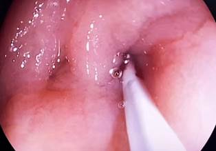
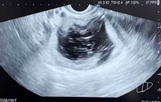
Figure 3 Irrigation and dilation of the jejunal loop through a nasojejunal tube visualized by endoscopic ultrasound. Image property of the authors.
Subsequently, deployment of a 10 × 15 mm Hot AXIOS lumen-apposing metal stent (Boston Scientific) was performed. The prosthesis was deployed (Figure 4) to create the gastroenteroanastomosis. A guidewire was then advanced through the prosthesis, and both the afferent and efferent loops were successfully identified (Figure 5).

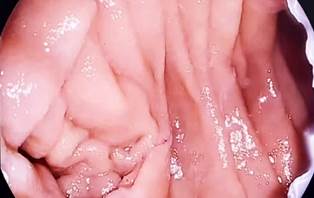
Figure 5 Gastroenteroanastomosis using a 10 × 15 mm lumen-apposing metal stent. Afferent and efferent loops. Image property of the authors.
The patient was transferred to the general ward after the procedure. She was reassessed at 24 hours, at which time oral intake was initiated with a liquid diet. At 48 hours, the diet was advanced to soft consistency, which was well tolerated without recurrence of obstructive symptoms. The patient was discharged at 72 hours without complications.
Discussion
Pyloric syndrome, or gastric outlet obstruction, is a disabling condition associated with high treatment costs. Although therapeutic approaches vary according to etiology, the use of self-expanding metal stents is widely adopted in both benign and malignant conditions to relieve obstruction. However, this strategy presents important limitations due to recurrence of luminal obstruction caused by tumor ingrowth or prosthesis migration15. Surgical gastroenterostomy represents another established alternative. Although it appears to effectively resolve obstruction with a lower risk of tumor ingrowth into the lumen, it is associated with higher complication rates, longer hospital stays, and consequently greater morbidity and healthcare costs16.
The development of endoscopic ultrasound over recent decades has enabled the emergence of several advanced endoscopic techniques, some of which remain under investigation. These advances have positioned EUS-GE as a safe, reproducible option associated with lower morbidity. Since early experimental animal studies conducted by Fritscher-Ravens et al.17 and Binmoeller Shah et al.18, multiple case series have reported favorable outcomes with EUS-GE, including those described by Khashab-Kumbhari et al.14. More recently, in 2025, two meta-analyses published by Rizzo et al.19 and Canakis et al.20 demonstrated that EUS-GE is a minimally invasive technique with a high success rate of up to 90%. Nonetheless, performance of this procedure requires centers with specialized expertise due to potential technical challenges, including intestinal loop identification, endosonographic localization, and precise puncture. Technological advances in dedicated devices, such as lumen-apposing metal stents, have facilitated the implementation of these techniques21.
Conclusions
Pyloric syndrome is a multifactorial condition caused by pyloric stenosis. It significantly affects patients’ quality of life and is associated with high nutritional risk. This condition predisposes individuals to a greater likelihood of complications, which may interact with surgical risk during therapeutic management.
Endoscopic management is currently the most widely accepted therapeutic approach and typically begins with endoscopic balloon dilation. However, as demonstrated in this case, although endoscopic ultrasound-guided gastroenteroanastomosis is a relatively recent technique with limited available literature, the present experience supports EUS-GE as a safe technical alternative associated with low morbidity for the management of benign pyloric syndrome. Multicenter randomized controlled trials are required to incorporate this technique into an initial management algorithm. Such incorporation should take into account patient-specific variables and institutional expertise.

