




 text in
  English (pdf)
 Article in xml format
 Article references
 Automatic translation
 Send this article by e-mail
 Cited by SciELO
  Cited by Google
  Similars in
SciELO
Similars in
SciELO  Similars in Google
 Permalink
INTRODUCTION
The prevalence of supernumerary teeth varies from 0.3 to 3.8%.1) It is defined as a number abnormality characterized by the excess of any tooth. When located in the midline palate between the two maxillary central incisors is termed mesiodens. And this is the most affected region with a prevalence of 0.15-1.9% in the general population.2)(3)(4
Even though this anomaly is highly prevalent, the etiology of supernumerary teeth has not been defined, but three theories try to explain this dental anomaly. The suggested theories include dichotomy of bud teeth, hyperactivity of the dental lamina5 and a combination of genetic and environmental factors; 1)(6 it can occur as single, multiple, unilateral or bilateral forms and in association with syndromes, such as Down syndrome or Cleidocranial dysplasia.7)(8 This alteration is more common in males than in females and is less frequent in the primary dentition.
Mesiodens are asymptomatic and may be discovered during routine radiological examination, when they affect the cortical bone causing increased bone volume or when they erupt in the oral cavity.
However, in some cases there can be a variety of complications including impaction, delayed eruption and ectopic eruption, crowding, diastema, axial rotation, and root resorption of adjacent teeth.7)(9)(10)(11 They can also cause crowding and may also be associated with pathologies like cyst.12 Less commonly, they may involve the permanent incisors, including root dilacerations and loss of tooth vitality.13
Therefore, early diagnosis of mesiodens is very important to prevent such complications, with clinical and radiographic examinations to locate the correct position of mesiodens and design surgical removal in the ideal time for intervention (immediate or delayed).
Supernumerary teeth can be classified based on position: mesiodens: occurring in the incisor region, paramolar: occurring besides a molar, and distomolar: occurring distal to the last molar.12
There are two types of treatment for this anomaly: clinical and radiological follow up, when the supernumerary tooth will not affect other structures; the other option is surgical removal, in which the supernumerary tooth is affecting neighboring structures or teeth, orthodontic indication, bulging cortical bone that creates discomfort to the patient or even when rash occurs in the oral cavity.
CASE REPORT
An 8-year-old girl was referred to the surgery department with complaints of swelling in the palate and pain when masticating. The intraoral examination showed swelling in the anterior palatal region without no mucosa color alteration. The patient had normal mixed dentition with no other abnormalities (Figure 1). There were no signs of syndromes.
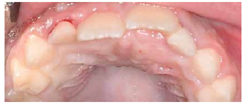
Figure 1 Clinical aspect of the oral cavity pre-surgery, with palatal swelling in the region of the incisors
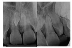
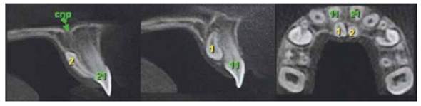
A periapical radiograph was requested and analyzed by means of the Clark radiographic technique (Figure 2) and for better visualization and planning a cone-beam computed tomography (CBCT) was taken, confirming the presence of two supernumerary teeth between central incisors (mesiodens) in inverted position. Also, there was not root resorption of the permanent roots of this region and no evidences of associated pathologies (Figure 3).
Therefore, a surgical procedure was planned. Pre-operative anti-inflammatory therapy (1 tablet of dexamethasone 4 mg) and anxiolytic (1/2 tablet of midazolam 7.5 mg) were used one hour in advance.
The surgery was performed with local anesthesia (lidocaine 2% epinephrine 1:100.000), using the infiltrative technique first in the vestibular region, later in the papilla between the central incisors and finally in the palatal region in the nasopalatine nerve. The access was performed by the palate due the image shown in the radiographic examination. Using a number 15 scalpel blade, an incision was made resulting in a flap without relaxing from right canine to left canine (Figure 4). After detaching the flap, visualization of the dental elements was not possible and ostectomy was necessary to expose a portion of the teeth (Figure 5). Using the opportunity of this surgical procedure, simple extraction of the primary upper lateral incisors was performed. The surgical area was sutured with silk thread 4.0 (Figure 6).
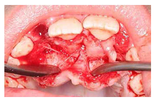
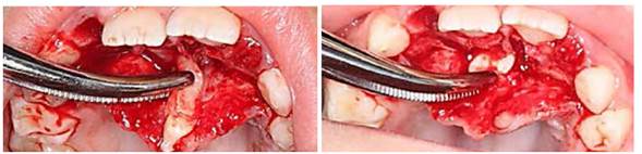
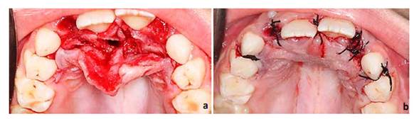
Figure 6 a. Aspect of surgical site after removal of supernumerary teeth. b. Surgical wound after simple suture with silk thread 4.0
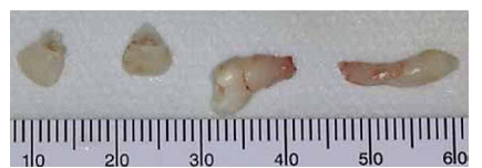
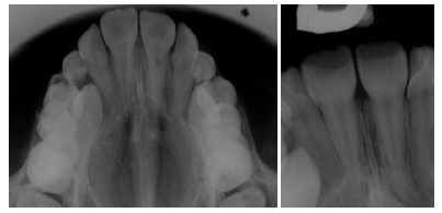
The patient was prescribed anti-inflammatories (1 dexamethasone 4 mg orally before going to bed). The sutures were removed 7 days later. A control image was requested to verify tooth eruption, adjacent structures compromise, and bone formation in the area (Figure 8).
DISCUSSION
Mesiodens are the most common among supernumerary teeth, located mesial to both central incisors; appearing peg shaped, in a normal or inverted position.14 Both dentitions are affected, but prevalence in the permanent dentition is higher than in the primary one.15 Many studies have shown that mesiodens affect boys more than girls (2:1);13)(16)(17) Huang et al found a sex ratio as 2.5:1 in favor of boys.18
This type of anomaly can be discovered as a result of patient's complaint or when they seek treatment for malocclusion or by bony swelling, most commonly in the anterior palatal region. The impacted mesiodens are often diagnosed by radiographic examination.17
Delayed, ectopic or asymmetric eruption of the central incisors should alert the clinician to the possibility of a mesiodens. The clinician should obtain accurate radiographs including panoramic, periapical and occlusal views.2 A radiological examination is critical for correct diagnosis and treatment planning. A thorough radiographic examination at various angles with the Clark radiographic technique using 2 positions may help estimating the exact location of the tooth, especially in cases of impacted mesiodens.19 For a precise view in the incisor region, an anterior occlusal or a periapical radiograph is also helpful, but nowadays CBCT shows the correct position and localization of mesiodens, but it is an expensive technique.
Mesiodens can cause a variety of complications, mainly when unerupted teeth affect the maxillary incisors, root resorption, axial rotation or displacement, diastema and dentigerous cyst formation.11)(17 In the case, the mesiodens was causing a bulge in the anterior region of the palate, causing difficult swallowing and a slight diastema between the permanents central incisors. Once a mesiodens has been diagnosed, treatment should be planned.
In children and adolescents, extraction has been recommended to prevent occlusal changes as well as cyst formation. Yague-García et al emphasized on early removal of supernumerary teeth in order to prevent complications.20
Therefore, after a clinical and radiographic examination, if extraction of a supernumerary is necessary, it should be performed at an appropriate time to promote self-eruption in early mixed dentition; this will result in better teeth alignment and can minimize the need for orthodontic treatment or any alteration in adjacent teeth.
In cases of unerupted supernumerary teeth with no pathology or possible harm to adjacent teeth not interfering with the orthodontic movement, they can be left in situ, but it is important to perform periodic radiographic control.
CONCLUSION
Among supernumerary teeth, mesiodens is the most prevalent mixed dentition. Given the large number of patients with this anomaly, knowing the signs and symptoms (swelling, changes in mucosa color, absence of teeth, difficulty in speech), as well as performing the appropriate and detailed image analysis, is critical for correct diagnosis (position and location) and for the surgical planning to be effective and faster, as this procedure in typically done in pediatric patients. After surgery, it is very important to follow the patient with clinical and radiographic examination to discard the possibility of damage to surrounding teeth and/or structures.

