


 English (pdf)
 Article in xml format
 Article references
 Send this article by e-mail
 Cited by SciELO
  Cited by Google
  Similars in
SciELO
Similars in
SciELO  Similars in Google
 Permalink
Introduction
UV radiation constitutes about 10% of solar radiation reaching the Earth's surface and poses significant risks to the skin due to its high energy and deep penetration 1. Within the UV spectrum, UVC radiation (λ = 100-290 nm)2 is entirely absorbed by the ozone layer, whereas UVB radiation (λ = 290-320 nm)2 accounts for 4-5% of the UV radiation that reaches the Earth’s surface. The majority, 95%, is UVA radiation (λ = 320-400 nm) (3 and can cause sunburn, DNA damage, and skin cancer 1. The remaining solar radiation includes visible light (50%) and infrared radiation (40%), which, along with artificial sources, contribute to photoaging, inflammation, overheating, hyperpigmentation, collagen degradation, and oxidative damage 1,4.
Photoprotection is crucial in preventing skin damage and associated pathologies, primarily caused by oxidative stress, sunburn, dryness, and other effects of sun exposure 2,3,5. There are two photoprotection mechanisms. The primary mechanism comprises organic or inorganic sun filters (sunscreens) that absorb or reflect radiation 2,5The secondary mechanism includes molecules that can neutralize reactive oxygen (ROS) and nitrogen (NOS) species when they are produced as a consequence of the transfer of high energy from light to skin biomolecules 2,3.
Although many sunscreens exist, some traditional synthetic organic UV-Vis filters can have adverse effects on both human health and the environment 6. Photoprotective metabolites such as mycosporine-like amino acids, phenolic compounds, flavonoids, sulfated polysaccharides, and fucoidans have been studied as photoprotective compounds through both primary and secondary mechanisms, implying fewer secondary effects 6-8. The structures of these marine-derived metabolites feature aromatic systems or long-conjugated double bonds with electron-donating or electron-withdrawing groups, making them structurally similar to synthetic filters. This allows them to absorb high-energy radiation, thus protecting the skin from UV radiation 9.
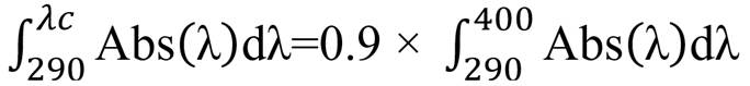
To assess photoprotection, assays are required to cover both the UVA and UVB spectra. While in vivo assays may be necessary for commercialization, in vitro assays are helpful for research purposes as they reduce time and test requirements, providing a good approach to the expected in vivo tests. The parameters used to evaluate photoprotection include: SPF (Sun Protection Factor) for UVB photoprotection, UVAr (UVA ratio) for protection against UVA radiation, and λc (critical wavelength) as a measure of photoprotection capability within the UVA and UVB spectrum. These parameters cover the bioactivity of the compounds thoroughly and can be simultaneously analyzed through in vitro tests using a single spectrophotometric measurement 10,11.
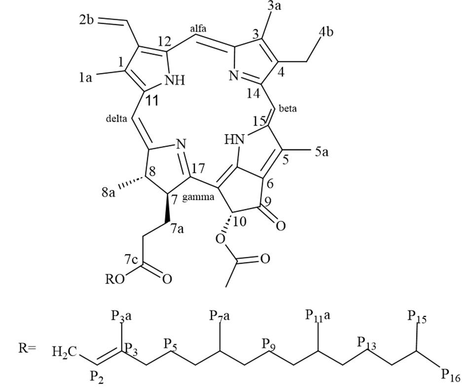
The Colombian Caribbean has 15 recognized species from the Dictyota genus. Their metabolic production consists mainly of diterpenes, which enables them to survive in different marine environments 12,13. Despite the potential applications of this algal biomass, such as antifouling, antiviral, and antimicrobial activities 8,14,15, research in the cosmetic industry with species that are part of Colombia’s marine biodiversity is still limited. This study presents the chemical characterization of pheophytin a isolated from the Caribbean brown algae Dictyota pinnatifida and the in vitro evaluation of its photoprotective activity.
Materials and methods
General experimental procedures
HPLC-grade methanol (MeOH) (99.9%), hexane (95%), silica gel 60 (70-230 MESH ASTM), and RP-18 (40-63 µm) were purchased from Merck Millipore, USA. Dichloromethane (DCM) (99.8%), and ethyl acetate (EtOAc) (99.8%) were purchased from Panreac AppliChem, USA. Ethanol (EtOH) (99.8%) and 2-hydroxy-4-methoxybenzophenone (BP-3) (98%) were purchased from Sigma Aldrich, USA. Ultrapure Type I Milli-Q water (H2O, membrane 2.2 μm) was used.
UV-Vis spectra of the crude extracts were recorded in a Shimadzu UV-1800 UV-Vis spectrophotometer using a quartz cell (1 cm). NMR analysis was performed in a 400 MHz Bruker Advance Neo device, using deuterated chloroform (CDCl3)(99.5%) from Merck Millipore, USA.
Algae Collection
A sample of Dictyota pinnatifida was collected in May 2015 by SCUBA diving in Providencia, Colombia. Sample identification was conducted by biologist Freddy Duque using morphological identification keys provided by De Clerk 16 and Litter and Litter 17. A sample has been registered for preservation and comparison under the code 596747 in the collection of the Instituto de Ciencias Naturales, Universidad Nacional de Colombia.
Extraction
172 g of algae was extracted three times with 300 mL of DCM/MeOH (1:1) for 24 hours each. Solvents were dried under vacuum to obtain crude extracts and then partitioned between DCM/H2O. The organic extract was fractionated by column chromatography (Silica Gel 60, 70-230 MESH ASTM, Merck) using a discontinuous gradient of increasing polarity (hexane, EtOAc, and MeOH), resulting in twelve fractions (F1 to F12). Fraction 5 was separated by column chromatography using RP-18 (40-63 µm, Merck) and a MeOH/H2O gradient from 3:7 to 1:0, resulting in nine subfractions (F5.1 to F5.9). Fraction F5.9 yielded 217 mg of pheophytin a.
Compound identification
1H- and 13C-NMR experiments were conducted on a Bruker Avance Neo 400Hz NMR instrument, using CDCl3 (99,5%, Merck). Compound identification was achieved through analysis of this information and comparison with previously reported spectra, as depicted in the Supporting Information 18-20.
Pheophytin a: dark-green oil. 13C-NMR (101 MHz, CDCl3): 189.8 (C-9), 173.1 (C-7c), 172.4 (C-18), 169.7 (C-10a), 161.4 (C-17), 155.8 (C-13), 151.1 (C-14), 149.8 (C-16), 145.3 (C-4), 143.0 (C-P3), 142.2 (C-11), 138.0 (C-15), 136.6 (C-12), 136.4 (C-2), 136.3 (C-3), 132.0 (C-1), 129.2 (C-2a), 129.1 (C-5), 129.1 (C-6), 122.9 (C-2b), 117.8 (C-P2), 105.4 (C-γ), 104.5 (C-β), 97.6 (C-α), 93.3 (C-δ), 64.8 (C-10), 61.6 (C-P1), 53.0 (C-10b), 51.3 (C-7), 50.2 (C-8), 39.9 (C-P4), 39.5 (C-P6), 37.5 (C-P10), 37.4 (C-P8), 37.4 (C-P12), 36.7 (C-P14), 32.9 (C-P7), 32.7 (C-P11), 31.3 (C-7a), 29.8 (C-7b), 28.1 (C-P15), 25.1 (C-P5), 24.9 (C-P9), 24.5 (C-P13), 23.2 (C-8a), 22.8 (C-P16), 22.7 (C-P15a), 19.8 (C-P7a), 19.8 (C-P11a), 19.6 (C-4a), 17.5 (C-4b), 16.4 (C-P3a), 12.2 (C-1a), 12.2 (C-5a), 11.4 (C-3a).
1H-NMR (400 MHz, CDCl3): δ 9.49 (s, H-β), 9.35 (s, H-α) , 8.54 (s, H-δ) , 7.96 (dd, J = 18.0, 11.6 Hz, H-2a), 6.27 (dd, J = 17.8, 1.5 Hz, H-2b trans), 6.25 (s, H-10), 6.16 (dd, J = 11.6, 1.6 Hz, H-2b cis), 5.11 (H-P2), 4.47 (H-P1a), 4.44 (H-8), 4.42 (H-P1b), 4.20 (H-7), 3.86 (s, H-10b), 3.67 (s, H-4a), 3.65 (s, H-5a), 3.38 (s, H-1a), 3.20 (s, H-3a), 2.61 (H-7a), 2.48 (H-7b), 2.33 (H-7a'), 2.18 (H-7b'), 1.86 (H-P4), 1.80 (d, J = 7.3 Hz, H-8a), 1.70 (H-4b), 1.60 (H-P5), 1.55 (H-P3a), 1.52 (H-P15), 1.32 (H-P7), 1.27 (H-P13), 1.24 (H-P8), 1.24 (H-P10), 1.24 (H-P12), 1.14 (H-P14), 1.10 (H-P9), 0.99 (H-P6), 0.87 (H-P7a), 0.87 (H-P15a), 0.84 (H-P11), 0.80 (H-P11a), 0.78 (H-P16).
In vitro photoprotection activity assays
2 mg/mL ethanolic solutions of pheophytin a and benzophenone-3 were prepared. From this, 30 and 10 ppm dilutions were further prepared in ethanol (> 99,8%, Sigma Aldrich). The absorbance for each solution was measured in a quartz cell (1cm) using a Shimadzu UV-1800 UV-Vis spectrophotometer, scanning between 290 and 400 nm in 1nm intervals.
Sun Protection Factor (SPF)
SPF, a measure of UVB photoprotection capacity, was determined using Mansur's spectrophotometric methodology 21 with some modifications as proposed by Nunes and coworkers 22. The spectrophotometric SPF was calculated based on absorbance readings in 5 nm increments, utilizing Mansur's equation (1):
 Eq (1)
Where CF stands for correction factor = 10, EE (λ) corresponds to the erythemal effect spectrum, and I (λ) stands for solar intensity spectrum, in which the EE X I product are constants (Table 1)23. Abs (λ) corresponds to the recorded absorbance at the studied wavelength.
Table 1 Constants for Mansur SPF determination21.
| Wavelength [nm] | EE X I |
| 290 | 0,015 |
| 295 | 0,082 |
| 300 | 0,287 |
| 305 | 0,328 |
| 310 | 0,186 |
| 315 | 0,084 |
| 320 | 0,018 |
UVA ratio (UVAr)
The UVA ratio, a quantitative measure assessing the balance between total UVA and UVB absorbances for evaluating UVA-photoprotection, was determined following the methodology outlined by Rojas and colleagues 24. Absorbances between 290 to 400 nm at 1 nm intervals were used, employing the following equation (2):
 Eq (2)
Here Abs (λ) corresponds to the recorded absorbance within the studied UVB (290-320 nm) and UVA range (320-400 nm) at 1 nm interval readings.
Critical wavelength (λ crit)
Critical wavelength evaluates the balance between UVA and UVB photoprotection, representing the overall photoprotection capacity within this spectral range. It corresponds to the wavelength at which 90% of the total absorbance curve is reached. Mathematically, it is determined using equation (3):
 Eq (3)
Abs (λ) represents the recorded absorbance between 290 to 400 nm in 1nm increments. Following Rojas and co-workers’ procedure24, the total area under the absorbance curve is designated as 100%, and the critical wavelength is determined by interpolation as the wavelength at which 90% of the curve is reached.
Results
F.5.9 (>200 mg) from the Dictyota pinnatifida sample was isolated as a dark green oil that was identified as pheophytin a (Figure 1) through 1H-NMR and 13C-NMR analyses, as well as two-dimensional COSY, HMBC, and HSQC experiments and comparisons with literature reports. Details on NMR spectra and full assignments are presented in the Supplementary Information.
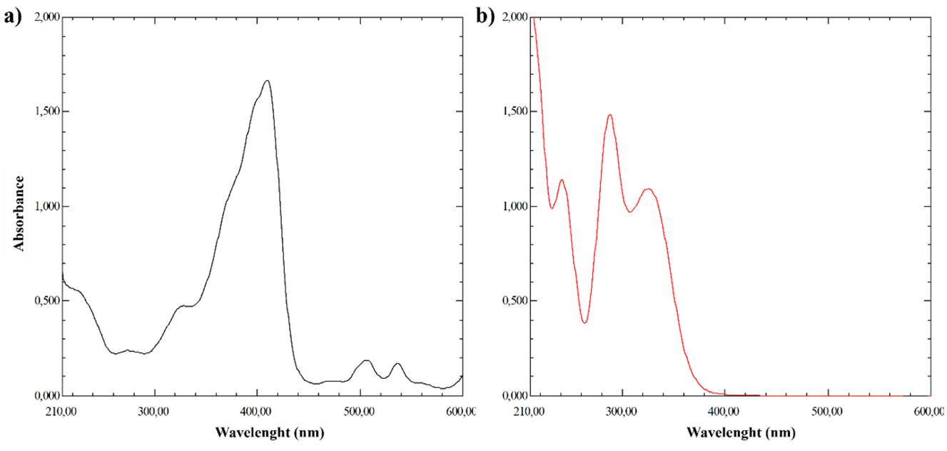
UV-Vis spectra of pheophytin a showed absorbance within the 210 and 600 nm range, with characteristic maxima absorbance peaks in the UVC, UVA, and visible regions (Vis) at 272, 326, 409, 506, and 536 nm (Figure 1a), characterizing this compound as UVA photoprotector. In contrast, benzophenone-3 used as reference material, has maxima absorbance peaks at 241, 288, and 325 nm, which corresponds to a UVB filter (Figure 1b).
SPF assessment for pheophytin a showed concentration-dependent values ranging from 1.4 and 2.9, UVAr between 4.6 and 6.8, and λc of 394 nm for 10 and 30 ppm solutions, respectively (Table 2). As a reference, the same determination was made for BP-3, a recognized UV filter, obtaining SPF values of 4.2 and 10.3, 0.9 and 1.1 for UVAr, and 353 nm and 349 nm for λc, in solutions of 10 and 30 ppm, respectively.
Table 2 Spectrophotometric SPF values obtained for pheophytin a
| Parameter (units) | Concentration (ppm) | BP-3 | pheophytin a |
| SPF ± SD | 30 | 10.310 ± 0.130 | 2.916 ± 0.355 |
| 10 | 4.208 ± 1.201 | 1.411 ± 0.138 | |
| UVAr ± SD | 30 | 1.054 ± 0.011 | 6.815 ± 2.296 |
| 10 | 0.915 ± 0.029 | 4.629 ± 1.347 | |
| λc ± SD (nm) | 30 | 349.674 ± 0.767 | 394.639 ± 0.493 |
| 10 | 353.539 ± 0.922 | 394.072 ± 0.912 |
BP-3: benzophenone -3 (standard); SPF: sun protection factor; UVAr: UVA ratio; λc: critical wavelength; SD: standard deviation (n=3; mean of three technical replicates).
Discussion
Colombia's sea harbors a diverse array of macroalgae; however, their chemical and cosmetic potential remains largely unexplored 25. One of the few chemical studies of Colombia’s macroalgae came from Dictyota pinnatifida. The metabolite composition includes prenylated germacrene and xeniane diterpenes. Of those compounds, a few exhibited inhibitory activities against Pseudomonas aeruginosa and Escherichia coli biofilms 26.
Dictyota pinnatifida study showed a significant amount (>200 mg) of a pigment that was isolated and identified as pheophytin a through 1D and 2D NMR analysis. The structure of this brown algae pigment includes multiple conjugated double bonds that allow it to absorb and stabilize UV and visible radiation. There are reports of similar structures in photosynthetic organisms with photoprotective effects, including different types of chlorophylls, carotenoids, and phycobilins 14. Some of these compounds are fucoxanthin, the main pigment in brown algae, chlorophylls a and b, differently distributed in brown and green algae, and phycobilins, principal pigments in red algae. Therefore, we anticipated that pheophytin a, a chlorophyll derivative, would exhibit mainly protective activity through primary photoprotection mechanism.
The UV-Vis spectra showed absorbance within the 210 and 600 nm range, confirming its potential as a photoprotector (Figure 2). This observation was confirmed through the in vitro photoprotective activity test in which SPF, UVAr, and λc values were determined (Table 2). SPF showed concentration-dependent-values ranging between 1.4 and 2.9 for 10 and 30 ppm solutions, respectively, lower than those determined for BP-3 used as a sunscreen standard. In comparison, UVAr and λc for pheophytin a were 6.815 and 394.6 nm for 30 ppm solutions, respectively, higher than those calculated for BP-3. These results showed that pheophytin a can be used as a sun filter for the UVA region, which could protect skin from sunburn, DNA damage, and skin cancer, among others.
Even though there are no in vitro tests to assess visible light protection, the absorbance maxima observed between 400 and 600 nm demonstrate that this pigment could be also used as a photoprotector for this region (infrared and visible light), making it suitable for skin-photoprotection applications preventing issues such as photoaging, inflammation, overheating, hyperpigmentation, collagen degradation, and oxidative damage, among others.
It is noteworthy that BP-3 and pheophytin a exhibit photoprotection in different UV regions; however, results demonstrate that rather than substituting, they can be used as complementary sunscreens in cosmetic formulas, protecting skin in the whole UV spectrum. Also, once pheophytin a preliminary photoprotection activity test has been addressed, further comparisons with dedicated UVA commercial filters, as well as stability, toxicity, and validated standard photoprotection activity tests must be assessed in the future to confirm this compound's suitability for cosmetic and other photoprotection-related applications.
Conclusions
The algae pigment pheophytin a was isolated from the Colombian Caribbean from Dictyota pinnatifida. This magnesium-free compound features a porphyrinic ring esterified with a phytol group, with a multiple conjugated double bond structure that makes it suitable as a photoprotector. In vitro evaluation of its photoprotective activity indicates potential for skin-photoprotection-related applications, particularly in UVA, visible, and infrared light. Further assessment through in vivo tests will be conducted. This is the first reported evaluation of pheophytin a photoprotective capacity, being relevant for the cosmetic industry that can explore its potential as an alternative or complement to traditional widespread sunscreens and as a more sustainable photoprotective ingredient.

