




 English (pdf)
 Article in xml format
 Article references
 Automatic translation
 Send this article by e-mail
 Cited by SciELO
  Cited by Google
  Similars in
SciELO
Similars in
SciELO  Similars in Google
 Permalink

BACKGROUND
Alzheimer disease (ad) is considered as the most common progressive neurodegenerative disorder and the most frequent form of dementia (Perneczky, 2019). According to the World Health Organization (WHO, 2012) Alzheimer disease is an important cause of morbidity and contributes significantly to the world statistics of mental and neurological disorders (Lopez & Kuller, 2019).
The efforts focused in the early diagnosis of ad are the best long terms strategies to delay the disease because they make possible for the patient to take advantage of the therapies available for the early phases (Karr et al., 2018). In that way, the seek for different tools that facilitate the diagnosis of ad is gaining relevance (Marandi & Gazerani, 2019; Zetterberg & Bendlin, 2021).
The cognitive function that is more affected for Alzheimer disease is memory. The study of ocular behavior patterns has evidenced that ocular movements in the patient with ad can reveal elements about previous experiences related with memory, without resorting to oral reports or requiring conscious memories from the person (Wilcockson et al., 2019). As stated by Crutcher, Calhoun‑Haney, Manzanares, Lah, Levey y Zola (2009) the monitoring of ocular movements enables the evaluation of the memory related with early detection of mild cognitive impairment as well as the evaluation of relevant aspects of that function for Alzheimer disease.
Recognition is one of the relevant aspects of the declarative episodic memory, which is related with the ability to remember facts or events in a conscious way, that can be studied through recordings of ocular movements. The importance of the study of recognition in patients with memory deficits, directly depends on the integrity of the medial temporal lobe that includes the hippocampal region, and area that is considerably compromised in patients with mnemonic alterations and neurodegenerative disorders such as ad (Monacis et al., 2019).
Additionally, the recognition memory is related to the functioning and integrity of the parietal medial posterior cortex and the prefrontal left cortex, as it is evidenced the activation of the Precuneus and the frontal lateral inferior left cortex in tasks related with the recuperation of the episodic memory (Ryan et al., 2022; Urgolites et al., 2018). Considering that this type of memory allows the association of current knowledge with previous experiences, it requires of two different processes: memory and familiarity. The memory implies the recognition that associates the facts with previous experiences, while familiarity is related with the sense of similarity of the visual stimulus (Schwedes & Wentura, 2019).
The posterior Precuneus and the hippocampal region activate during the recuperation of mental images. In that way, it is expected that the subjects with ad, who present a significant deterioration of the parietal areas, present difficulties in the regeneration or recuperation of the contextual associations of mental images, particularly when the evocation or recuperation of the information must be in latency periods longer than a minute (Serra et al., 2020). In this case, the ability to code the information can be preserved but the effectivity in the evocation diminishes as latency increases.
Furthermore, from the studies of neuroimage it has been proved that the recovery of the recognition memory implies the activity of the left prefrontal cortex, because this region facilitates and guides the access to knowledge of the semantics of information, maintains the signals of recuperation and participated in the selection of relevant information (Lundstrom et al., 2005; Neri et al., 2021).
Crutcher, et al. (2009) proposes that the evaluation of the element of recognition that was mentioned before can be developed from the paradigm of the visual paired‑comparison task. This task can be considered as a memory test that is sensitive to the detection of memory disorders and deficits in prodromal stages of ad. In addition to that, the authors affirm that the task is highly sensitive to minimum damages in the hippocampus, as it was observed in studies developed with rats, monkeys and human beings.
The vpc task is a task of recognition memory that evaluated the preference of the individual through the proportion of observation time that the subject has in relation to a new image, in comparison to an image that has been seen previously. For this task, the analysis of ocular movements is oriented to the percentage and length of the fixations. In healthy individuals, the percentage of fixations and its length increases in a disproportioned way until they reach approximately 70% upon new aspects (Zola et al., 2013). In individuals with memory deficits the proportion of fixations is distributed in an almost equitable way between the new stimulus and the previous ones (Crutcher et al., 2009).
Different studies (Bueno et al., 2019; Marandi & Gazerani, 2019; Opwonya et al., 2022) mentioned suggest that the analysis of ocular movements can reach an important value in the evaluation of behavioral measures. The effects of the alterations in the cognitive function of the memory over the behavior of ocular movements are promising alternatives for the diagnose and characterization of disorders as Alzheimer disease (Marandi & Gazerani, 2019; Zetterberg & Bendlin, 2021). Therefore, this project has as main aim to evaluate the recognition memory through oculomotorius behavior in visual paired‑comparison task on patients with Alzheimer disease and a control group Spanish speaker.
METHODS
Participants
Two groups were evaluated: ad and control. A total of 30 subjects (23 women, 7 men, Mage = 72,8 years old, range 63 – 83) participated. 15 of them belong to the group ad and 15 to the control group. One of the participants in the ad group had a gds 4, the rest of the participants had a diagnosis of ad correspondent to gds 5.
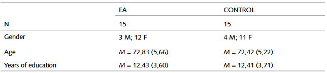
The participants who presented non‑corrected visual or hearing alterations or who had glasses in bad condition that could prevent the development of the task and the comprehension of simple instructions were excluded of the sample. Additionally, the participants who were in an advanced stage of de disease (gds 6) at the moment to develop the tests were not considered.
The participants were evaluated and diagnosed in consensus with possible ad by the interdisciplinary group for the study of dementias of Universidad Nacional de Colombia. Participants completed a neuropsychological test MoCA (Nasreddine et al., 2005) and agreed voluntarily to participate in the study through an informed consent in agreement with the regulations of the ethics committee of the Medical Department of the Universidad Nacional de Colombia.
Equipment and Stimuli
The oculomotorius behavior was registered using the Eye‑Tracking Tobii Tx300. This device recognizes and storages information about ocular movements with a frequency of 300Hz (data collected per eye per second). The stimulus was presented in a 23 inches lcd screen with 1920×1080 of resolution. The mathematic algorithm of speed umbral filter (i‑vt) incorporated in the software Tobii Studio was used to differentiate the ocular fixation of saccadic movements through speed umbral. An umbral of ocular speed of de 30º/s was stablished to define the start and the end of a fixation.
Procedure
Initially, the cognitive screening assessment MoCA was applied to all the participants of the ad group and the control group.
In each session, the participant was sitting in front of the Eye‑Tracker in a static chair located 29 inches from the screen under constant illumination conditions. After that, a short calibration session of nine points located at ≤18º was developed. After the calibration, it was presented to the participants the visual paired‑comparison task. The participants had four rehearsals to ensure the comprehension of the task. Additionally, to guarantee that the memory deficit, characteristic of ad, did not intervene in the successful execution of the task, the instructions were repeated to all the participants in the control group and the ad group before each activity.
The task had two phases: familiarization and test. During the familiarization phase, two identic images located one next to the other were presented for 5000 milliseconds. After a period of 2000 milliseconds or 2 minutes, the test phase where to images were presented in the screen for 5000 milliseconds started (Zola et al., 2013). In the test phase, the screen showed an image identic to the one presented in the familiarization phase and a new one. The participants were asked to observe the images on the screen as if they were watching tv.
RESULTS
For each one of the groups, all the variables related with the parameters of ocular movements showed a normal or normalized distribution (p>0.05) and for the average comparison test six variables presented different variances. For those variables, the comparison test t’‑Student for independent sample was used, while the other variables were analyzed with the t‑Student comparison test for independent variables because they presented homogenous variances. All the date of ocular movement and fixation were extracted from the Tobii Studio software and analyzed in spss.
During the familiarization phase, two identic stimuli were presented to the subjects during 5000ms, followed by the test phase with a delay interval of 2000ms or two minutes.
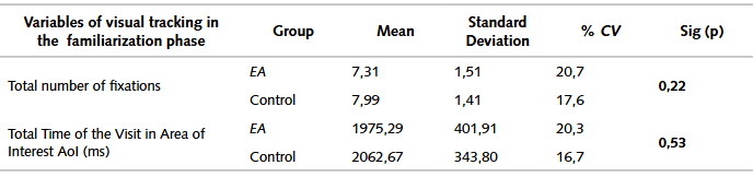
The analysis through the identification of interest areas in the familiarization phase, evidenced that there are not significate differences between the control group and the ad group in any of the observed variables (total length of the visit and number of fixations), which suggests that the two groups do not differ in the amount of time they spent looking at the familiarization images, regardless the variations in the delay interval. Results indicate that the participants with ad as well as the control subjects show a similar number of fixations during the familiarization phase.
During the test phase, the original image, which was previously presented during the familiarization phase, was projected for the participants along with a new image during a time slot of 5000ms.
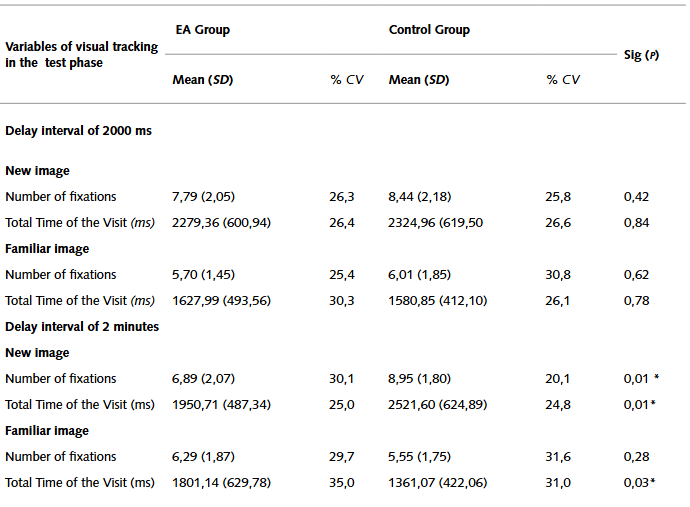
It was interesting for this research the percentage of time or duration of the visit over the area of interest of the new image in comparison with the familiar image, considering the two delay intervals (2000ms and two minutes). The two groups of participants spent similar amounts of time looking at the new image when the delay interval of the presentation of the familiarization phase and the test phase was 2000ms, showing that there are not significant differences between the two groups in any of the analyzed variables.
On the other hand, for the delay interval of two minutes, the groups differ in the percentage of time they spend observing the new image (p<0,01), as the participants in the control group look at the image longer than the ad group. In the same way, significant differences are presented in the average of time of duration of the visit over the familiar image between the two groups, because the ad group looks at the familiar image in the test phase longer than the control group.
Likewise, differences in the average of fixation developed over the interest area during the test phase in the delay interval of two minutes (P<0,01) were identified. The participants in the AD group present a lower number of fixations over the new image in comparison with the control group.
Discussion and Conclusions
In first place, it is necessary to remember that the recognition memory is related with the declarative memory and depends on the integrity of the medial temporal lobe, the hippocampus and the diencephalic structures (Ryan et al., 2020). Additionally, Zola (2000) highlights that the results obtained in tests on rats, monkeys and human beings with bilateral lesions of the hippocampus and structures related with the temporal medial lobe, evidence that the development in the visual paired‑comparison task (cvp) depend of the declarative memory.
It suggests that this kind of memory can be evaluated through the visual paired comparison, as the task is sensitive to the minimum damage of the involved structures and even detects deterioration of the memory associated with the mild cognitive impairment (Lagun, et al., 2011).
In patients diagnosed with ad, one of the most affected aspects is the declarative memory, understood as the capacity to consciously remember facts and events which entails the recognition memory, because this alteration is related with the level of cerebral atrophy, particularly the medial temporal lobe, the entorhinal cortex, and the hippocampus; alterations that are evident in patients with this disease (Pandey & Ramakrishnan, 2020).
In the same way, the execution of the cvp task can be measured through the register of ocular movements with higher detail, in a quantitative and objective way, in comparison with the presentation of the task that requires the verbalization of the participant’s answers (Haque, et. al., 2019). Since the last century, studies as the one developed by Daffner et al (1992) have taken advantage of the techniques of register of ocular movements with individuals who were diagnosed with ad, however, there have not been many projects in Spanish population this field reported so far.
This study evaluated the recognition memory using the cvp task through the register of oculomotorius behavior. To begin with, the results evidence that there is no relationship between the performance of the participants in the cvp task and the total score in moca. It is possible that this happens because the task is particularly focused in the measurement of the explicit memory (Crutcher, et al., 2009), while moca evaluates seven different cognitive areas: visuospatial/ executive, nomination, memory, attention, language, abstraction and orientation (Roalf, et al., 2013).
From the analysis of two aspects, the duration of the visit and the average of fixations during the familiarization phase and the test phase, it is possible to affirm that there is no evidence of statistically significant differences during the familiarization phase of the task in the two group. As expected, there are significant differences between the two groups in terms of the amount of time that the participants spend looking at the new image when the interval of time is two minutes. In this case, the participants in the ad group show a decrease in the average length of the visit to the new image. These findings are in agreement with the information obtained by Lagun et al (2011).
Similarly, the results are congruent with the research by Crutcher (et al., 2009) at certain extent. Even though their study was developed with three different groups: control, Parkinson disease, and dcl, differences were found in the dlc group, associated with Alzheimer disease, and the control group during the test phase in relation to the duration of the visit over the new image and the two‑minute delay.
In that way, in accordance with the mentioned studies (Crutcher et al., 2009; Lagun et al., 2011) the control group as well as the ad group evidenced an equivalent performance in recognition memory, related with the increase in the visualization time of the new image, in regard to the familiar image, when the delay time was just two seconds. There were not differences in the average of fixations in any of the presented conditions.
To sum up, results suggest that an interval of delay of two minutes between the presentation of the familiarization phase and the test phase, is enough to prove the recognition memory as the subjects of the ad group are not able to remember that the familiar image had been observed before (Crutcher et al., 2009).
The proposal by De Chastelaine et al. (2016) and Dörfel, et al. (2009) can be considered as a possible pathophysiological explanation for this phenomenon. The findings of the present study can be the consequence of the degeneration of areas of the parietal medial posterior cortex, of the Precuneus, of the left prefrontal cortex and the hippocampal region, which are characteristics of early stages in ad.
Furthermore, in agreement with the model of Knudsen (2007), this kind of task and the findings evidence that the participants with AD have difficulties in the processes that contribute to spatial attention and its functional components. The commitment of the patients with ad diagnosis in the domain of the episodic memory can affect the construction of neuronal representations related with this element. That is why the competitive selection of the representation with a higher intensity does not happen just over the new image but over both. The familiar image as well as the new one, enter in the circuit that underlies the working memory, directing the ocular movements and the voluntary visual attention in a similar way for both types of images.
In consequence, the cvp task reveals the relations between spatial attention, working memory, visuospatial control, ocular movements and episodic memory in patients with neurodegenerative alterations. Baddeley (2000) affirms that recent research in the field of cognitive neuroscience consider that the working memory and long term memory are related, as the subcortical current originated in the hippocampus and the anterior cingulum, implied in the representation of the world and the construction of spatial‑temporal coordinates, are extended to the prefrontal cortex to direct the attention to internal and external events (Eriksson et al., 2015; Wanke & Schwabe, 2020).
All things considered, it is precise to highlight that the task of recognition memory and its evaluation though the cvp task, developed through the analysis of ocular movements is an useful screening tool for the differentiation of people with ad, even in early stages of the disorder and in healthy individuals. Although the cvp task is not part of the neuropsychological protocols of evaluation, its implementation can contribute to the identification in deficits in the recognition memory and the comprehension of processing models that involve spatial attention, working memory and episodic memory.

