




 English (pdf)
 Article in xml format
 Article references
 Automatic translation
 Send this article by e-mail
 Cited by SciELO
  Cited by Google
  Similars in
SciELO
Similars in
SciELO  Similars in Google
 Permalink

Introduction
A 74-year-old male patient was referred to the Dermatology Department at the Italian Hospital, specifically to the wound care section, following hospital discharge. The referral was prompted by two rounded ulcers on the posterior aspect of the right leg. These ulcers caused mild pain, with a numerical verbal rating scale of 3/10, and displayed a fibrin-necrotic base.
The patient had recently been discharged after being hospitalized for congestive heart failure. During the hospital stay, he developed bilateral lower limb edema, which resulted in skin overstretching and the formation of blisters. These blisters later progressed into clinically established ulcers, indicating a clear morphological correlation.
Due to the patient's complex medical condition and the need to continue anticoagulation, a decision was made to postpone the biopsy that was originally planned to be performed in the operating room. Instead, a non-surgical wet debriding treatment using hydrogel was initiated to manage the ulcers effectively.
Within a week, the patient's condition worsened, with increased swelling of the right lower limb, significant pain, and signs of cutaneous superinfection, leading to an enlargement of the ulcers. As a result, the patient was readmitted to the hospital for a biopsy, tissue Gram- culture, venous and peripheral arterial evaluation, and pain management.
Medical history
The patient had insulin-requiring diabetes mellitus, dyslipidemia, hypertension, gout, chronic kidney disease (CKD) stage 3b (creatinine clearance 30 ml/min), with associated secondary hyperparathyroidism in CKD, heart failure, established peripheral artery disease, chronic venous insufficiency of lower limbs, and coronary artery disease. He had undergone myocardial revascularization surgery in 2007, pacemaker implantation for arrhythmia in 2007, and bioprosthetic aortic valve replacement in 2017.
Chronic medication
The patient was receiving acenocoumarol, bisoprolol, simvastatin, aspirin, insulin (Levemir and Novorapid), furosemide, omeprazole, spironolactone, allopurinol, tamsulosin, calcitriol, raquiferol, chiacaps, and Omega 100.
Results received
Histopathological examination
Microscopy: Epidermis with areas of hyperparakeratosis. Hyalinized dermis with neovascularization. In medium-sized vessels located in the deep dermis and subcutaneous tissue, intimal hyperplasia and concentric calcium deposits are observed in the media layer (Von Kossa positive). Calcium deposits were also identified at the peri-eccrine level.
Diagnosis: Calcific arteriolopathy.
Tissue Gram-culture: Multiresistant Pseudomonas aeruginosa (600,000 CFU), sensitive only to colistin; gentamicin-resistant Enterococcus faecalis; and ampicillin + ciprofloxacin-resistant Escherichia coli.
Right lower limb angiography: Occlusion of the anterior tibial artery from its origin was noted, unrelated to the angiosome of the lesions. Revascularization was not required.
Venous Doppler ultrasound: Acute venous thrombosis was ruled out.
In this context, the following measures were implemented:
A 7-day course of intravenous antibiotic treatment with colistin + ampicillin sulbactam, adjusted for renal function.
Intravenous pain management.
Post-angiographic cilostazol initiation.
Referral to hematology to transition anticoagulation from dicumarinic drugs to new-generation medications, leading to the suspension of acenocoumarol and the initiation of apixaban.
Endocrinology and metabolic osteopathy assessment for calcitriol suspension.
Strict nephrological follow-up with drug adjustments and hydro-saline management based on renal function.
Local moist ulcer dressings with saline solution hygiene, collagenase treatment for local necrotic tissue debridement, and a combination cream (fusidic acid + betamethasone) for healthy perilesional skin. Surgical debridement was deferred due to the patient's ongoing pain.
Given the patient's good overall clinical progress, a decision was made to discharge the patient from the hospital with outpatient follow-up by various specialties.
A comprehensive wound care approach was initiated, including pain management with pregabalin 75 mg and regulated paracetamol. Local wound care involved the daily application of an ointment containing sodium thiosulfate, along with general skin care and compression using elastic bandages.
The ointment used was composed of the following ingredients:
- Sodium thiosulfate solution - 25 % (approximately 25 ml)
- Pure zinc oxide -15 grams
- Cornstarch -15 grams
- Glycerin - 5 ml
The compounding pharmacist developed this magistral preparation using zinc oxide as an antiseptic and astringent, and cornstarch as an absorbent. The goal was to maximize the absorption of the sodium thiosulfate solution into the ointment, as the powdered form of the drug was unavailable locally. Glycerin was added for its humectant property to preserve the skin's moisture. Finally, the saturated sodium thiosulfate solution (approximately 25 ml) was incorporated. This resulted in a non-occlusive paste that could be easily washed away with water. It's worth noting that zinc oxide is a heavy compound that tends to settle during storage, requiring the preparation to be homogenized each time it was used.
A joint evaluation involving the Dermatology, Nephrology, and Hematology services took place, and it was decided to postpone the initiation of intravenous sodium thiosulfate due to the positive clinical progress of the ulcers with topical management, improvement in pain with oral treatment, and infection control, all within the context of the patient's multiple comorbidities mentioned previously. Complete wound healing was achieved seven months after the etiological diagnosis (Figures 1, 2 and 3).
Discussion
Calcific uremic arteriolopathy, also known as calciphylaxis, is a rare disease with high morbidity and mortality rates, characterized by the deposition of calcium in the medial layer of arterioles [1]. This deposition promotes the formation of microthrombi, leading to ischemia and subcutaneous necrosis. Frequently, these ulcers become infected, posing a significant risk of developing sepsis, the primary cause of death in this population [2].
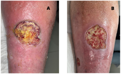
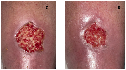
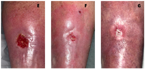
Source: Own elaboration.
Figure 3 E) Fifth month of follow-up F) Sixth month of follow-up G) Seventh month of follow-up with complete epithelialization
Calcific arteriolopathy most commonly affects patients with advanced chronic kidney disease (calcific uremic arteriolopathy). However, not all cases of calcific arteriolopathy occur in renal patients; it has also been reported in individuals with normal kidney function (non-uremic calcific arteriolopathy), particularly in the context of oncological diseases or the use of specific drugs [2,3].
Sodium thiosulfate has been proposed as a potential treatment for calcific arteriolopathy, administered intravenously, orally, or through intralesional injection. However, there have been few reports of its use as a topical ointment achieving complete lesion resolution [4-6].
The mechanism of action of sodium thiosulfate is still under discussion. Some research suggests that it acts as a calcium chelator, while others point to direct extracellular effects, inhibiting vascular calcification independently of its calcium-binding properties and occurring selectively in injured blood vessels [7]. From a clinical standpoint, calcific uremic arteriolopathy is a highly painful condition, and sodium thiosulfate has demonstrated significant pain improvement in patients treated intravenously [8].
As far as we know, this is the third reported case of topical treatment using different formulations based on sodium thiosulfate for patients diagnosed with atypical ulcers due to calciphylaxis. Experience with the topical use of this drug is derived from its application as an antidote for cutaneous extravasations and in the treatment of calcinosis cutis [9,10]. Regarding the local mechanism of action, it is believed that the benefits are mainly attributed to its anti- inflammatory and antioxidant effects, and it may also increase the solubility and accelerate the dissolution of the deposited calcium [11-13].
Given that the use of warfarin (a vitamin K antagonist) and vitamin D increases the risk of calciphylaxis, and the patient's vitamin D levels were adequate, a decision was made to discontinue calcitriol and to switch anticoagulation to apixaban [1].
In this patient, although the levels of calcium and phosphorus were normal, the level of intact parathyroid hormone (PTH) was moderately elevated: 220 pg/ml for a creatinine clearance of 39.4 ml/min. However, it is important to note that while elevated serum calcium and phosphate levels increase the risk of calciphylaxis in patients with kidney disease, it has been reported that 86 % and 40 % of patients with severe kidney disease and calciphylaxis, respectively, have normal to low serum calcium and phosphate levels.
The patient meets criteria for polypharmacy, receiving 3-5 medications over an extended period. Despite efforts to deprescribe annually, the process has been challenging due to multiple comorbidities, each justifying prescriptions correlating with associated chronic conditions.
It is worth highlighting that, upon achieving complete healing of the ulcerated calciphylaxis lesion, a significant improvement in glomerular filtration was observed. The patient's serum creatinine decreased to 1.5 mg/dL, with a glomerular filtration rate (GFR) of 60 mL/min (Table 1). This improvement was confirmed through three consecutive measurements of renal filtration parameters. This notable improvement in GFR was interpreted as the positive impact of resolving the systemic inflammatory state associated with the resolution of the calciphylaxis lesion, which is an external and localized manifestation of an underlying systemic process.
Table 1 Main laboratory values at the beginning (baseline) and the end of their treatment (final values)
Regarding the advanced local wound care at our institution, we prioritize the TIME framework and implement a multimodal treatment approach addressing the triggering cause and precipitating factors (pain-inflammation-edema-infection-arterial status) [14-17]. The local management using sodium thiosulfate ointment for occlusive moist wound healing proved to be a valid and effective option, considering the patient's multiple comorbidities and positive clinical progress. In our patient's case, the presence of gait instability and the associated risk of falls suggested a manifestation of the instability-falls syndrome, which, when considered alongside the patient's medical history, aligned with stage 4 of the Clinical Frailty Scale, commonly referred to as vulnerability. We believe that the approach proposed effectively prevented the syndrome from advancing further, thereby decreasing the chances of complications such as immobility, sepsis originating from skin wounds, and multiorgan failure. These complications significantly contribute to the mortality rate among patients diagnosed with calciphylaxis.
Conclusion
This report describes the successful healing of a cutaneous ulcer secondary to calciphylaxis in a patient with non-dialytic chronic kidney disease. Additionally, there was a significant improvement in the glomerular filtration rate following a comprehensive multidisciplinary approach, allowing for the effective use of topical sodium thiosulfate treatment without the need for intravenous therapy.
It is essential to emphasize that calcific arteriolopathy is a condition that requires interdisciplinary management for favorable outcomes. The focus should be on identifying potential triggers, correcting metabolic imbalances, addressing any associated medications (such as anticoagulants), and effectively managing pain and superimposed infections. In this context, collaborative work among different medical teams was prioritized to achieve disease control.

