


Services on Demand
Journal
Article
 English (pdf)
 Article in xml format
 Article references
 Send this article by e-mail
Indicators
-
 Cited by SciELO
 -
 Access statistics
Related links
-
 Cited by Google
 -
 Similars in
SciELO
Similars in
SciELO -
 Similars in Google
Share

 Permalink
Revista Colombiana de Ciencias Pecuarias
Print version ISSN 0120-0690
Rev Colom Cienc Pecua vol.27 no.3 Medellín July/Sept. 2014
ORIGINAL ARTICLES
Ulcerative gastric lesions in Brasileiro de Hipismo horses¤
Presencia de lesiones ulcerativas gástricas en caballos Brasileiro de Hipismo
Presença de lesões ulcerativas gástricas em cavalos Brasileiro de Hipismo
José R Martinez Aranzales1*, MVZ, MS, PhD; Cyril A De Marval2, MV, MS; Geraldo E Silveira Alves3, MV, MS, PhD.
1 Grupo Centauro, Escuela de Medicina Veterinaria, Facultad de Ciencias Agrarias, Universidad de Antioquia UdeA, Calle 70 No. 52-21, Medellín, Colombia.
2 RCAT-PMMG – Regimento de Cavalaria Alferes Tiradentes – Policia Militar, Belo Horizonte, Minas Gerais, Brasil.
3 Universidade Federal de Minas Gerais (UFMG), Escola de veterinária, Departamento de Clínica e Cirurgia Veterinárias, Av. Antônio Carlos 6627, Pampulha, MG 31270-901. Belo Horizonte, Brasil.
* Corresponding author: José R Martínez Aranzales. Grupo Centauro, Escuela de Medicina Veterinaria, Facultad de Ciencias Agrarias, Universidad de Antioquia UdeA, Calle 70 No. 52-21, Medellín, Colombia. E-mail: jrramonmvz@yahoo.com.
(Received: February 25, 2013; accepted: March 3, 2014)
Summary
Background: Background: equine gastric ulcer syndrome (EGUS) has a high and variable occurrence in horses of different ages and activity levels. Objective: the aim of this study was to determine the presence and severity of ulcers in Brasileiro de Hipismo (BH) horses owned by the military police. Methods: a group of 80 horses (38 males and 42 females, aged between 3.3 and 29.2 years) performing different activities underwent clinical and gastroscopic evaluation. Descriptive statistics for the presence of ulcers and chi-square (X2) analysis with Fisher exact test were conducted to determine a possible association between EGUS and type of activity, sex or age. Results: occurrence of at least one ulcer was detected in 45% of the horses, and most of them (40%) were located in the squamous mucosa of the Margo plicatus. No significant association (p>0.05) was observed between any of the factors evaluated and the presence of EGUS. Conclusion: the BH horses owned by cavalry's military have low incidence of EGUS despite being subjected to stress, which confirms a multifactorial nature of gastric lesions in this subpopulation.
Key words: equine, gastric mucosa, gastroscopy, ulcer.
Resumen
Antecedentes: Antecedentes: el Síndrome de Úlcera Gástrica Equina (EGUS) presenta una alta y variable ocurrencia en equinos de diferentes edades y tipo según la actividad que desarrollan. Objetivo: determinar la presencia e intensidad de las lesiones ulcerativas gástricas en caballos de la raza Brasilero de Hipismo (BH) al servicio de la policía militar. Métodos: un grupo de 80 equinos (38 machos y 42 hembras) con edades entre 3,3 y 29,2 años, en actividades de adaptación, entrenamiento, patrullaje e inactivos fueron evaluados por examen físico y gastroscópico. Se realizaron análisis por estadística descriptiva para porcentaje de ocurrencia de úlceras y análisis de Chi-cuadrado (X2) con test exacto de Fisher para verificar posible asociación entre EGUS, actividad, género y edad. Resultados: se determinó una ocurrencia de úlcera del 45%, siendo la mucosa escamosa (40%) la más comprometida en todo el Margo plicatus, sin embargo, no hubo asociación significativa (p>0,05) entre los factores evaluados y la presencia de EGUS. Conclusión: los caballos estudiados presentaron baja incidencia de EGUS a pesar de estar sometidos a situaciones de estrés, confirmando la naturaleza multifactorial de las lesiones gástricas en esta subpoblación de equinos.
Palabras clave: equino, gastroscopia, mucosa gástrica, úlcera.
Resumo
Antecedentes: a Síndrome da Ulcera Gástrica Equina (EGUS) apresenta uma ocorrência alta e variável em equinos de diferentes faixas etárias e classes segundo a atividade que desenvolvem. Objetivo: determinar a presença e intensidade das lesões ulcerativas gástricas em cavalos da raça Brasileiro de Hipismo (BH) a serviço da polícia militar. Métodos: um grupo de 80 equinos (38 machos e 42 fêmeas), com idade entre os 3,3 e 29,2 anos, desenvolvendo atividades de adaptação, treinamento, patrulhamento e inativos foram avaliados por exame físico e gastroscópicos. Analise por estatística descritiva para taxas de ocorrências de ulceras e analise de Qui-quadrado (X2) com teste de exato Fisher foram realizados para verificar possível associação entre EGUS, atividade, sexo e idade. Resultados: foi determinada uma ocorrência de úlcera de 45%, sendo a mucosa escamosa (40%) mais comprometida ao longo do Margo plicatus. Entretanto, não houve associação significativa (p>0,05) entre os fatores avaliados e a presença de EGUS. Conclusão: os cavalos estudados da raça BH apresentaram incidência baixa da EGUS apesar de submetidos a situações de estresse, sinalizando para a natureza multifatorial das lesões gástricas nesta subpopulação de equinos.
Palavras chave: equinos, gastroscopia, mucosa gástrica, úlcera.
Introduction
Equine gastric ulcer syndrome (EGUS) is a common pathology in horses of different ages and it is associated with multiple risk factors. EGUS is considered a cause of reduced performance in equine athletes and other equines assigned to a variety of activities (Nieto et al., 2009; Orsini et al., 2009). Inadequate environmental, nutritional, and management factors may contribute to gastric ulcerative processes, which typically define the multifactorial origin of EGUS. In this respect, risk factors inherent to the animal and the handling system, such as strenuous exercise, inadequate feeding, stress, confinement, use of non-steroidal anti-inflammatory drugs (NSAIDs), and certain bacterial infections have all been associated with the development of EGUS (Lorenzo-Figueras et al., 2002; Videla and Frank, 2009). The intensity or combination of these factors may determine the severity of the ulceration.
EGUS occurrence has been extensively studied and its prevalence ranges between 51 and 90% for competition breeds and high performance horses (Murray et al., 1996; Nieto et al., 2004; Bezdekova et al., 2005; Jonsson and Egenvall, 2006; Tanzali et al., 2011). Prevalence lower than 58% has been described for leisure, bull catching and show horses, as well as for brood mares (Murray et al., 1989; Wiedner et al., 2008). However, other studies reported higher incidences for similar activities. On the other hand, prevalence above 50% has also been reported for horses out of competition and in apparently normal condition (Videla and Frank, 2009).
Brasileiro de Hipismo (BH) horses are characterized by larger than average size, docile temperament, and skills in saddling, jumping and endurance riding. These horses are also used in military police regiments. Horses used for urban military patrolling are subjected to physical activities depending on their training and age, which generates various chronic stressors over the years. These conditions may cause changes in their gastric health.
Studies on EGUS occurrence in BH horses performing activities typical of a military regiment (such as adaptation, training and urban patrolling) could not be found in the literature. The aim of this study was to evaluate the presence and intensity of ulcerative gastric lesions in BH horses serving in the military police. In particular, we analyzed possible associations between EGUS and factors such as type of activity, sex and age of the animals.
Materials and methods
A sample of 80 BH horses (38 castrated males and 42 mares, all aged between 3.3 and 29.2 years) belonging to the Alferes Tiradentes Cavalry Regiment (RCAT) of Minas Gerais Military Police (PMMG) in Belo Horizonte, Brazil, was evaluated by physical and gastroscopy examination. Equines were divided into four groups of 20 animals, according to activity: G1: animals in adaptation (pre-training); G2: animals in dressage (horses in military training); G3: animals in urban patrolling (horses in military exercise); and G4: inactive and retired animals (because of disabling medical conditions or age). The age, weight and body scores are presented in Table 1.
Animal handling and environmental particularities were characterized for each group. Animals in adaptation, dressage and patrolling were kept in confinement and fed Tifton hay (Cynodon dactylon) at approximately 1.5% body weight (bw), a commercial concentrate ration offered at 1% bw and split into three daily meals, water and mineral salt was offered ad libitum. Animals underwent physical activity on alternate days, depending on the group. The activity per working day lasted two hours for G1 horses, three hours for G2, and six hours for G3. The G4 horses remained on picket and were fed under the same scheme as the other groups. All animals were vaccinated and subjected to regular control for endo and ectoparasites.
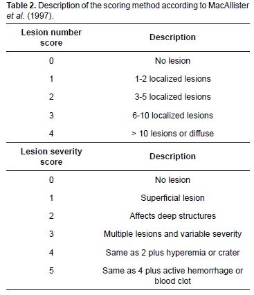
All horses underwent a 12-14h fasting period for solids and 4h for liquids before gastroscopy. After sedation (detomidine, 10 μg/kg/IV) and placement of full-mouth speculum (type Haussmann) animals were examined by systematic visualization of the gastric mucosa through a flexible videoendoscope (PortaScope®, 1800PVS, Bradenton, FL, USA) (12 mm diameter, 300 cm length) introduced via the nasal passage. The stomach was inflated with air to improve visibility during endoscopy and water jets were squirted to clean food remnants and improve visibility of the gastric surface. The gastric lesions found were video recorded and scored according to the number of lesions (0-4) and intensity (0-5) as recommended by MacAllister et al. (1997) (Table 2). All examinations were conducted by the same team and interpreted using a single-blind method by an endoscopist. This study was approved by the Comitê de Ética em Experimentação Animal da Universidade Federal de Minas Gerais under protocol number 234/09.
Statistical analysis
Descriptive analysis was used to determine the occurrence rate of ulcers, which were scored according to number (''number score'') and intensity (''severity score''). Chi-square (X2) analysis and Fisher exact test were used to determine significant differences (p<0.05) in the possible association between presence of ulcers and potential risk factors: age, sex and type of activities.
Results
All animals were apparently healthy upon physical exam. Gastroscopies were performed uneventfully. The fasting period was appropriate for complete visualization of the non-glandular and most of the glandular mucosa. Although gastric content was found in a small area of the antral region, it was not enough to compromise the inspection. The entire length of the esophagus, Margo plicatus, and pyloric region was visualized.
Gastroscopy findings are summarized in Table 3 and Figures 1 and 2. A total of 36/80 (45%) horses had gastric ulcers, 32/80 (40%) of which in the non-glandular mucosa. The score for number of lesions ranged between one (1) and four (4), with the latter having few animals affected (Figure 3). The score for severity of ulcers ranged between one (1) and three (3). Three out of 80 horses (3.7%) had ulcerative lesions in the glandular mucosa with grades one (1) and two (2) for score number and intensity, respectively. Only one horse presented grade 1 lesions in both types of mucosa and these were mainly located in the lesser and greater curvature of both mucosal regions adjacent to the Margo plicatus. No lesions were found in the esophagus or pyloric area.
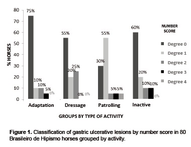
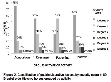
The endoscopy findings by groups showed a greater number of horses (14/20) in G3 suffering from ulcerative lesions, with number score from one (1) to four (4) and severity score from one (1) to three (3). G1 had the lowest number of animals affected (5/20) with a number score equal or lower than three (≤ 3). G1 and G4 had 9/20 and 8/20 animals with ulcers, respectively. G4 had more animals with severity scores 2 and 3. No significant associations were found (p>0.05) between gastric ulcers and type of activity (p=0.086), sex (p=0.10), or age (p=0.07).
Discussion
Studies on the prevalence and incidence of gastric ulcerative lesions in equines have highlighted some of the limitations of gastroscopy to estimate the degree of mucose damage in EGUS (Andrews et al., 2002). In spite of the fasting period, presence of remaining food in few places of the glandular area did hamper the evaluation of possible lesions in that area. Such difficulty was not encountered in the non-glandular mucosa. In this study we found low presence of lesions in the mucose area, which is in agreement with Buchanan and Andrews (2003), Videla and Frank (2009), and Tamzali et al. (2011).
The interpretation of lesion number and intensity in terms of clinical relevance is another source of discrepancy in the statistics of EGUS prevalence and incidence for some horse populations. Jeune et al. (2009) and Luthersson et al. (2009a) consider grades 0 and 1 as normal, although ulcer intensity is not necessarily correlated with clinical signs (Murray et al., 1989). Moreover, changes in the weather, breed, handling, and activities performed by the horses have contributed to those discrepancies (Jonsson and Egenvall, 2006).
The general incidence of EGUS in our study was 45%, a percentage that falls within the estimated rates for horses considered not overly stressed or in low physical demand, such as leisure, show, and exhibition horses (Murray et al., 1989; Bezdekova et al., 2005; McClure et al., 2005; Wiedner et al., 2008; Luthersson et al., 2009b). This incidence could fall to 19% if we neglected the relevance of the injuries classified as score 1; therefore, the incidence would be even lower than expected for this category.
It has been suggested that military cavalry horses are exposed to many stressors, as evidenced by Leal et al. (2011), who found differences in plasma cortisol concentration. This leads to behavioral changes in animals in confinement or in military regiment (Rezende et al., 2006; Muñoz et al., 2009), evidencing a close relation between stereotypy and gastric health (Moeller et al., 2008; Wickens and Heleski, 2010; Hemmann et al., 2012). This is why the hypothesis of high EGUS occurrence in the studied population was suggested.
The low incidence of ulcerations in this study suggests the predominance of gastric mucosa protective factors over other harmful factors, such as the stress derived from physical activities. Possibly, the feed handling provided to these horses counteracted the caustic action of gastric juice, as shown by De Graaf-Roelfsema et al. (2010). They concluded that stress from exercise is not important in EGUS development compared to environmental changes and feeding management since no increase in EGUS incidence was observed for properly fed horses under strenuous training. However, the multifactorial nature of this syndrome may involve other factors that were not considered.
Dietary management contributes positively to practical prevention strategies, treatment, and decreased recurrences of EGUS (Reese and Andrews, 2009). In this study, dietary management favored a relatively low incidence of ulceration, despite the excess concentrate feed/body weight ratio usually recommended (0.5 kg/100 kg; Nadeau et al., 2000). This indicates that, in addition to the ratio between fiber and concentrate, the frequency and type of feeding has a strong influence in gastric health (De Graaf-Roelfsema et al., 2010), and three meals per day was apparently adequate. Additionally, it is important to take into consideration that dietary fiber helps maintain a healthy gastric mucosa, therefore, providing greater amounts of forage to horses with EGUS or putting them out to pasture should be part of any therapeutic strategy.
Studies with different breeds in Brazil have shown a 47.6% prevalence in asymptomatic adult horses in stalls and pasture (Fernandes et al., 2003). Belli et al. (2005) reported 75% prevalence in hospitalized horses with presumptive diagnosis of EGUS. A preliminary study on horses performing common activities reported EGUS prevalence between 46.5 and 77.91%, showing a significant association between presence of gastric lesions and type of activity (Berger et al., 2009). Regarding Brasileiro de Hipismo horses kept at military regiments, prevalence studies identifying and comparing factors that induce gastric lesions could not be found. The EGUS incidence found in this study is low compared with other horse breeds.
Regarding location of ulcerative lesions, we found that the non-glandular mucosa was the most compromised area, particularly in proximity of the Margo plicatus, coinciding with existing literature (Hammond et al., 1986; Vatistas et al., 1999; Murray et al., 1989; Luthersson et al., 2009b; Tamzali et al., 2011). On the other hand, the low presence of ulcers in the glandular and both mucosae simultaneously contrasts with a report by Luthersson et al. (2009a). This suggests a difference in the efficiency of mucosae's self-protection mechanisms, which are more relevant than harmful mechanical factors, such as type of physical activity, regarding the appearance of gastric ulcers. Lesions in the glandular mucosa may be related to the use of NSAIDs. The medical records did not show any recent use of these drugs in the studied animals, which might explain the low presence of lesions in this gastric region.
The lack of association between lesion incidence and the studied factors (i.e., type of activity, sex and age) agrees with results from other studies (Bell et al., 2007; Tamzali et al., 2011). Nevertheless, some other studies have shown their involvement, thus they are considered to be risk factors for the onset of this syndrome (Lorenzo-Figueras and Merrit, 2002; Orsini et al., 2009). Geriatric and castrated male horses have shown greater susceptibility to EGUS (Rabuffo et al., 2002). This was not evidenced in the present study, in which all males were castrated and did not show greater occurrence when compared to females. Regarding age, specimens 3 to 29 years old were evaluated, and our results are in disagreement with other reports (Chameroy et al., 2006; Jonsson and Egenvall, 2006). There was a tendency for more animals with number scores between grades 2 and 3 in the retired horses, in agreement with Luthersson et al. (2009b). These findings demonstrate the influence of factors related to management and the environment in the incidence of the syndrome.
The majority of gastropathies in the patrolling horses can be explained by the stress suffered during transportation to patrol urban areas. In addition, these horses were used to patrol a large urban area (330 km2) three times a week, a factor that may also predispose them to develop gastric ulcers (McClure et al., 2005). However, there was no statistical association between the studied variables. In spite of the fact that the retired horses remained unleashed, apparently it did not benefit the gastric mucosa integrity in comparison with the partially confined animals, which is contrary to the results of other studies (Husted et al., 2009; Jeune et al., 2009). This could be explained by the cumulative effect of age and previous medical occurrences that disabled these horses for regular activities.
The activities endured during adaptation, training and working by cavalry military horses do not seem to be related with the occurrence of gastropathies. This also suggests a concomitant involvement of other potentially ulcerogenic factors and the need to control them (Luthersson et al., 2009b; De Graaf-Roelfsema et al., 2010; Flores et al., 2011). Because of the lack of information on gastric changes in Brasileiro de Hipismo horses, it is important to undertake further research to elucidate the involvement of factors inherent to the individual and the environment, as well as the relationship between welfare and gastric health in animals showing low EGUS prevalence without clinical signs.
Acknowledgments
The authors acknowledge Mr. Walmir Santos Viana for allowing us to conduct this study at RCAT-PMMG in Belo Horizonte. Funding for this study was provided by CNPq, CAPES / PEC-PG – Brazil, the University of Antioquia (Colombia), and UFMG (Brazil).
Notes
¤ To cite this article: Martinez JR, De Marval CA, Silveira GE. Ulcerative gastric lesions in Brasileiro de Hipismo horses. Rev Colomb Cienc Pecu 2014; 27:211-219
References
Andrews F, Reinemeyer CR, McCracken MD, Blackford JT, Nadeau JA, Saabye L, Sötell M, Saxton. Comparison of endoscopic, necropsy and histology scoring of equine gastric ulcers. Equine Vet J 2002; 34:475-478. [ Links ]
Bell RJ, Mogg T, Kingston J. Equine gastric ulcer syndrome in adult horses: A review. N Z Vet J 2007; 55:1-12. [ Links ]
Belli CB, Fernandes WR, Silva LC. Estudo gastroscópico em equinos adultos com suspeita de ulceração gástrica. Revta Bras Ciênc Vet 2005; 12:92-98. [ Links ]
Berger H, Silva R, Klemm M, Orsolini A. Gastric Ulcers in Brazilian performance horses. Proceedings of the 11th International Congress of the World Equine Veterinary Association (WEVA) 2009; Guarujá, SP, Brazil, (Abstract). [ Links ]
Bezdekova B, Jahn P, Vyskocil M, Plachy J. Gastric ulceration and exercise intensity in Standarbred Racehorses in Czech Republic. Acta Vet Brno 2005; 74:67-71. [ Links ]
Buchanan BR, Andrews FM. Treatment and prevention of equine gastric ulcer syndrome. Vet Clin North Am Equine Pract 2003; 19:575-597. [ Links ]
Chameroy KA, Nadeau JA, Bushmich SL, Dinger JE, Hoagland TA, Saxton AM. Prevalence of non-glandular gastric ulcers in horses involved in a university riding program. J Equine Vet Sci 2006; 26:207-211. [ Links ]
De Graaf-Roelfsema E, Keizer HD, Wijnberg ID, Van Der Kolk JH. The incidence and severity of gastric ulceration does not increase in overtrained Standardbred horses. Equine Vet J 2010; 42:58-61. [ Links ]
Fernandes WR, Belli CB, Silva LC. Achados gastroscópicos em equinos adultos assintomáticos. Arq Bras Med Vet Zootec 2003; 55:405-410. [ Links ]
Flores RS, Byron CR, Kline K. Effect of feed processing method on average daily gain and gastric ulcer development in weanling horses. J Equine Vet Sci 2011; 31:124-128. [ Links ]
Hammond CJ, Mason DK, Watkins KL. Gastric ulceration in mature Thoroughbred horses. Equine Vet J 1986; 18:284-287. [ Links ]
Hemmann K, Raekallio M, Kanerva K, Hanninen L, Pastell M, Palviainen M, Vainio O. Circadian variation in Ghrelin and certain stress hormones in crib-biting horses. Vet J 2012; 193:97-102. [ Links ]
Husted L, Sanchez L, Baptiste K, Olsen S. Effect of a feed/fast protocol on pH in the proximal equine stomach. Equine Vet J 2009; 41:658-662. [ Links ]
Jeune SS, Nieto JE, Dechant JE, Snyder JR. Prevalence of gastric ulcers in Thoroughbred broodmares in pasture: A preliminary report. Vet J 2009; 181:251-255. [ Links ]
Jonsson HA, Egenvall A. Prevalence of gastric ulceration in Swedish Standardbred in race-training. Equine Vet J 2006; 38:209-213. [ Links ]
Leal BB, Alves GES, Douglas RH, Bringel B, Young RJ, Haddad JP, Viana WS, Faleiros RR. Cortisol circadian rhythm ratio: A simple method to detect stressed horse at higher risk of colic? J Equine Vet Sci 2011; 31:188-190. [ Links ]
Lorenzo-Figueras M, Merritt A. Effects of exercise on gastric volume and pH in the proximal portion of the stomach of horses. Am J Vet Res 2002; 63:481-1487. [ Links ]
Luthersson N, Nielsen KH, Harris P, Parkin TDH. The prevalence and anatomical distribution of equine gastric ulceration syndrome (EGUS) in 201 horses in Denmark. Equine Vet J 2009a; 41:619-624. [ Links ]
Luthersson N, Nielsen KH, Harris P, Parkin TDH. Risk factors associated with equine gastric ulceration syndrome (EGUS) in 201 horses in Denmark. Equine Vet J 2009b; 41:625-630. [ Links ]
MacAllister CG, Andrews FM, Deegan E, Ruoff W, Olovson SG. A scoring system for gastric ulcers in the horse. Equine Vet J 1997; 29:430-433. [ Links ]
McClure SR, White G, Sifferman R, Bernard W, Doucet M, Vrins A, Holste J, Fleishman C, Alva R, Cramer L. Efficacy of omeprazole paste for prevention of gastric ulcers in horses in race training. J Am Vet Med Assoc 2005; 226:1681-1684. [ Links ]
Moeller B, McCall C, Silverman S, McElhenney W. Estimation of saliva production in crib-biting and normal horses. J Equine Vet Sci 2008; 28:85-90. [ Links ]
Muñoz L, Torres J, Sepulveda O, Rehhof C, Ortiz R. Frecuencia de comportamientos anormales estereotipados en caballos chilenos estabulados. Arch Med Vet 2009; 41:73-76. [ Links ]
Murray MJ, Schusser GF, Pipers FS, Gross SJ. Factors associated with gastric lesions in Thoroughbred horses. Equine Vet J 1996; 28:368-374. [ Links ]
Murray MJ, Grodinsky C, Anderson AC, Radue PF, Schmidt GR. Gastric Ulcers in horses: a comparison of endoscopic findings in horses with and without clinical signs. Equine Vet J 1989; 7:68-72. [ Links ]
Nadeau JA, Andrews FM, Mathew AG, Argenzio AG, Blackford JT, Sohtell M, and Saxton AM. Evaluation of diet as a cause of gastric ulcers in horses. Am J Vet Res 2000; 617:84-90. [ Links ]
Nieto JE, Snyder JR, Vatistas NJ, Jones JH. Effect of gastric ulceration on physiologic responses to exercise in horses. Am J Vet Res 2009; 70:787-795. [ Links ]
Nieto JE, Snyder JR, Beldomenico P, Aleman M, Kerr JW, Spier SJ. Prevalence of gastric ulcers in endurance horses: A preliminary report. Vet. J 2004; 167:33-37. [ Links ]
Orsini JA, Hackett ES, Grenager N. The effect of exercise on Equine Gastric Ulcer Syndrome in the Thoroughbred and Standardbred athlete. J Equine Vet Sci 2009; 29:167-171. [ Links ]
Rabuffo TS, Orsini JA, Sullivan E, Engiles J, Norman T, Boston R. Associations between age, sex and prevalence of gastric ulceration in Standardbred racehorses in training. J Am Vet Med Assoc 2002; 221:1156-1159. [ Links ]
Reese RE, Andrews FM. Nutrition and Dietary Management of Equine Gastric Ulcer Syndrome. Vet Clin North Am Equine Pract 2009; 25:79 – 92. [ Links ]
Rezende MJ, McManus C, Martins R, Oliveira L, Garcia J, Louvandini H. Comportamento de cavalos estabulados do exército Brasileiro em Brasília. Ciênc Anim Bras 2006; 7:327-337. [ Links ]
Spiers VC. The alimentary tract. In: Robinson NE, editors. Clinical Examination of Horses. Philadelphia: WB Saunders; 1997. p.261-298. [ Links ]
Tamzali Y, Marguet C, Priymenko N, Lyazrhi F. Prevalence of gastric ulcer syndrome in high-level endurance horses. Equine Vet J 2011; 42:1-4. [ Links ]
Vatistas NJ, Snyder JR, Carlson G, Johnson B, Arthur RM. Cross sectional study of gastric ulcers of the squamous mucosa in Thoroughbred racehorses. Equine Vet J 1999; 29:34-39. [ Links ]
Videla R, Frank A. New perspectives in Equine Gastric Ulcer Syndrome. Vet Clin North Am Equine Pract 2009; 25:283-301. [ Links ]
Wickens CL, Heleski CR. Crib-biting behavior in horses: A review. Appl Anim Behav Sci 2010; 128:1-9. [ Links ]
Wiedner EB, Schmitt DL, Kiso W, Kinchen K, Lindsay WL. Gastroscopy of 30 circus horses. Proc. 10th International Congress of World Equine Veterinary Association (WEVA), Moscow, 2008; p.489. [ Links ]
