


Services on Demand
Journal
Article
 English (pdf)
 Article in xml format
 Article references
 Send this article by e-mail
Indicators
-
 Cited by SciELO
 -
 Access statistics
Related links
-
 Cited by Google
 -
 Similars in
SciELO
Similars in
SciELO -
 Similars in Google
Share

 Permalink
Revista de la Universidad Industrial de Santander. Salud
Print version ISSN 0121-0807
Rev. Univ. Ind. Santander. Salud vol.45 no.3 Bucaramanga Sep./Dec. 2013
Amphotericin B: an antifungal drug in
nanoformulations for the treatment
of paracoccidioidomycosis
Mônica Pereira Garcia1*, Maria de Fátima Menezes Almeida Santos1, Camila Arruda Saldanha1, Diêgo Cesar Iocca1, Ricardo Bentes Azevedo1
1. Department of Genetics and Morphology, Institute of Biological Science, University of Brasília, Brasília, DF, Brazil.
Correspondence: Mônica Pereira Garcia, Universidade de Brasília - Campus Universitário Darcy Ribeiro, Instituto de Ciências Biológicas, Departamento de Genética e Morfologia, CEP: 70910-900 Brasília - DF, Brasil. mgarcia@unb.br.
Recibido: Noviembre 12 de 2012 Aprobado: Septiembre 1 de 2013
Forma de citar: Pereira Garcia M, Meneses Almeida Santos MF, Saldanha Arruda C, Iocca DC. Amphotericin B: an antifungal drug in nanoformulations for the treatment of paracoccidioidomycosis. rev.univ.ind.santander.salud 2013; 45 (3): 45-53
ABSTRACT
The use of magnetic nanoparticles (MNPs) in drug delivery vehicles must address issues such as drugloading capacity, desired release profile, aqueous dispersion stability, biocompatibility with cells and tissue, and retention of magnetic properties after interaction with macromolecules or modification via chemical reactions. Amphotericin B (AmB) is still the first choice for the treatment of severe paracoccidioidomycosis, an important systemic fungal infection caused by Paracoccidoides brasiliensis. Unfortunately, AmB causes acute side effects (mainly urinary problems) following intravenous administration, which limits its clinical use. The use of magnetic nanoparticles stabilized with biocompatible substances, together with the possibility of their conjugation with drugs has become a new nanotechnological strategy in the treatment of diseases for drug delivery to specific locations, such as the lungs in paracoccidoidiodomycosis. This review provides an overview of the disease, its etiologic agent and treatment with emphasis on the main strategies to improve the use of AmB in nanoformulations.
Keywords: Paracoccidioides brasiliensis; amphotericin B; magnetite nanoparticles; magnetic fluid; drug delivery complex
La anfotericina B: una droga antifúngica en nanoformulaciones
para el tratamiento de la paracoccidioidomicosis
RESUMEN
El uso de nanopartículas magnéticas (MNPS) en los vehículos de suministro de fármacos debe abordar cuestiones como la capacidad de carga de las drogas, el perfil deseado de liberación, estabilidad de la dispersión acuosa, biocompatibilidad con las células, tejidos y la conservación o la modificación de las propiedades magnéticas después de la interacción con macromoléculas y/o reacciones químicas. La anfotericina B (AnB) continua siendo la primera opción para el tratamiento de la paracoccidioidomicosis grave, una importante infección sistémica causada por el hongo Paracoccidioides brasiliensis. Sin embargo, la AnB causa efectos secundarios agudos (principalmente problemas urinarios) tras la administración intravenosa, limitando su uso clínico. El uso de nanopartículas magnéticas estabilizadas con sustancias biocompatibles y conjugadas con fármacos, se ha convertido en una nueva estrategia nanotecnológica para el tratamiento de enfermedades en sitios específicos, como los pulmones en paracoccidoidiodomycosis. En esta revisión se hace una descripción general de la enfermedad, su agente etiológico y su tratamiento con énfasis en la principales estrategias para mejorar el uso de AnB en nanoformulaciones.
Palabras clave: Paracoccidioides brasiliensis, anfotericina B, nanoparticulas de magnetita; fluido magnético; entrega controlada de medicamentos
Introduction
The paracoccidioidomycosis (PCM) is a systemic mycosis autochthonous from South and Central America, endemic in rural populations, but with heterogeneous distribution (with low and high endemicity).
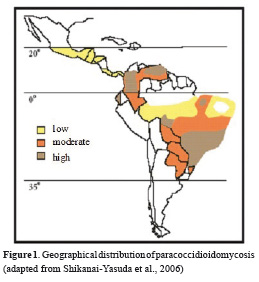
This mycosis affects mainly men between 30 and 60 years old. It is believed that 50% of the inhabitants of endemic areas in countries such as Venezuela, Colombia, Argentina and Brazil have been exposed to the etiologic agent of this mycosis. However, only 2% of individuals develop some clinical manifestation of the disease 1,2. It is noteworthy that when not diagnosed and treated appropriately, the PCM can disseminate affecting progressively the lungs, and may reach other organs. Even if the patient presents improvements due to the treatment, it can develop pulmonary fibrosis, the most serious sequel resulting from lung granulomatous processes, leading to limitations of the individual activities 3.
The etiologic agent of the PCM is Paracoccidioides brasiliensis, a saprobiotic fungus that in nature presents itself as filamentous between 25 °C and 30 °C, i.e. a multicellular mycelium containing propagules called conidia infectors. Once inhaled by mammals, the propagules turn into yeast forms of fungus that will be their parasitic form in host tissues, thus behaving as a thermo-dimorphic fungus 1,4.
Treatment of paracoccidioidomycosis
Unlike other pathogenic fungi, the P. brasiliensis is sensitive to most antifungal drugs therefore various antifungal agents are used in the treatment of PCM 5. The choice of drug to be used is in accordance with the state of the patient, and requires, besides a long period of treatment, patient monitoring in order to evaluate the efficacy and his tolerance to the antifungal 6. Among the most popular drugs, stand out Itraconazole and Amphotericin B 1.
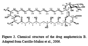
Amphotericin B is a polyene antibiotic and is produced by the actinomycete Streptomyces nodosus. It has been the drug of choice for the treatment of most systemic mycoses 7,8.
AmB mechanism of action occurs by its hydrophobic interaction with ergosterol, the most abundant sterol in P. brasiliensis cell membranes. These interactions induce the formation of aqueous pores in the fungi membranes and thus triggering death 9,10. Although Amphotericin B preferentially binds to the ergosterol, it can also bind to cholesterol11, the most abundant sterol founded in mammals' cells membranes 9,12, and for that reason its administration in humans must be controlled. This drug has broad-spectrum antifungal action, potent fungicidal activity and rare episodes of resistance, which contributes to the clinical success of this drug13. Nevertheless, AmB induces severe side effects in humans and is considered one of the most toxic antibiotics for humans7,13, often leading to patient hospitalization during the administration of this drug for adequate monitoring 1,14,15. The most frequent side effects are nausea, vomiting, fever, headache, hypotension, liver damage, anemia and especially nephrotoxicity 7,13.
The incidence of nephrotoxicity induced by AmB is very high, affecting 49 to 65% of the patients 16. Nephrotoxicity is due to the binding of AmB to cholesterol in the nephron convoluted tubules cell membranes14,15,17, creating an ion channel in these membranes that allows the flow of ions and small intracellular molecules. When these ions exit, especially potassium, impairment of cell metabolism occurs 18. The accumulation of potassium ions in the blood, hyperkalemia, can induce severe cardiac arrhythmia 19. The serum levels of the drug contribute to increase such toxicological effects, since the greater the amount of free flowing drug, more damage will cause to the kidneys. Nephrotoxicity is the cause of prolonged hospitalization and mortality rates, especially in patients requiring hemodialysis.
In order to reduce the adverse effects of Amphotericin B in treatment of PCM, new strategies are being developed in the formulation of this drug, specially using nanotechnology.
Nanotechnology as a tool for drug delivery
Nanotechnology is referred as the manipulation of matter with at least one dimension sized from 1 to 100 nanometers. Nanobiotechnology is the science that investigates the interactions between those nanoscale materials and biological systems. Thus, nanostructured materials, nanoparticles in particular, exhibit new thermal, mechanical, magnetic and optical properties such as small size, large surface area to mass ratio, and high reactivity delivery, that allow for their widespread application in biomedicine and many industrial sectors 20.
A major contribution of nanobiotechnology for new formulations of conventional drugs is the possibility of creating functional systems of drug delivery at the nanoscale so that their kinetic properties and dynamics can be modified to optimize its pharmacological response 21. Among the benefits of a nanoscale system for drug delivery is the bioavailability enhancement of associated drugs and/or improvement of drugs distribution and targeting in tissues. This controlled drug delivery allows decreasing in the number of drug applications in patients, and also reduces nanoparticle uptake by the reticuloendothelial system (RES).
Nanoparticles are typically defined as solids with less than 100 nm in all three dimensions. Most often, they are particles having diameters about 10 nm or less and this size is similar to most biological molecules and structures 22. Thereby, nanoparticles can be useful in biomedical research and applications. Some nanoparticles commonly consist of magnetic elements such as iron, nickel and cobalt and their chemical compounds. These nanoparticles can be synthesized and modified with various chemical functional groups and conjugated with biological molecules or structures, such as drugs of interest, opening a wide range of potential applications in biomedicine 20. Metal and magnetic nanoparticles have been continuously used and modified to enable their use as a drug delivery system.
It is worth mentioning that liposomes also are used in drug delivery. Liposomes are spherical vesicles made from phospholipids bilayer. Thus lipid-soluble drugs can be incorporated into their lipid phase, whereas water-soluble drugs can be entrapped into their aqueous phase. Their advantage is the easily manufacture process, they are no covalent aggregates, have almost no toxicity, biodegradable, among others. Therefore, nanoparticles and liposomes can be applied to facilitate the administration of antimicrobial drugs, thereby overcoming some of the limitations in traditional antimicrobial therapeutics. According to Zhang et al, antimicrobial drugs encapsulation enhances therapeutic effectiveness and minimizes side effects of the drugs 23.
Antimicrobial drug associated to metal nanoparticles
Metal nanoparticles exhibit excellent bactericidal action against Gram-positive and Gram-negative bacteria. This effect has been attributed to small size and high surface to volume ratio, which allows them to interact closely with microbial membranes and is not merely due to the release of metal ions in solution 24. Silver nanoparticles (AgNPs), for example, have the ability to anchor to the bacterial cell wall and subsequently penetrate it, thereby causing structural changes, like the permeability, in the membrane and cell death 25. Thus, AgNPs can kill antibiotic-resistant microbes. Several studies have demonstrated antimicrobial effects of silver nanoparticles against Escherichia coli, Staphylococcus aureus, Salmonella typhimurium, Pseudomonas aeruginosa, Bacillus subtilis and yeast strains 26-29. It is noteworthy that AgNPs exhibited no antibacterial activity in the presence of serum proteins. However, as demonstrated by Gnanadhas and co-workers AgNPs capped with citrate or poly (vinylpyrrolidone) exhibited antibacterial activities in vivo against Salmonella infection compared to uncapped AgNPs 30. It is worth mentioning that there are numerous consumer products utilizing the antimicrobial properties of AgNPs, such as cosmetics, water filters, and food packaging containers. Although the most common metal nanoparticles used as the antimicrobial agent are AgNPs, gold nanoparticles (AuNPs) are being used effectively against strains of Gram-positive and Gram-negative bacteria like Micrococcus luteus, Pseudomonas aeruginosa Escherichia coli and Staphylococcus aureus. It also exhibit antifungal activity against Aspergillus niger and Fusarium oxysporum 31. Additionally AuNPs dispersed on zeolites eliminate Escherichia coli and Salmonella typhi in 90 minutes 32.The authors showed that AgNP were intrinsically antibacterial, whereas AuNP were antimicrobial only when ampicillin was bound to their surface. The antimicrobial activity of AuNPs can be attributed to the ability of interaction with functional groups on bacterial cell and inactive bacteria; it cause structural changes, degradation and cell death 33. Furthermore AuNPs also may act as drug carriers34.
Antimicrobial tests have also shown that copper nanoparticles can have antimicrobial activity; surfaces of the copper nanoparticles interact directly with the outer bacterial membrane causing it to rupture and thus killing bacteria 24. The authors clearly demonstrated that copper nanoparticles synthesized in green synthesis method exhibit more antibacterial activity against Escherichia coli than copper sulphate solution and pure ginger extract. The encapsulation of antimicrobial drugs in nanoparticle systems enhances therapeutic effectiveness and minimizes side effects of antimicrobial agents.
Magnetic nanoparticles and antimicrobial activity
Magnetic nanoparticles (MNPs) commonly consist of magnetic elements such as iron, nickel, cobalt and manganese or zinc, and most often by ferrite as magnetite (Fe3O4) or maghemite (γ-Fe2O3) 35. Such nanoparticles when dispersed in colloidal solutions constituted magnetic fluids (MFs), stable suspensions of magnetic nanoparticles with a diameter generally ranging between 5 and 15 nm, in inorganic or organic solvent carrier. In these solutions, the particle-liquid interactions are strong enough that their magnetic behaviors are transmitted to the liquid as a whole 36. Magnetic nanoparticles can bind to drugs, proteins, enzymes, antibodies, or nucleotides and can be directed to an organ. Although MNPs are mostly used on cancer research and treatment, new antibiotic coated magnetic nanoparticles intended as magnetically controllable pharmaceutical agents for the recovery of bacteria loaded tissues and organs. It is reasonable to assume that the conjugation of antimicrobial drugs to magnetic nanoparticles can combine the best properties of both, generating an improved antimicrobial nanoparticle, and enhancing therapeutic effectiveness of antimicrobial drugs in the treatment of infectious diseases.
Antimicrobial activity of MNPs was described by Grumezescu and collaborators, they reveal the synergistic effect of the synthesized water dispersible magnetic nanocomposites on the activity of different antibiotics against Gram-positive and Gram-negative bacterial strains37. Similarly, Dong and co-workers have shown that the combination of barbituric acidbased N-halamine with magnetic nanoparticles exhibit higher biocidal activity than the bulk powder barbituric acid-based N-halamine besides facilitates the repeated antibacterial applications 38. Nevertheless, magnetic nanoparticles are recognized by macrophages of the mononuclear phagocyte system and are eliminated from the body. In order to improve biocompatibility, to reduce toxicity and to ensure non-immuno-genicity, particles have been encapsulated (e.g., with chitosan, dextran, lactic acid).
Another important factor is that it consists of iron, which acts to maintain the operation of essential metabolic pathways present in living organisms in general 39,40. For pathogenic microorganisms, especially P. brasiliensis, the ability to acquire iron is crucial for the establishment of infection, so that the ability to capture this element from the host is considered a virulence factor 41. Moreover, studies show that P. brasiliensis, both the yeast and mycelial forms, has a metabolic demand for iron42. Cano and colleagues demonstrated that the restriction of iron was one of the mechanisms by which inhibit the transformation of yeast in the form of conidia in activated macrophages, subsequently yeast growth within macrophages 43. Similarly, studies with chloroquine, a drug which affects the metabolism of iron in macrophages decreases the intracellular survival of the yeast P. brasiliensis in macrophages by interfering with the acquisition of iron by the fungus 44-46.
Other studies also demonstrate antimicrobial effect of zinc oxide (ZnO) nanoparticles. Vani et al 47 reports that they can be applied effectively for the control of microorganisms and the prevention of infections caused by Staphylococcus aureus. The antibacterial activity of ZnO nanoparticles also has been studied by Jones et al 48. According to the authors these nanoparticles have a potential application as bacteriostatic agent in visible light and may be used to control the spread and infection of a variety of bacterial strains.
Anfotericin formulations
The burden of invasive fungal infections (IFIs) has increased in the last years, especially from the increasing prevalence of individuals with immunosuppression, causing high morbidity and mortality, partly among ill patients 49,50. Amphotericin B deoxycholate (AmB-D) has been the cornerstone for the treatment of IFIs over the past four decades. Its broad-spectrum fungicidal activity has been showing efficient in candidiasis, cryptococcosis, aspergillosis, histoplasmosis, blastomycosis, coccidioidomycosis, zygomycosis, sporotrichosis, fusariosis, and phaeohyphomycosis. Fungi resistant to AmB are rare, including Trichosporon spp., Aspergillus terreus, Scedosporium spp. and Malassezia furfur 51.
However, as already written, conventional AmB-D is associated with adverse effects in 50-90% of cases, including acute infusion reactions, electrolyte imbalances, and dose-dependent nephrotoxicity 46. The infusion reactions are probably linked with the induction of pro-inflammatory cytokines demonstrated to be produced by AmB and the release of TNF-alpha from macrophages 52. Nephrotoxicity is defined in most studies as duplication of baseline creatinine levels. It is associated with vasoconstriction causing ischemic injury and direct interaction with epithelial cell membranes causing tubular dysfunction 53.
Given the above, extensive efforts were made to reformulate AmB in the last 15 years. AmB has strong lipophilic properties that led to the encapsulation of the drug into lipossomes or binding to lipid complexes. These lipid formulations of AmB are an attempt to enhance efficacy by increased dosing and to improve the safe profile, reducing the adverse effects 52.
Three lipid formulations of AmB are licensed and available. They are: 1) amphotericin B lipid complex (ABLC), composed of amphotericin B complexed with two phospholipids in a 1:1 drug-to-lipid molar ratio. The two phospholipids, l-α-dimyristoylphosphatidylcholine and l-α-dimyristoylphosphatidylglycerol, are present in a 7:3 molar ratio. ABLC has a ribbons-shaped complex with length range from 1.6 to 11.1 nm. The commercial product is Albelcet®. 2) amphotericin B colloidal dispersion (ABCD), consists of a 1:1 (molar ratio) complex of amphotericinB and cholesteryl sulphate. Upon reconstitution it forms a colloidal dispersion of microscopic uniform disc-shaped particles with diameter range from 120 to 140 nm and thickness of 4 nm. The commercial product is Amphotec® and Amphocil®. 3) Liposomal amphotericin B (L-AmB), consists of a 1:9 (drug-to-lipid molar ratio) of amphotericin B with hydrogenated soy phosphatidylcholine, distearoyl, hosphatidylglycerol, cholesterol, sucrose, and disodium succinate hexahydrate as buffer. It consists of unilamellar bilayer liposomes with amphotericin B intercalated within the membrane. Due to the nature and quantity of amphophilic substances used, and the lipophilic moiety in the amphotericin B molecule, the drug is an integral part of the overall structure of the liposomes. L-AmB is sphere-shaped with diameter range from 45 to 80 nm. The commercial product is Ambisome® 54,55.
These lipid formulations differ in several aspects, in their lipid composition, shape, physicochemical properties and pharmacokinetic parameters. They share different accumulation rates to various tissue components.
ABLC, because of its size, is taken up rapidly by macrophages and becomes sequestered in tissues of the mononuclear phagocyte system such as the liver and spleen, so it has lower circulating amphotericin B serum concentrations when compared to AmB-D 56. Lung levels are considerably higher than those achieved with other lipid-associated preparations, suggesting a potential formulation for the treatment of fungal respiratory infections, such as paracoccidioidomycosis. The recommended therapeutic dose of ABLC is 5 mg/ kg/day 57.
ABCD complexes remain largely intact, after intravenous injection, and are rapidly removed from the circulation by macrophage. The peak plasma level (Cmax) achieved is lower than that attained by AmB-D. ABCD exhibits dose-limiting, infusion-related toxicities; consequently, the administered dosages should not exceed 3-4 mg/kg/ day 56.
L-AmB avoids substantial recognition and uptake by the reticuloendothelial system due to its small size and negative charge. Therefore, a single dose of L-AmB results in a much higher Cmax/MIC value than AmB-D and a much larger area under the concentration-time curve. Tissue concentrations in patients receiving L-AmB tend to be lower in kidneys and lung and highest in the liver and spleen. Recommended therapeutic dosages are 3-6 mg/kg/day 56-58.
Wade and coworkers, in a well-designed head-to-head observational study, compared the nephrotoxicity and other adverse events among patients receiving liposomal amphotericin B or amphotericin B lipid complex. A total of 327 hospitalized patients were analyzed, they differed in terms of age, gender, race, and urgent/emergent admission status, but all of them were infected with Aspergillus, Candida, and/or Cryptococcus, were older than 18 years, with evidence of renal impairment or with increase risk of nephrotoxicity from AmB. They observed that those receiving ABLC demonstrated approximately threefold greater odds of developing nephrotoxicity compared to patients who received L-AMB, as well as the ABLC therapy was associated with significantly higher rates of infusion reactions 50.
In clinical studies it has been proved that the cost of treatment with lipid formulations of AmB can be overweighed by the cost of nephrotoxicity. Interpreting the findings of all studies is further complicated by differences in study populations with respect to age, disease state, infectious organism, risk factors for IFIs, exposure to nephrotoxic agents, and different definitions of nephrotoxicity 58.
In summary, all of the lipid formulations have demonstrated equivalent efficacy and reduced toxicity compared to AmB-D, but by a mechanism that is not yet exactly known. It is assumed that they offer less free drug that is able to bind to the kidney epithelial cells as mammalian cells are affected only by high free amphotericin B concentrations.
A formulation of poly (lactic-co-glycolic acid) (PLGA) and dimercaptosuccinic acid (DMSA) polymeric nanoparticles loaded with AmB-D (Nano-AmB) was tested in mice infected with P. brasiliensis. At 30 days post-infection, the animals were treated with Nano-AmB (6 mg/kg of encapsulated AmB-D, intraperitoneally (ip), interval of 72 h) or AmB-D (2 mg/kg, ip, interval of 24 h) during 30 days. Nano-AmB showed a marked antifungal efficacy. No renal or hepatic biochemical abnormalities, as well as no genotoxicity and cytotoxicity effects, were found in the animals treated with Nano-AmB. Thus, Nano-AmB comprises an AmB formulation able to lessen the number of drug administrations, once it showed a favorable extended dosing interval 59.
Alternatively, our group have been developed a new formulation of AmB associated with maghemite-based magnetic fluid stabilized with bilayer of lauric acid (BCL-AmB).

It is a very stable nanomaterial (over 240 days) with average size value of 13 nm. BCL-AmB presented antifungal activity against P. brasiliensis with a higher MIC value compared to AmB-D, and presented no cytotoxicity to the human urinary cells while inducing low cytotoxicity to the peritoneal macrophages. In vivo studies showed that BCL-AmB was effective against the acute form of PCM experimental, but not the chronic infection, and did not induce clinical, biochemical and histolopathological alterations (Paper in preparation).
Lung tissue samples removed from P. brasiliensis-infected and BCL-AmB treated mice were deposited onto Surface Enhanced Raman Scattering (SERS) active substrates for recording the Raman spectra. The results revealed spectral changes in relative intensities which are associated to the oxidation state of both the protein b588 and the myeloperoxidase enzyme, and so consistent with the oxygenation process of neutrophils's heme groups triggered by fungal infection 60.
It is possible to consider that the fungus infection in animals treated with Free AmB and BCL-AmB is much less than in animals treated with PBS, and both treatments led to similar therapeutic outcomes. We claim that the therapeutic approach using BCL-AmB has advantages over the conventional one, since the AmB content administered in BCL-AmB is 40% lower than the content administered in Free AmB, and it is well know that adverse effects reduce as the AmB doses reduce also. In addition, the magnetic drug carrier (BCLAmB) administration was performed in intervals threetimes (72 hours) longer than free AmB (24 hours) 60.
Therefore, it is reasonable to believe that AmB when coupled to magnetic nanoparticles stabilized with bilayer lauric acid, by having similar antifungal activity and do not induce adverse effects at therapeutic doses in acute infection and also allows reduction of the number of applications, can be an alternative nanotool to the treatment of acute form of PCM, but further studies must be done to improve effectiveness in chronic infection.
CONCLUSION
In conclusion, the use of magnetic nanoparticles stabilized with biocompatible substances, together with the possibility of their conjugation with drugs has become a new nanotechnological strategy in the treatment of diseases for drug delivery to specific locations, such as the lungs in paracoccidoidiodomycosis.
CONFLICT OF INTEREST
The authors of this paper have no conflict of interest.
AGRADECIMIENTOS
Al Doctor Mario F. Escobar, Coordinador Medicina Interna, Clínica Universitaria el Bosque; al Doctor Carlos Castro, Coordinador Asignatura Bioquímica Médica. Facultad de Medicina. Universidad el Bosque; y al Departamento de Audiovisuales Universidad el Bosque.
REFERENCIAS
1. Brummer E, Castaneda E, Restrepo A. Paracoccidioidomycosis: an update. Clin Microbiol Rev. 1993;6(2):89. [ Links ]
2. Coutinho ZF, Silva D, Lazéra M, et al. Paracoccidioidomycosis mortality in Brazil (1980- 1995). Cad Saúde Pública. 2002;18(5):1441-1454. [ Links ]
3. Cock AM, Cano LE, Vélez D, Aristizábal BH, Trujillo J, Restrepo A. Fibrotic sequelae in pulmonary paracoccidioidomycosis: histopathological aspects in BALB/c mice infected with viable and non-viable Paracoccidioides brasiliensis propagules. Rev Inst Med Trop S Paulo. 2000;42(2):59-66. [ Links ]
4. Gomes GM, Cisalpino PS, Taborda CP, De Camargo ZP. PCR for diagnosis of paracoccidioidomycosis. J Clin Microbiol. 2000;38(9):3478-3480. [ Links ]
5. Shikanai-Yasuda MA, Telles Filho FQ, Mendes RP, Colombo AL, Moretti ML. Guideliness in paracoccidioidomycosis. Rev Soc Bras Med Trop. 2006;39(3):297-310. [ Links ]
6. Hahn RC, Morato Conceição YT, Santos NL, Ferreira JF, Hamdan JS. Disseminated paracoccidioidomycosis: correlation between clinical and in vitro resistance to ketoconazole and trimethoprim sulphamethoxazole. Mycoses. 2003;46(8):324-329. [ Links ]
7. Lemke A, Kiderlen A, Kayser O. Amphotericin B. Appl Microbiol Biotechnol. 2005;68(2):151-162. [ Links ]
8. Ruijgrok EJ, Fens MHAM, Bakker-Woudenberg IAJM, Van Etten EWM, Vulto AG. Nebulized amphotericin B combined with intravenous amphotericin B in rats with severe invasive pulmonary aspergillosis. Antimicrob Ag Chemother. 2006;50(5):1852-1854. [ Links ]
9. Ghannoum MA, Rice LB. Antifungal agents: mode of action, mechanisms of resistance, and correlation of these mechanisms with bacterial resistance. Clin Microbiol Rev. 1999;12(4):501-517. [ Links ]
10. Gruszecki WI, Gagos M, Herec M, Kernen P. Organization of antibiotic amphotericin B in model lipid membranes. A mini review. Cell Mol Biol Lett. 2003;8(1):161-170. [ Links ]
11. Yilma S, Cannon-Sykora J, Samoylov A, et al. Large-conductance cholesterol-amphotericin B channels in reconstituted lipid bilayers. Biosens Bioelectron. 2007;22(7):1359-1367. [ Links ]
12. Baginski M, Sternal K, Czub J, Borowski E. Molecular modelling of membrane activity of amphotericin B, a polyene macrolide antifungal antibiotic. Acta Biochim polonica-english edit. 2005;52(3):655. [ Links ]
13. Saravolatz LD, Ostrosky-Zeichner L, Marr KA, Rex JH, Cohen SH. Amphotericin B: time for a new "gold standard". Clin Infect Disease. 2003;37(3):415. [ Links ]
14. Deray G. Amphotericin B nephrotoxicity. J Antimicrob Chemother. 2002;49(suppl 1):37-41. [ Links ]
15. Atkinson Jr AJ, Bennett JE. Amphotericin B pharmacokinetics in humans. Antimicrob Ag Chemother. 1978;13(2):271-276. [ Links ]
16. Baley JE, Meyers C, Kliegman RM, Jacobs MR, Blumer JL. Pharmacokinetics, outcome of treatment, and toxic effects of amphotericin B and 5-fluorocytosine in neonates. J Pediatr. 1990;116(5):791-797. [ Links ]
17. Fukui H, Koike T, Saheki A, Sonoke S, Seki J. A novel delivery system for amphotericin B with lipid nano-sphere (LNS®). Intern J Pharmaceut. 2003;265(1):37-45. [ Links ]
18. Santin F, Moutinho FQ, Amaral AS, Takahira RK. Laboratorial accompaniment of kidney funtion of health dogs experimentally treated with therapeutic dosages of amphotericin B. Ciên Rur. 2006;36(6):1816-1823. [ Links ]
19. Groot O, Trof R, Girbes A, Swart N, Beishuizen A. Acute refractory hyperkalaemia and fatal cardiac arrest related to administration of liposomal amphotericin B. Neth J Med. 2008;66(10):433-437. [ Links ]
20. Mody V, Siwale R, Singh A, Mody H. Introduction to metallic nanoparticles. J Pharm Bioallied Sci. 2010;2(4):282-289. [ Links ]
21. Chellat F, Merhi Y, Moreau A, Yahia LH. Therapeutic potential of nanoparticulate systems for macrophage targeting. Biomaterials. 2005;26(35):7260-7275. [ Links ]
22. Willard MA, Kurihara L, Carpenter EE, Calvin S, Harris VG. Chemically prepared magnetic nanoparticles. Int Mater Rev. 2004;49;3-4. [ Links ]
23. Zhang L, Pornpattananangku D, Hu CM, Huang CM. Development of nanoparticles for antimicrobial drug delivery. Curr Med Chem. 2010;17(6):585-94. [ Links ]
24. Subhankari I and Nayak PL. Antimicrobial Activity of Copper Nanoparticles Synthesised by Ginger (Zingiber officinale) Extract. World J Nano Sci Technol. 2013;2(1):10-13. [ Links ]
25. Prabhu S and Poulose EK. Silver nanoparticles: mechanism of antimicrobial action, synthesis, medical applications, and toxicity effects. Intern Nano Lett 2012;2(1):32. [ Links ]
26. Park K. Nanotechnology: What it can do for drug delivery. J Control Release. 2007;120(1-2):1-3. [ Links ]
27. Kim JS, Kuk E, Yu KN, Kim JH, Park SJ, Hu Lee J, Kim SH, Park YK, Park YH, Hwang CY, Kim YK, Lee YS, Jeong DH. Antimicrobial effects of silver nanoparticles. Nanomedicine. 2007;3(1):95-101. [ Links ]
28. Tamboli DP, Lee DS. Mechanistic antimicrobial approach of extracellularly synthesized silver nanoparticles against gram positive and gram negative bacteria. J Hazard Mater. 2013;260:878- 84. [ Links ]
29. Agnihotri S, Mukherji S, Mukherji S. Immobilized silver nanoparticles enhance contact killing and show highest efficacy: elucidation of the mechanism of bactericidal action of silver. Nanoscale. 2013;5(16):7328-40. [ Links ]
30. Gnanadhas DP, Ben Thomas M, Thomas R, Raichur AM, Chakravortty D. Interaction of silver nanoparticles with serum proteins affects their antimicrobial activity in vivo. Antimicrob Agents Chemother. 2013;57(10):4945-55. [ Links ]
31. Smitha SL, Gopchandran KG. Surface enhanced Raman scattering, antibacterial and antifungal activetriangular gold nanoparticles Spectrochimica Acta Part A: Molecular and Biomolecular Spectroscopy. 2013;102:114-119 [ Links ]
32. Brown AN, Smitha K, Samuelsb TA, Lub J, Obareb SO, Scotta ME. Nanoparticles Functionalized with Ampicillin Destroy Multiple-Antibiotic- Resistant Isolates of Pseudomonas aeruginosa and Enterobacter aerogenes and Methicillin-Resistant Staphylococcus aureus. Appl Environ Microbiol. 2012;78(8):2768. [ Links ]
33. Editor Mahendra Rai, Nelson E. Duran. Metal Nanoparticles in Microbiology; Springer-Verlag Berlin and Heidelberg GmbH and Co. KG Germany, 2011. [ Links ]
34. Kumar S, Kumar N, Bhanjana G, Thakur R, Dilbaghi N. Enhanced Antimicrobial Activity Of Antibiotics Mixed With Metal Nanoparticles. AIP Conference Proceedings 1393, 203, 2011. [ Links ]
35. Sun S, Zeng H, Robinson DB, et al. Monodisperse MFe2O4 (M= Fe, Co, Mn) nanoparticles. J Am Chem Soc. 2004;126(1):273-279. [ Links ]
36. Rosensweig R. Directions in ferrohydrodynamics. J Appl Physi. 1985;57(8):4259-4264. [ Links ]
37. Grumezescu AM, Andronescu E, Holban AM, Ficai A, Ficai D, Voicu G, Grumezescu V, et al. Water dispersible cross-linked magnetic chitosan beads for increasing the antimicrobial efficiency of aminoglycoside antibiotics. Intern J Pharmaceut. 2013;454:233- 240. [ Links ]
38. Dong A, Sun Y, Lan S, Wang Q, Cai Q, Qi X, et al. Barbituric acid-based magnetic N-Halamine nanoparticles as recyclable antibacterial agents. ACS Appl. Mater. Interfaces 2013; 5:8125-8133. [ Links ]
39. Schaible UE, Kaufmann SHE. Iron and microbial infection. Nature Rev Microbiol. 2004;2(12):946- 953. [ Links ]
40. Sheftel A, Stehling O, Lill R. Iron-sulfur proteins in health and disease. Trend Endocrinol Metab. 2010;21(5):302-314. [ Links ]
41. Kornitzer D. Fungal mechanisms for host iron acquisition. Curr Opin Microbiol. 2009;12(4):377- 383. [ Links ]
42. Arango R, Restrepo A. Growth and production of iron chelants by Paracoccidioides brasiliensis mycelial and yeast forms. Med Mycol. 1988;26(2):113-118. [ Links ]
43. Cano LE, Gomez B, Brummer E, Restrepo A, Stevens DA. Inhibitory effect of deferoxamine or macrophage activation on transformation of Paracoccidioides brasiliensis conidia ingested by macrophages: reversal by holotransferrin. Infec Immun. 1994;62(4):1494-1496. [ Links ]
44. Dias-Melicio LA, Moreira AP, Aparecida Calvi S, de Campos Soares AMV. Chloroquine inhibits Paracoccidioides brasiliensis survival within human monocytes by limiting the availability of intracellular iron. Microbiol Immun. 2006;50(4):307-314. [ Links ]
45. Dias-Melicio LA, Calvi SA, Peraçoli MTS, Soares AMVC. Inhibitory effect of deferoxamine on Paracoccidioides brasiliensis survival in human monocytes: reversal by holotransferrin not by apotransferrin. Rev Inst Med Trop S Paulo. 2005;47(5):263-266. [ Links ]
46. Dias-Melicio LA, Calvi SA, Bordon AP, Golim MA, Peraçoli MTS, Soares AMVC. Chloroquine is therapeutic in murine experimental model of paracoccidioidomycosis. FEMS Immunol Med Microbiol. 2007;50(1):133-143. [ Links ]
47. Vani R, Raja SB, Sridevi TS, Savithri K, Devaraj SN, Girija EK, Et al. Surfactant free rapid synthesis of hydroxyapatite nanorods by a microwave irradiation method for the treatment of bone infection. Nanotechnology. 2011;22(28):285701. [ Links ]
48. Jones N, Ray B, Ranjit KT, Manna AC. Antibacterial activity of ZnO nanoparticle suspensions on a broad spectrum of microotganisms. FEMS Microbiol Lett. 2008;279(1):71-76. [ Links ]
49. Rüping MJ, Vehreschild JJ, Cornely OA. Patients at high risk of invasive fungal infections: when and how to treat. Drugs. 2008;68(14):1941-62. Review. [ Links ]
50. Wade RL, Chaudhari P, Natoli JL, Taylor RJ, Nathanson BH, David L. Horn nephrotoxicity and other adverse events among inpatients receiving liposomal amphotericin B or amphotericin B lipid complex. Diagn Micr Infec Dis. 2013;76:361-367. [ Links ]
51. Kleinberg M. What is the current and future status of conventional amphotericin B? Intern J Antimicrob Agents. 2006;27S:S12-S16 [ Links ]
52. Antoniadou A, Dupont B. Lipid formulations of amphotericin B: where are we today?. J Mycol Med. 2005;15:230-238 [ Links ]
53. Deray G, Mercadal L, Bagnis C. Nephrotoxicity of amphotericin B. Nephrologie. 2002;23(3):119-22. [ Links ]
54. Patel R. Amphotericin B colloidal dispersion. Expert Opin Pharmacother. 2000;1(3):475-488. [ Links ]
55. Moen MD, Lyseng-Williamson KA, Scott LJ. Liposomal Amphotericin B. Drugs. 2009;69(3):361- 392. [ Links ]
56. Hamill RJ. Amphotericin B Formulations: A Comparative Review of Efficacy and Toxicity. Drugs. 2013;73(9):919-934. [ Links ]
57. Garnacho-Montero J, Olaechea P, Alvarez-Lerma F, Alvarez-Rocha L, Blanquer J, Galvan B, et al. Epidemiology, diagnosis and treatment of fungal respiratory infections in the critically ill patient. Rev Esp Quimioter. 2013;26(2):173-188 [ Links ]
58. Loo AS, Muhsin SA, Walsh TJ. Toxicokinetic and mechanistic basis for the safety and tolerability of liposomal amphotericin B. Expert Opin Drug Saf. 2013;12(6):881-895. [ Links ]
59. Amaral AC, Bocca AL, Ribeiro AM, Nunes J, Peixoto DLG, Simioni AR, et al. Amphotericin B in poly(lactic-co-glycolic acid) (PLGA) and dimercaptosuccinic acid (DMSA) nanoparticles against paracoccidioidomycosis. J Antimicrob Chemother. 2009;63:526-533. [ Links ]
60. Santos CMB, Silva SW, Saldanha CA, Santos MFMA, Garcia MP, Azevedo RB, Morais, PC. SERS as a valuable tool for detection and treatment follow-up of fungal infection in mice lungs: use of Amphotericin B and its nanoencapsulation onto magnetic nanoparticles. J Raman Spectros. (2013); 44: 695-702. [ Links ]
