


Services on Demand
Journal
Article
 Article in xml format
 Article references
 Send this article by e-mail
Indicators
-
 Cited by SciELO
 -
 Access statistics
Related links
-
 Cited by Google
 -
 Similars in
SciELO
Similars in
SciELO -
 Similars in Google
Share

 Permalink
Revista Med
Print version ISSN 0121-5256On-line version ISSN 1909-7700
rev.fac.med vol.15 no.2 Bogotá July/dic. 2007
ARTÍCULO
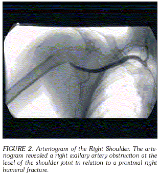
AXILLARY ARTERY INJURY SECONDARY TO HUMERAL NECK FRACTURE. CASE REPORT LESIÓN DE LA ARTERIA AXILAR SECUNDARIA A FRACTURA DEL CUELLO DEL HÚMERO. REPORTE DE CASO OSCAR JAVIER MANRIQUE M.Da*, RUBÉN PERALTA Ph.D.b Y MARGARITA APONTE M.D.c a Department of Surgery, Massachusetts General Hospital, Harvard Medical School, Boston, MA. * Correspondencia: omanrique@partners.org. Dirección postal: 31 Grove St. Unit B1; Boston, Massachusetts 02114, USA Phone: 617 459 8095. Abstract Fractures of the proximal humerus account over 75% of all humeral fractures in patients older than the age of 40 [1]. After the age of 50, women have a much higher incidence of these fractures than men, due to a higher incidence of osteoporosis. If the patient is less than 50 years old, high-energy trauma is the most common etiology. However, if the patient is over the age of 50, low energy mechanisms are more common [1]. Although axillary artery injury occurs frequently with dislocations of the shoulder and fractures of the clavicle, such injury is not commonly associated with fractures of the proximal humerus [2-5]. We present a patient with an axillary artery injury associated with a comminuted fracture of the proximal humerus. Key words: axillary artery, shoulder fractures. Las fracturas de húmero proximal corresponden al 75% de las fracturas en pacientes mayores de 40 años. Después de los 50 años y por su alta incidencia de osteoporosis, las mujeres tienen una mayor frecuencia de fracturas que los hombres. Y aunque en pacientes menores de 50 años el trauma de alto impacto es la causa principal de fractura, en pacientes que sobrepasan esa edad los mecanismos de bajo impacto son la causa más común. Las lesiones de la arteria axilar ocurren frecuentemente con dislocaciones de hombro y con fracturas de clavícula, no asociándose usualmente a fracturas del humero proximal. Presentamos el caso de una paciente con lesión de la arteria axilar asociada a una fractura conminuta de humero proximal. Usualmente el diagnóstico se sospecha por el cuadro clínico, teniendo en cuenta que durante el examen físico inicial, la circulación colateral del hombro puede demostrar pulsos normales, a pesar de que haya una lesión en la arteria axilar. Para tratar este tipo de lesiones el diagnóstico temprano es un elemento clave y para disminuir complicaciones futuras la circulación se debe restablecer en las primeras seis a ocho horas. De ahí la importancia de mantener un alto índice de sospecha cuando existen fracturas cercanas a grandes vasos y dada la anatomía vascular alrededor de la región proximal del humero, se debe descartar una lesión de la arteria axilar. Palabras clave: arteria axilar, fracturas proximales de húmero. Case Report A 69 year old female, fell at home from standing height, injuring her right shoulder. At the time of her fall, she described loss of sensation and motor function of her right hand. She initially presented to an outside hospital, and was then transferred to our institution for management of a right proximal humerus fracture-dislocation. It was noted by emergency medial personel during her transport that her right brachial and radial pulses were initially weak and subsequently became absent. Upon arrival at our institution, the right upper extremity had a palpable deformity and ecchymosis was present over the anterior aspect of her shoulder. The patient was unable to move her right hand fingers. She had decreased sensation in the median ulnar and radial nerve distribution. No brachial, ulnar or radial pulses were palpable or sensed by Doppler. Radiographic examination of her right shoulder revealed a comminuted fracture of the right proximal humerus (Figure 1). An angiogram was performed, showing occlusion of the right axillary artery at the level of the fracture, with reconstitution of the proximal brachial artery at the junction of the proximal/mid humeral diaphysis. No ulnar or radial arteries were visualized in the distal forearm (Figure 2). The patient was then taken to the operating room emergently.
Recibido: Marzo 26 de 2007. Aceptado: Mayo 7 de 2007.
b Department of Surgery, University of Massachusetts, Worcester, MA.
c Department of Obstetrics and Gynecology, McGaw Medical Center, Northwestern University.
Resumen

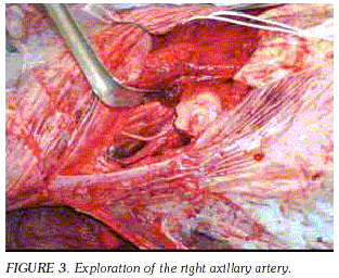
Under general anesthesia, exploration of the axillary artery was performed through an infraclavicular approach, with division of the pectoralis major and minor muscles. Exploration revealed a palpable axillary artery above the site of fracture. The axillary artery was tethered at the level of the subscapular artery. A longitudinal incision was made in the fascia overlying the right brachial artery. This artery was dissected in a retrograde fashion up to the region of injury; no pulse was palpated distally to the injury (Figure 3).
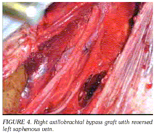
An axillary-brachial bypass graft was performed to reestablish flow on the right arm with a reversed left saphenous vein interposition graft (end to end anastomosis) (Figure 4). Prior to completing the anastomosis, forward and backward flushing maneuvers were performed and anastomosis was completed. Restoration of blood flow was confirmed with the appearance of palpable radial and ulnar pulses. Beacuse of the emergent management, no fasciotomy was indicated at that point.
Given the degree of fracture displacement and comminution, as well as concerns about protecting the vascular repair, the Orthopaedic Trauma Service then elected to perform a hemiarthroplasty rather than attempt open reduction and internal fixation of the humeral head. The incision used for axillary artery exploration and repair was extended proximally and distally, a lateral skin flap was developed, and a standard deltopectoral interval was exploited to access the glenohumeral joint. Greater and lesser tuberosity fragments were identified and tagged along with their rotator cuff attachments, and an Anatomic (Zimmer, Warsaw, Indiana) humeral component was cemented into 30° retroversion using standard cement technique. Prosthetic height was referenced off of the pectoralis major insertion. The greater and lesser tuberosities were reduced and held with heavy, nonabsorbable suture. Defects were filled with local graft taken from humeral head fragments. Full passive range of motion was achieved without impingement upon the vascular repair. The incision was closed in layers over a drain. Post-operatively the patient was permitted passive range of motion with external rotation limited to 30° to protect repair of the lesser tuberosity. In follow-up, the patient has healed her surgical incision without complication and has maintained excellent perfusion to the extremity.
Discussion
Injury to the axillary artery as a consequence of fracture of the neck of the humerus is an infrequent complication [3-5]. The diagnosis is usually suggested by the clinical picture. The most common signs of arterial axillary injury are absence of peripheral pulses, and clinical evidence of extremity ischemia including temperature discrepancy, pallor, cyanosis, paresthesias, sensory loss, and increasing paralysis. However, because of the good collateral circulation around the shoulder, the first clinical examination may demostrate a normal pulse of the extremity, depsite the presence of an arterial injury. When disruption of the intima and subsequent subintimal dissection and thrombosis occurs, secondary occlusion of the damaged artery can result. Thus, such delayed thrombosis may lead to ischemia only after hours or days of the initial injury [5].
There are several mechanisms by which the axillary artery can be injured in association with this type of fracture, and the regional anatomy that may predispose the artery to injury merits discussion. In the setting of a displaced fracture of the humeral surgical neck, the pull of the pectoralis major typically translates the diaphyseal component medially. The surgical neck of the humerus marks the level at which the anterior and posterior humeral circumflex vessels branch off of the axillary artery, passing anterior and posterior to the humeral diaphysis respectively, effectively tethering the artery to the humerus at this point. Additionally, if the humeral head dislocates as a result of the injury, it most commonly does so in an anterior, inferior direction, crowding the axilla and compressing the latter's neurovascular structures. Aside from laceration of the artery by bony fragments, there may be overstretching of the artery, which can lead to rupture, especially in an atheromatous artery [6,7]. Another important mechanism associated with fractures is intimal disruption and thrombosis [8]. With this mechanism of injury, the artery is stretched across the bony fragment and the adventitia remains intact while the fragile intima tears, leading to thrombosis. A patient with such an injury most commonly presents with an absent or diminished radial or brachial pulse[3]. Early recognition of these clinical signs is essential to avoid irreversible ischemic damage. Prompt arteriography is mandatory characterize fully any suspected arterial injury. Preoperative angiography is not only important to make accurate localization of the injury but can also provide important information regarding collateral flow. Nevertheless, certain emergent cases may necessitate intervention without the benefit of preoperative mapping.
Early diagnosis and treatment remains the cornerstone of successfully treating these injuries. Circulation must be restored within six to eight hours to decrease future complications [3]. In general, one must keep a high index of suspicion whenever a fracture is near to a major vessel. In particular, the vascular anatomy in the region of the humeral surgical neck can predispose the axillary artery injury. The presence of peripheral pulses does not guarantee that significant arterial injury has not occurred and appropriate imaging studies should be obtained without delay if injury is suspected.
References
1. Clark, M.J.K.a.W.A., Orthopedic Therapy of the Shoulder. 2nd edition ed. Lippincott Williams & Wilkins: 1995 [ Links ]
2. Gates J.D, J.B. Knox, Axillary artery injuries secondary to anterior dislocation of the shoulder. J Trauma, 1995. 39(3): 581-3. [ Links ]
3. Byrd R.G, R.P, Byrd Jr, T.M. Roy, Axillary artery injuries after proximal fracture of the humerus. Am J Emerg Med, 1998. 16(2): 154-6. [ Links ]
4. Laverick, M.D. Management of blunt injuries of the axillary artery and the neck of the humerus: case report. J Trauma, 1990. 30(3): 360-1. [ Links ]
5. Jensen B, Jacobsen J, Andreasen H. Late appearance of arterial injury caused by fracture of the neck of the humerus. J Trauma, 1987. 27(12): 1368-9. [ Links ]
6. Cleveland J, J. Ellis, J. Dague, Complete disruption of axillary artery caused by severe atherosclerosis and trivial nonpenetrating trauma. J Trauma, 1979. 19(8): p. 635-6. [ Links ]
7. Theodorides, T. and C. de Keizer, Injuries of the axillary artery caused by fractures of the neck of the humerus. Injury, 1976. 8(2): 120-3. [ Links ]
8. Shuck, J.M., G.E. Omer, Jr., and C.E. Lewis, Jr., Arterial obstruction due to intimal disruption in extremity fractures. J Trauma, 1972. 12(6): 481-9. [ Links ]

