SciELO - Scientific Electronic Library Online

 
vol.88 número218Frequency-based methods for the detection of damage in structures: A chronological reviewIn vitro evaluation the toxicity in mixture of glyphosate and methyl metsulfuron against strains of Trichoderma spp índice de autoresíndice de assuntospesquisa de artigos
Home Pagelista alfabética de periódicos  

Serviços Personalizados

Journal

Artigo

Indicadores

Links relacionados

  • Em processo de indexaçãoCitado por Google
  • Não possue artigos similaresSimilares em SciELO
  • Em processo de indexaçãoSimilares em Google

Compartilhar


DYNA

versão impressa ISSN 0012-7353versão On-line ISSN 2346-2183

Dyna rev.fac.nac.minas vol.88 no.218 Medellín jul./set. 2021  Epub 29-Mar-2022

https://doi.org/10.15446/dyna.v88n218.92405 

Articles

Plant electrophysiology: bibliometric analysis, methods and applications in the monitoring of plant-environment interactions

Electrofisiología vegetal: análisis bibliométrico, métodos y aplicaciones en el monitoreo de las interacciones planta-ambiente

Silvia L. Chaparro-Cárdenas a   b  
http://orcid.org/0000-0002-2589-259X

Julian Andres Ramirez-Bautista a  
http://orcid.org/0000-0002-6472-5751

Wilson Gamboa-Contreras a  
http://orcid.org/0000-0001-5526-3156

Andrés Leonardo Moreno-Chacón c  
http://orcid.org/0000-0002-1402-6178

Frank Carlos Vargas-Tangua a  
http://orcid.org/0000-0002-6611-0144

a Departamento de Investigación, Fundación Universitaria de San Gil-Unisangil, San Gil, Colombia. schaparro@unisangil.edu.co, jramirez@unisangil.edu.co, wgamboa@unisangil.edu.co, fvargas@unisangil.edu.co

b Centro de Investigación en Ciencia Aplicada y Tecnología Avanzada-Instituto Politécnico Nacional, Querétaro, México. schaparro@unisangil.edu.co

c Universidad Pontificia Bolivariana, Bogotá, Colombia. andresleonardo.moreno@upb.edu.co


Abstract

Plants have mechanisms to perceive and transmit information between their organs and tissues to respond quickly to abiotic and biotic external stimuli from the environment, producing different electrical potential types. It is reported on the generation and conduction of electrochemical impulses within different tissues and organs of the plant, which have been acquired using different methods to use them in various applications in the improvement of the agro-industrial sector and the development of different types of phytosensors. In this review paper, the various studies that have been carried out since Pfeffer, Burdon-Sanderson, Darwin, Haberlandt, and Bose discovered electrical activity in plants until today are reported. Plants provide mechanisms to perform biosensors based on responding to environmental changes, opening a great path for the design of low-cost and highly sensitive sensors and sensor networks, the current trend is towards experimental analysis using various stimuli.

Keywords: vegetal electrophysiology; electric potentials; action potentials; variation potentials; plant electrical measurements

Resumen

Las plantas tienen mecanismos de percepción y transmisión de información entre sus órganos y tejidos para responder rápidamente a estímulos externos abióticos y bióticos, produciendo diferentes tipos de potenciales eléctricos. Se ha reportado en literatura sobre la generación y conducción de impulsos electroquímicos dentro de diferentes tejidos y órganos de la planta, los cuales han sido adquiridos mediante diferentes métodos para utilizarlos en diversas aplicaciones, donde se cuenta la mejora del sector agroindustrial y el desarrollo de diferentes tipos de fitosensores. En este artículo de revisión, se reportan los diversos estudios que se han llevado a cabo desde que Pfeffer, Burdon-Sanderson, Darwin, Haberlandt y Bose descubrieron la actividad eléctrica en las plantas hasta el día de hoy. Las plantas brindan mecanismos para realizar biosensores basados en la respuesta a los cambios ambientales, abriendo un gran camino para el diseño de sensores y redes de sensores de bajo costo y alta sensibilidad, la tendencia actual apunta hacia el análisis experimental utilizando diversos estímulos.

Palabras clave: electrofisiología vegetal; potenciales eléctricos; potenciales de acción; potenciales de variación; medidas eléctricas de plantas

1. Introduction

Electrical signals are present in many physiological activities. But this study started long ago, when plants as Mimosa pudica, Drosera, and Dionea muscipula, attracted the attention of researchers as Pfeffer, Burdon-Sanderson, Darwin, Haberlandt, and Bose [1-4]. Who discovered that the tactile movements of these plants activated action potentials that were propagated from the stimulation site to motor organs, where the movement of sensitive cells occurs, in response to the stimuli, similar to nerves in animals [5-7]. Still, this electrical behavior is also important in non-sensitive upper plants [8,9]. These signaling mechanisms may include hormones (e.g. abscisic acid, salicylic acid, ethylene, jasmonic acid, etc), chemicals (e.g. Ca2+, K+, sugars, proline, amino acids, polyamines, reactive oxygen species (ROS), etc.) or physical events (e.g. propagation of electrical or hydrostatic pressure waves, changes in osmotic pressure, etc.) [10,11].

In plant cells, ion channels mainly relate to three physiological functions: cell osmoregulation, cell signaling by amplification, and electrical signals propagation, that include the control of membrane potential. Depending on the opening or closing of ion channels, the plasma membrane can be depolarized, repolarized or hyperpolarized; developing unique electrical properties based mainly on the transport of H+, K+ and anions [12-15]. Growth (the result of cell division) and differentiation (due to asymmetricity) are the two key developmental processes in all organisms. In fact, it is known that cell polarity and uneven differentiation as a result of uneven organization of cellular components precede differentiation [14,15].

Plants have been documented to develop electrical signals in the processes of respiration and photosynthesis [16] phloem transport [17], acid rain [18], irradiation with various lengths of wave [19], and the rapid deployment of plant defenses throughout the plant [20]. The bio-electrochemical system in plants regulates not only responses to biotic and abiotic stress, but also photosynthetic, growth, development and adaptation processes [20]. In this context, several agents including phytohormone gradients, light, pH, temperature and electro-magnetic fields can perturbate previously homogeneous systems and induce their subsequent heterogeneity and polarity.

There are different types of electrical signals in plants, such as local electrical potential (LED), action potential (AP), variation potential (VP, or slow wave - SW) [21,22][8]. APs after a stimulus-induced by non-damaging attacks such as illumination, cold, mechanical, and electrical stimuli, reaches a certain threshold regardless of the stimulus's force, which leads to membrane depolarization [23,22]. VPs are slower signals related to stimulus strength with variable shape, amplitude, and time, lasting periods of 10s to 30m, induced by damaging stimuli such as burns and cuts [24]. LEP is a sub-threshold response induced by change in environmental factors (e.g. soil, water, fertility, light, air temperature and humidity). Although LEP is only locally generated and is not transferred to other parts of a plant, it has tremendous impact on the physiological status of the plant [23,22]. In contrast, both AP and VP can transmit from the stimulated site to other parts of the plant.

Approaches used to measure electrical activity in plants mainly include intracellular and extracellular measurements [2,22]. Intracellular measurements directly and individually record the value of cell membrane potential; the procedure generally consists of inserting a microelectrode into a cell's cytoplasm carefully using micromanipulators. Whereas extracellular measurements are based on the acquisition of the total sum of signals produced by the depolarization-repolarization process for large groups of cells. Extracellular measurements are widely used in animal electrophysiology (electrocardiograms (ECG), surface electromyography (SEMG) and electroencephalograms (EEG)) [25,26].

Studies have been carried out on electrical signals in plants under various conditions (laboratory, greenhouse, etc.). For example, in [22], the authors report the development and testing of an electrophysiological sensor for use in greenhouse conditions, without a Faraday cage, and the automatic classification of these signals using supervised machine learning allowing the detection of these physiological modifications due to environmental conditions. In [27], a system for constant monitoring of cucumber plants' electrical signals in a greenhouse was developed, showing that the electrical signals in plants respond to environmental changes. On the other hand, [9] determined the electrical responses, both VP and AP, of tomato plants wounded by flame.

This review covers from a historical background of the plant electrophysiology up to the uses that researchers have given to this behavior, together with an understanding of the mechanism of generation and conduction of the electrical signal in plants. Using a bibliometric analysis, the plant electrophysiology trend is towards the application in agriculture and environmental monitoring, promoting the development of mixed societies of biological and artificial components, which take advantage of the intrinsic detection capacity of plants.

2. Bibliometric analysis

A systematic review of research that recorded the analysis of the electrical activity in plants under conditions of biotic and abiotic stress was performed, emphasizing the type of electrode used, the potential measured, and the study's application. The search strategy was developed by identifying the following relevant terms: Plant electrophysiology, plant electrical measurements, and pollution, plants as environmental biosensors, and electrical potentials in plants used in the electronic databases: EBSCO, IEEE, ScienceDirect, Scopus and Web of Science. The inclusion criteria emphasized recording electrical activity in plants in specific applications such as monitoring and study systems, plant disease development, pollution detection, and other applications. Exclusion criteria included mathematical modeling investigations and behavioral simulation.

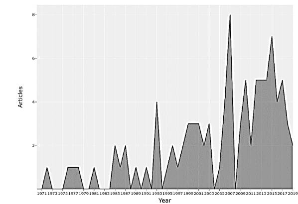
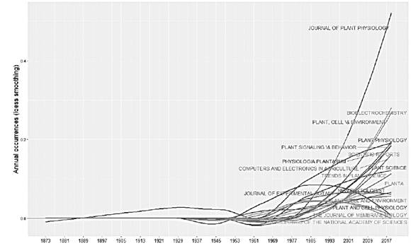
Plants are structures with ideal adaptive capacities where electrical processes play an important role. The objective of this research is to obtain a better understanding of the behaviors, interactions and communications between the plants, as well as the applications that have been reported. Plants are exposed daily to a wide variety of environmental factors, which induce disturbances in them, including variation in light, air and soil pollution, temperature, water deficiency, mechanical damage, application of nutrients, insect attacks and pathogens, etc. The electrical response that plants have to these biotic and abiotic stimuli allows the development of biosensors for application in different areas such as agriculture, environmental monitoring, detection of geological events, detection of pollutants, to name a few. Research has been carried out for a long time in the field of plant electrophysiology, although studies have been reported since 1873, in recent years annual scientific production has increased, having an annual growth rate of 2%, as shown in fig. Motivating the appearance of new scientific journals and their growth over time (Figs. 1 and 2).

Source: The Authors

Figure 1 Annual scientific production in plant electrophysiology. 

Source: The Authors

Figure 2 Source dynamics growth per year on plant electrophysiology. 

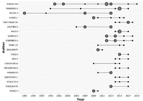
The study of the electrophysiology of plants has been opening the way in many different fields to the intrinsic behavior of plants, increasing the number of authors over time, indicating that it is an area that is constantly growing. Considering the number of articles published, the Fig. 3 shows the scientific production of the 20 main researchers.

Source: The Authors

Figure 3 Production of the top-authors over time. 

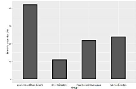
This scientific production has been concentrated mainly in 3 areas and some pilot studies. As can be seen in the Fig. 4, the reported works are concentrated in greater quantity in experimental analyzes applying controlled stimuli, occupying 42% of the studies. On the other hand, the detection of pests and pollutants with 22% and 24% respectively, show to be areas of interest for the researchers considering the enormous potential that exists in using the plants as biosensors of the environment regardless of whether the application is agriculture or environmental analysis (Fig. 4).

Source: The Authors

Figure 4 Scientific production trends 

In response to environmental changes, plants permanently adjust their metabolic and physiological processes. They continually collect and systematize information about their environment. This is the result of an incredibly complex set of molecular, biochemical and physiological mechanisms for signal perception and activation of multiple cross-signaling pathways. Plants mainly generate various types of intracellular and extracellular signals in the form of AP and VP, in response to environmental changes. This information together with computerized methods offers the possibility of obtaining low-cost biosensors that can be used as a network in different applications, as studied above. These trends make it possible to obtain mixed societies of biological and artificial organisms in agriculture and mainly in environmental monitoring.

Many authors through laboratory studies have demonstrated the potential of using plants as sensors to detect what biotic and abiotic stresses are for plants. In agriculture it is possible to detect by monitoring its electrical activity the application of chemical products, which would bring sustainable and ecological agriculture, optimizing inputs as well as water resources. Monitoring of the environment and pollution is another interesting application since it is possible to use plants as low-cost sensor networks to analyze environmental behavior from homes to correlate it with diseases and air purification needs. Considering that plants have pathways for transmitting electrical signals to respond quickly to environmental stressors.

3. Electrical activity in plants

Plant cells also produce and transfer bioelectrical signals as extracellular signals in response to changes in their environmental conditions [22]. Also, investigations confirmed that the neurotransmitters found in higher plants (e.g. acetylcholine ergic) participated in the bioelectrical activity of higher plants to regulate the membrane permeability of plant cells and the physiological processes in higher plants [5-7]. Accordingly, electrical signals are probably the initial response of the plant to an exterior stimulus. This type of response may trigger physiological variation (e.g. elongation growth, respiration, moisture absorption, substance unloading at the phloem, reduction of the turgor pressure and variation of photosynthesis and transpiration, gas exchange, and activation and transcription of the protease inhibitor gene), and thus may mediate the interrelationships between each organ and tissue inside the plant as well as between itself and the external environment [28].

During plant growth, electrical signals in plants can show different characteristics due to weak light, high humidity, and low potassium [29]. This suggests the potential use of these electrical signals in applications that indicate the physiological state of plants for the adjustment and control of biotic and abiotic conditions. Electrical signals in plants can carry local stimulation information to other cells, tissues, and organs so that they respond appropriately. Therefore, electrical signals are very important and have important physiological effects on plants [27,28].

3.1 Variation Potentials (VP)

VPs are induced in the xylem parenchyma cells by a local change in a hydraulic wave or a chemical stimulus. They are characterized by amplitudes and velocities that decrease with increasing distance from the generation site and xylem tension. Their ionic base differs from the underlying APs in that they are induced by a transient quenching of a P-type ATPase-H+ and a turgor-dependent activation of Ca2+ mechanosensitive channels [2,28]. The VP are mainly caused by injuries such as burns [30] or hot water treatment [31]. This potential does not follow the all-or-nothing principle, are correlated with stimulus strength, and last a few seconds to 30 minutes [28]. Additionally, VP have a slower transmission rate than APs; most of the plant AP studied so far have a velocity in the range of 0.005-0.2 m*s-1 [32], while the velocity of the VP are in the range of 0.001-0.01 m*s-1 and their amplitude decreases with increasing distance from the stimulation site along their path [28].

3.2 Action Potentials (AP)

An AP generally travels with constant speed and magnitude [33]. APs are generally triggered by noninvasive stimuli [32]. They are transmitted along the phloem over long distances [34] and regulate the rapid movements of the leaves in plants sensitive to touch such as Mimosa [29] and Venus [35]. Furthermore, cold shock [36], as well as in some specific physiological functions [37]. As in animal neurons, APs follow the all-or-nothing principle [38].

APs in higher plants are the information carriers in intracellular and intercellular communication during biotic and abiotic environmental changes [18,39]. Plants possess most of the chemistry of the neuromotor system in animals, that is, neurotransmitters, such as acetylcholine, cellular messengers such as calmodulin, cellular motors, for example, actin and myosin, voltage-gated ion channels, and contact sensors, light, gravity and temperature. Although this cellular equipment has not reached the same great complexity as the case of the nerves, a very simple neural network has been formed within the phloem that allows plants to successfully communicate over long distances. The reason that plants have developed pathways for the transmission of electrical signals through oscillations in their molecule, ion, and voltage fluxes probably lies in the need to respond quickly to environmental stressors.

The lengthening or growth of the roots and of the leaves has been associated with electrical signaling and, therefore, with the displacement of ions, such as Ca2+, Cl- and H+, through the membranes [40]. Ion flux waves constitute an alternative long-distance and rapid signaling network in plant that integrates and responds to a number of external and environmental signals, which are often related to stress signaling, e.g. ROS [41]. The quality of light, that is, of the spectrum as a whole, which wavelengths of irradiance are perceived, influences the type of electrical signals that are developed [41]. For example, blue light (400-500 nm) induced rapid AP in soybeans, while 500-630 nm did not induce any membrane potential [42], leading to the conclusion that irradiation of blue light induces positive phototropism.

4. Methods to measure electrical activity

4.1 Plant intracellular measurement

Intracellular measurement directly records a cell's plasma membrane potential, usually using a glass microelectrode with a tip diameter lower than 1 µm. Different methods have been used such as a glass microelectrode with 500 mM KCl and Ag/AgCl immersed in the bath solution using a micromanipulator [24], microelectrode filled with 100 mM KCl was inserted into a mesophyll cell [43,30], 3 M KCl filled glass microelectrodes Ag/AgCl [44], electrodes made from capillary glass [45,26]. In [38], they fabricate glass microelectrode from microcapillaries (WPI) were with tip diameters less than 1 µm and were back-filled with 100 mol m~3 KG. In [46] is reported a glass micropipette pulled on a vertical puller, with aperture diameter at the tip of 0.05 µm, filled with a 1% aqueous solution of LYCH and back-filled with 3 M LiC1. In this method, the microelectrode is inserted into the cell, through a micromanipulator with a stereomicroscope, and the reference electrode is placed in the bath solution. The ingredients and concentrations of the bath solution vary depending on the plant cells' conditions to be analyzed.

4.2 Plant extracellular measurement

Measurements of extracellular potential allow detecting differences in the stable electrical potential of groups of cells for extended periods (several days), this technique can be carried out mainly by two methods, through surface recording or measurements with inserted electrodes [47,48]. In many cases inside of a Faraday cage and using several filters with a recording system composed of preamplifiers, A/D converter and devices for recording, sampled at 400 Hz with a gain of 4 and several filters are applied: low pass at 30 Hz and band-stop at 50-60 Hz and 100 Hz.

The surface recording method is non-invasive and has been reported to be physically stable, generally consisting of electrodes attached to the plant's surface, wrapped in cotton to provide adequate contact [49,50]. Different electrodes, electrode wetting solutions, and bonding methods have been reported.

Electrodes as: Ag/AgCl electrode [24][51], nonpolarizable reversible Ag/AgCl electrodes prepared from Teflon coated silver wire [52-54], graphite patch electrodes [55].

The union of the electrode with the plant has been analyzed in different ways as: Ag/AgCl electrodes connect to the plant surface using an aqueous conductive gel similar to that of ECG [56,57], Ag/AgCl connected with conductive gel and the reference electrode a solution (1 mM KCl, 0.5 mM CaCl2 and 0.1 mM NaCl) surrounding the roots [58-60][43], Ag/AgCl-wire (0.4mm in diameter) wrapped in cotton moistened with 01% (w/v) KCI solution [38][47], Ag/AgCl electrodes covered by a conductive hydrogel [61], felt-tip calomel electrodes attached to de plant with 1 mM KCI ionic bridges [62,63][9], Ag/AgCl electrodes moistened with 100 mM KCl agar [64], Ag/AgCl electrodes prepared with 0.2 mm silver wire and 0.05mm elastic cooper wire [65], Non-polarizable Ag-AgCl electrodes connected by an agar-coated cotton thread protruding from a glass pipette containing 0.1M KCl gelled with 1% agar [66]. Besides, the bath electrode method has been used in [27,67,68,46], where is used a cotton thread soaked in an experimental solution (KCl 0.1 mM, MgCl2 0.1 mM, CaCl2 0.5 mM, Na2SO4 0.05 mM), or an Ag/AgCl-pellet electrode impaled into agar with KCl.

The electrode insertion method commonly uses thin metal wires that, when inserted into the shoot or the plants' leaf vein, come into contact with the internal tissue incorporating large groups of cells. This method produces wounds in the plants that can alter the external stimulus measurements. Different electrodes have been reported as thin metal tips made of platinum (Pt) or Ag/AgCl wires have been reported as thin metal tips made of platinum (Pt) or Ag/AgCl wires [49,69-71,46], metal probe [72], Ag/AgCl electrodes from Teflon-coated silver wires [73], silver wires (0.2 mm in diameter) [62,63,9], microelectrode filled with 100 mM KCl [64], wires (50/im diameter 10% iridium-platinum) [66], reversible Ag/AgCl electrodes prepared with AgCl on 5mm long silver wire tips without Teflon coating in a 0.1 MKCl aqueous solution [61], Ion-selective microelectrodes describe by [74]. In [16], two types of electrodes were used: glass microelectrodes from boro-silicate glass (Hilgenberg, Malsfeld, Germany) filled with 3 mol/L KCl solution, and conventional Ag/AgCl electrodes made of silver wires 0.2 mm in diameter coated with AgCl. [7] used custom-made electrodes of silver-coated copper filament diameter less than 0.5 mm of a coaxial cable (2.79 mm diameter). In [75-77] used three stainless steel needle electrodes of 0.35 mm in diameter and 15 mm in length (base, in the middle and on top of the stem), similar to those used in Electromyography. Solid stainless steel has been used in [78] stainless steel 304 SS type 316 rod, 3.18 cm long and 0.2 cm diameter, and in [79] 15mm electrodes wrapped with a cable secured an epoxied cap of thermo-sheath. In other studies, use nonpolarizable reversible Ag/AgCl-electrodes with a diameter of 0.25 mm [20], 0.14 mm [80], and 0.35mm [15].

Also, the aphid technique has been reported. This method consists of using the stylet of an aphid when it pierces the sap in the phloem to attach a microelectrode to it, using a micromanipulator. The stylet works as a salt bridge between the cytoplasm and the microelectrode [48,81-84].

Other techniques often used for acquiring the electrical potentials are:

Patch-clamp recording technique involves sealing glass capillaries to the protoplast membranes' surface and then opening a small hole with a short voltage pulse. The plasma membrane's inner surface then contacts the solution in the glass tube, allowing the movement of ions to be traced using their electronic charges [48]. This technique records the ion channel as Outward rectifying K+ channel [85,86], Inward rectifying K+ channel [87,88], Fast anion channel [89] and Slow anion channel [90]. In [91] prepared the patch pipettes from Kimax-51 glass capillaries and coated with silicone, the pipette solution contained 150 mm CsCl, 1 mm MgCl2, 10 mm EGTA, 1 mm MgATP, and 10 mm HEPES/Tris, pH 7.4.

The non-invasive microelectrode vibrating probe technique measures ionic or molecular activity without invading the cell, using a reference electrode and an ion selectivity microelectrode, with an electrolyte and a liquid ion exchanger. By vibrating the electrode determines the voltage across the ion concentration gradient [48]. With this technique, in [92] investigated in the initial phases of plasma membrane depolarization, which ion act as the depolarizing agent, besides they determined the kinetics of H+, Ca2+, K+, and Cl- fluxes and the changes in their concentrations near bean mesophyll and attached epidermis.

Optical measurements using voltage-sensitive dye (VSD) in this technique are captured with high-resolution cameras. The optical signals emitted by the VSD attached to the plant cells are analyzed. This technique has been applied by [13]. For membrane staining have used different voltage-sensitive dye, VSD bis-(1,3-dibutylbarbituric acid)-trimethine oxonol (DiBAC4(3)) [24], RH-414 [-N-(3-triethylammoniumpropyl)-4-(4-(4-diethylamino)phenyl)butadienyl) pyridinium- dibromide] [93].

5. Applications

The electrical activity in plants not only appears as a reaction to the impact of chemical compounds such as herbicides, air and soil pollution, stimulants of plant growth, salts, water, etc.; but also, with physical factors such as electromagnetic fields, mechanical injuries, changes in temperature, among others. The behavior of electrical activity in plants has been an area of particular interest in recent and ancient studies. The applications that previous researchers have worked on are presented in groups below.

5.1 Monitoring and study systems

Monitoring the plant's response to environmental changes is an active field in plant electrophysiology. Several studies have been conducted that considers temperature, humidity, light, and other factors. Special attention has been paid to the studies of Mimosa pudica (sensitive plant), Drosera (sundew), Dionaea muscipula (flytrap) the significant action potential that generates when stimulated [94,95,4,66].

Studies have found patterns of electrical potentials in plants under water stress, in [96-98,15,55,22] was observed an increase in the electrical signal in non-irrigated fruit plants, which changes when starting irrigation, producing a negative signal slope until reaching stability under normal conditions of irrigation. The roots can feel the soil's drying and send signals to the leaves to coordinate the processes over a long distance, especially in large plants where water transport is slower [99] (Fig. 5).
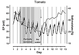
Source: [22].

Figure 5 Changes of electrical potential resulting from irrigating. a) Experiment with tomato plant reported in 2019. 

Thermal stress causes changes in the magnitude of the electrical signal; the decrease in electrical signals' magnitude has been evidenced by the reduction of temperature [27]. Furthermore, it has been found that the speed of propagation of action potentials against thermal stress is comparable to that produced by various species of mammals [52].

Light is another environmental factor that induces changes in the electrical potential of plants. Low illumination causes a decrease in the electrical activity of plants [27,100]. Dark light transitions trigger potential membrane changes in the leaf mesophyll, as well as at the root level [101], thereby modulating K+ ion fluxes, Ca2+, Cl- and H+. In the case of roots, the light-induced electrical signal was faster than the hydraulic signal (reduction of xylem pressure), indicating a predominant role of electrical signaling in ion fluxes and nutrient absorption at the root level [101].

The mechanical damage caused by pruning, wind, or insects can be related to the intensity and duration of the said stimulus, as reported by [102,56], who have conducted experiments with Persea Americana (avocado) and Vitis vinifera (grape) plants.

Other reported studies have developed new algorithms to compare with a neural network, deep learning, and SVM to identify and classify action potentials when there is an electrical stimulus [103].

These studies allow to know the plants' behavior, develop instruments for continuous monitoring, and evaluate in real-time the electrical response of the plants in stress situations under field conditions for modifying environmental conditions, maximizing productivity and quality.

5.2 Plant disease development

The existence of variation of the membrane potential upon the attack of herbivores has been analyzed in some studies. Insects cause mechanical damage to plants by chewing them. This action has been studied by [104] finding that plant-insect interactions produce depolarization in the plant; this electrical signal travels rapidly throughout the plant, from the origin of the stimulus, causing a plant response. Plants differentiate mechanical wounding from herbivory through recognition of compounds present in insect oral secretions; a faster depolarization response has been shown to exist when damage is done by insect herbivory [105-107].

Studies have shown the possibility of monitoring the development of virus infections in plants. In [108], a relationship between the progression of tobacco ringspot virus (TRSV) infection in Vigna sinensis plants with the frequency of hyperpolarized and depolarized transmembrane potential was evidenced. The study showed that the response to the metabolic inhibitor sodium azide varied between control plants (H) and cells infected with TRW. Results that can be contrasted with the studies of [109-111].

In the study carried out by [112], it was observed that when causing mechanical damage to a lima bean leaf, a strong depolarization response occurs in the bite area, followed by a transient hyperpolarization and, finally, a constant depolarization in the rest of the sheet.

5.3 Pollution detection

Signals of electrical activity in plants have also been used to monitor environmental conditions and provide information related to pollution. In [113] the authors propose a method of segmentation of time-series measurements to analyze electrical signals from ligustrum and buxus plants in the detection of critical levels of ozone (O3) plant's response to air pollution with O3, see in Fig. 6.

Source: [113].

Figure 6 Response of the ligustrum plant signal to ozone exposure 

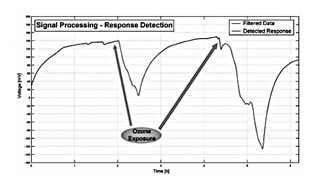
These results can be contrasted with those obtained by [114]. The study used a correlation-based approach to analyze electrical signals from Ligustrum texanum and Buxus macrophilla. The behavior of the biosensor was similar, and in the study a decrease in the electrical signal was observed in the face of ozone pollution.

Other contaminants such as sulfuric (H2SO4) and nitric acids (HNO3) can also be sensed using action potentials. These two acids that predominate in acid rain were analyzed by [115,20], in the study, effects were found in variations of action potentials induced by the application of H2SO4 or HNO3 in soybeans by acidification of the soil or spray on the leaves. The duration of action potentials reported, after the application of acid HNO3 and H2SO4, was 0.2 and 0.02 s.

The response of the plant with environmental pollutants as H2SO4, O3 and sodium chloride (NaCl), was also analyzed by [75], in the study the authors used tomato (Solanum lycopersicum) and cucumber (Cucumis sativus) plants; the work focused on the classification of the electrical signal extracted upon the stimuli H2SO4, O3 and NaCl in two different quantities (5 mL and 10 mL), using a machine learning approach, they obtained an average classification of 70% precision. The figure shows the behavior of the plants that the authors obtained with the study. In a similar study reported by [116], a curve fitting approach is shown in extracting signal characteristics to classify H2SO4, O3 and NaCl stimuli. In the study, the researchers obtained a classification accuracy of 98% (Fig. 7).
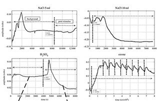
Source: [75].

Figure 7 Plant response upon 3 stimuli, vertical dotted lines mark indicates the application of stimulus. 

The electrical response of the plant to pollutant gas was reported by [117], in the study the authors observed a decrease in the electrical activity of the plant, when it is placed in the presence of formaldehyde gas, the electrical change is attributed to the effect of plant's air purification.

On the other hand, uncoupler carbonyl cyanide-p-trifluoromethoxyphenyl hydrazone (FCCP) have been analyzed by several authors, and they have found that the FCCP induces ultra-fast action potentials and decreases the resting potential in a soybean [42,20,118].

The behavior of the electrical activity of plants upon sources of contamination was also studied by [80], they observed that there are fast-acting potentials and decreases the potential for variation to zero in soybeans, when exposed to pentachlorophenol (PCP), which is a contaminant used as an herbicide, insecticide, and fungicide.

5.4 Other applications

In other reported studies, the polarity of the plant tissue has been identified, in order to obtain a sustainable energy source. In the study of [72], they identified polarity based on plant growth and noted that by assembling branch sections in series or parallel arrangements, it is possible to increase the voltage or current depending on the arrangement. The power generation of the plant was also the object of study of [119], in the reported work, a self-powered wireless system was developed, which collects the energy of the plant itself for its operation and transmits the signal generated by the plant with stimuli of electricity and water, the study was carried out on avocado plants.

As reported in [120], electrical activity has potential use in the detection of seismic events, in the experiments of [121] and [122] using silk trees (Albizzia julibrissima) and Ulmus kaeki, respectively, it was possible to detect abnormal bioelectric potentials in trees before earthquakes, the hypothesis that is handled is that plants can perceive underground electromagnetic emissions or geochemical reactions.

6. Conclusion and future research

Plants have pathways for the transmission of electrical signals to respond to environmental stressors, the relationship between biotic and abiotic stimuli and the electrical activity of plants is still under study and takes more strength over the years, since it is an important factor for the development of biosensors that use plants as a detection device. For this reason, researchers have reported various types of sensors to acquire the electrical signal, as well as algorithms capable of recognizing behavior patterns and opening the way to applications in many fields.

The use of computerized methods interconnected with plants using intracellular or extracellular acquisition systems enables the development of biosensors for rapid and real-time monitoring of the environment; in agriculture control factors that influence the harvest; detect contaminants and pesticides; climate change detection; There are many applications that offers the electrical behavior of plants when stimulated.

It is evident that there is great potential for future hybrid applications between biological and artificial organisms, in which the environment will benefit since it will be possible from an optimal and sustainable agricultural production, through the pollution monitoring for improving the quality of life, to obtaining applications in bioelectricity, bioelectronics and biometrics.

The study of the mechanisms of electrical signals in higher plants in response to environmental changes during growth and development is still a subject under construction. Despite the large number of reports describing electrical signals in higher plants, there are few quantitative mechanism models that allow a reliable prediction of the measured shapes of electrical signals in higher plants. It is necessary to develop mathematical models to provide an explanation for electrical phenomena in plants. A more complete understanding of electrogenic ion transport systems involving their density and the corresponding voltage-dependent kinetics is required. New measurement methods (for example, the non-invasive microelectrode vibrating probe technique, the patch clamp technique, as well as modern modeling and stimulation methods) can provide support for the development of an accurate kinetic model of the signal’s plants.

References

[1] Volkov, A.G., Plant electrophysiology: signaling and responses. Plant Electrophysiology. Signaling and Responses, 2012, pp. 1-377. DOI: 10.1007/978-3-642-29110-4 [ Links ]

[2] Fromm, J. and Lautner, S., Electrical signals and their physiological significance in plants. Plant, Cell and Environment, 30(3), pp. 249-257, 2007. DOI: 10.1111/j.1365-3040.2006.01614.x [ Links ]

[3] Burdon-Sanderson, J., Electrical phenomena which accompany irritation of the Leaf of Dionaea muscipula. Proc R Soc London, 21, pp. 495-496, [online]. 1872. Available at: http://rspl.royalsocietypublishing.org/subscriptionsLinks ]

[4] Stahlberg, R., Historical overview on plant neurobiology. Plant Signaling and Behavior, 1(1), pp. 6-8, 2006. DOI: 10.4161/psb.1.1.2278. [ Links ]

[5] Yan, X., Wang, Z., Huang, L., Wang, C., Hou, R., Xu, Z. and Qiao, X., Research progress on electrical signals in higher plants. In Progress in Natural Science, 19(5), pp. 531-541, 2009. DOI: 10.1016/j.pnsc.2008.08.009. [ Links ]

[6] Simons, B.P., The role of electricity in plant movements. New Phytologist, 87(1), pp. 11-37, 1981. DOI: 10.1111/j.1469-8137.1981.tb01687.x. [ Links ]

[7] Tran, D., Dutoit, F., Najdenovska, E., Wallbridge, N., Plummer, C., Mazza, M., Raileanu, L.E. and Camps, C., Electrophysiological assessment of plant status outside a Faraday cage using supervised machine learning. Scientific Reports, 9(1), art. 17073, 2019. DOI: 10.1038/s41598-019-53675-4. [ Links ]

[8] Chatterjee, S.K., An approach towards plant electrical signal based external stimuli monitoring system. University of Southampton (91), Southampton, U.K., 2017. [ Links ]

[9] Stanković, B. and Davies, E., Both action potentials and variation potentials induce proteinase inhibitor gene expression in tomato. FEBS Letters, 390(3), pp. 275-279, 1996. DOI: 10.1016/0014-5793(96)00672-2. [ Links ]

[10] Spoel, S.H. and Dong, X., Making sense of hormone crosstalk during plant immune responses. Cell Host and Microbe, 3(6), pp. 348-351, 2008. DOI: 10.1016/j.chom.2008.05.009. [ Links ]

[11] Zhang, W., He, S.Y. and Assmann, S.M., The plant innate immunity response in stomatal guard cells invokes G-protein-dependent ion channel regulation. Plant Journal, 56(6), pp. 984-996, 2008. DOI: 10.1111/j.1365-313X.2008.03657.x. [ Links ]

[12] Barbier-Brygoo, H., Vinauger, M., Colcombet, J., Ephritikhine, G., Frachisse, J.M. and Maurel, C., Anion channels in higher plants: Functional characterization, molecular structure and physiological role. Biochimica et Biophysica Acta - Biomembranes, 1465(1-2), pp. 199-218, 2000. DOI: 10.1016/S0005-2736(00)00139-5. [ Links ]

[13] Qin, Y., Huang, L., Liu, A., Zhao, D.J., Wang, Z.Y., Liu, Y.M. and Mao, T.L., Visualization of synchronous propagation of plant electrical signals using an optical recording method. Mathematical and Computer Modelling, 58(3-4), pp. 661-669, 2013. DOI: 10.1016/j.mcm.2011.10.036. [ Links ]

[14] Beilby, M.J. Action potential in charophytes. International Review of Cytology, 257, pp. 43-82, 2007. DOI: 10.1016/S0074-7696(07)57002-6. [ Links ]

[15] Oyarce, P. and Gurovich, L., Electrical signals in avocado trees responses to light and water availability conditions. Plant Signaling and Behavior, 5(1), pp. 34-41, 2010. DOI: 10.4161/psb.5.1.10157. [ Links ]

[16] Koziolek, C., Grams, T.E.E., Schreiber, U., Matyssek, R. and Fromm, J., Transient knockout of photosynthesis mediated by electrical signals. New Phytologist, 161(3), pp. 715-722, 2004. DOI: 10.1111/j.1469-8137.2004.00985.x. [ Links ]

[17] Fromm, J. and Bauer, T., Action potentials in maize sieve tubes change phloem translocation. Journal of Experimental Botany, 45(4), pp. 463-469,1994. [ Links ]

[18] Shvetsova, T., Mwesigwa, J. and Volkov, A.G., Plant electrophysiology: FCCP induces action potentials and excitation waves in soybean. Plant Science 161(5), p’p. 901-909, 2001. DOI: 10.1016/S0168-9452(01)00484-8 [ Links ]

[19] Volkov, A.G. and Ranatunga, D.R.A., Plants as environmental biosensors. Plant Signaling & Behavior, 1(3), pp. 105-115, 2006. [ Links ]

[20] Volkov, A.G., Green plants: electrochemical interfaces. Journal of Electroanalytical Chemistry, 483(1), pp. 150-156, 2000. DOI: 10.1016/S0022-0728(99)00497-0. [ Links ]

[21] Pereira, D.R., Papa, J.P., Saraiva, G.F.R. and Souza, G.M., Automatic classification of plant electrophysiological responses to environmental stimuli using machine learning and interval arithmetic. Computers and Electronics in Agriculture, 145(August 2017), pp. 35-42, 2018. DOI: 10.1016/j.compag.2017.12.024. [ Links ]

[22] Tran, D., Dutoit, F., Najdenovska, E., Wallbridge, N., Plummer, C., Mazza, M., Raileanu, L.E. and Camps, C., Electrophysiological assessment of plant status outside a Faraday cage using supervised machine learning. Scientific Reports, 9(1), pp. 1-9, 2019. DOI: 10.1038/s41598-019-53675-4. [ Links ]

[23] Dziubińska, H., Trȩbacz, K. and Zawadzki, T., Transmission route for action potentials and variation potentials in Helianthus annuus L. Journal of Plant Physiology, 158(9), pp. 1167-1172, 2001. DOI: 10.1078/S0176-1617(04)70143-1. [ Links ]

[24] Zhao, D.J., Chen, Y., Wang, Z.Y., Xue, L., Mao, T.L., Liu, Y.M., Wang, Z.Y. and Huang, L., High-resolution non-contact measurement of the electrical activity of plants in situ using optical recording. Scientific Reports, 5(March), pp. 1-14, 2015. DOI: 10.1038/srep13425. [ Links ]

[25] Karlsson, L., Instrumentation for measuring bioelectrical signals in plants. Review of Scientific Instruments, 43(3), pp. 458-464, 1972. DOI: 10.1063/1.1685661. [ Links ]

[26] Frachisse‐Stoilsković, J.M. and Julien, J.L., The coupling between extra‐ and intracellular electric potentials in Bidens pilosa L. Plant, Cell & Environment, 16(6), pp. 633-641, 1993. DOI: 10.1111/j.1365-3040.1993.tb00481.x. [ Links ]

[27] Wang, Z.Y., Leng, Q., Huang, L., Zhao, L.L., Xu, Z.L., Hou, R.F. and Wang, C., Monitoring system for electrical signals in plants in the greenhouse and its applications. Biosystems Engineering, 103(1), pp. 1-11, 2009. DOI: 10.1016/j.biosystemseng.2009.01.013. [ Links ]

[28] Stahlberg, R., Cleland, R.E. and Van Volkenburgh, E., Slow wave potentials - A propagating electrical signal unique to higher plants. In: Baluška, F., Mancuso, S. and Volkmann, D. (eds), Communication in Plants. Springer, Berlin, Heidelberg. 2006, pp. 291-308. DOI: 10.1007/978-3-540-28516-8_20. [ Links ]

[29] Gallé, A., Lautner, S., Flexas, J., Ribas-Carbo, M., Hanson, D., Roesgen, J. and Fromm, J., Photosynthetic responses of soybean (Glycine max L.) to heat-induced electrical signalling are predominantly governed by modifications of mesophyll conductance for CO2. Plant, Cell and Environment, 36(3), pp. 542-552, 2013. DOI: 10.1111/j.1365-3040.2012.02594.x. [ Links ]

[30] Sambeek, J.W., Van and Pickard, B.G., Mediation of rapid electrical, metabolic, transpirational, and photosynthetic changes by factors released from wounds. I. Variation potentials and putative action potentials in intact plants. Canadian Journal of Botany, 54(23), pp. 2642-2650, 1976. DOI: 10.1139/b76-284. [ Links ]

[31] Baluška, F., Mancuso, S. and Volkmann, D., Communication in plants: neuronal aspects of plant life. Springer, 2006. [ Links ]

[32] Fromm, J. and Lautner, S., Electrical signals and their physiological significance in plants. Plant, Cell & Environment, 30(3), pp. 249-257, 2007. DOI: 10.1111/j.1365-3040.2006.01614.x. [ Links ]

[33] Fromm, J. and Eschrich, W., Transport processes in stimulated and non-stimulated leaves of Mimosa pudica - I. The movement of 14C-labelled photoassimilates. Trees, 2(1), pp. 7-17, 1988. DOI: 10.1007/BF00196974. [ Links ]

[34] Fromm, J., Control of phloem unloading by action potentials in Mimosa, (n.d). [ Links ]

[35] Volkov, A.G., Plant electrophysiology: signaling and responses. In Plant Electrophysiology: signaling and responses, Springer-Verlag Berlin Heidelberg, 2012, 11 P. DOI: 10.1007/978-3-642-29110-4. [ Links ]

[36] Lautner, S., Erhard, T., Grams, E. and Matyssek, R., Characteristics of electrical signals in poplar and responses in photosynthesis. American Society of Plant Biologists, 138(August), pp. 2200-2209, 2005. DOI: 10.1104/pp.105.064196.2200. [ Links ]

[37] Fromm, J. and Spanswick, R., Characteristics of action potentials in willow (Salix viminalis L.). Journal of Experimental Botany, 44(7), pp. 1119-1125, 1993. DOI: 10.1093/jxb/44.7.1119. [ Links ]

[38] Fromm, J. and Spanswick, R., Characteristics of action potentials in willow (Salix viminalis L.). Journal of Experimental Botany, 44(7), pp. 1119-1125, 1993. DOI: 10.1093/jxb/44.7.1119. [ Links ]

[39] Sukhov, V., Sukhova, E. and Vodeneev, V., Long-distance electrical signals as a link between the local action of stressors and the systemic physiological responses in higher plants. Progress in Biophysics and Molecular Biology 146, pp. 63-84, 2019 DOI: 10.1016/j.pbiomolbio.2018.11.009. [ Links ]

[40] Ober, E.S. and Sharp, R.E., Electrophysiological responses of maize roots to low water potentials: relationship to growth and ABA accumulation. Journal of Experimental Botany , 54(383), pp. 813-824, 2003 DOI: 10.1093/jxb/erg060. [ Links ]

[41] Marten, I., Deeken, R., Hedrich, R. and Roelfsema, M.R.G., Light-induced modification of plant plasma membrane ion transport. Plant Biology, 12(Suppl.1), pp. 64-79, 2010. DOI: 10.1111/j.1438-8677.2010.00384.x. [ Links ]

[42] Volkov, A.G.V, Abady, A.L., Homas, D.J.T. and Hvetsova, T.S., Green plants as environmental biosensors: electrochemical effects of carbonyl cyanide 3-Chlorophenylhydrazone on soybean. Analytical Chemistry, 17, pp. 359-362, 2001. [ Links ]

[43] Sukhov, V., Sherstneva, O., Surova, L., Katicheva, L. and Vodeneev, V., Proton cellular influx as a probable mechanism of variation potential influence on photosynthesis in pea. Plant Cell and Environment, 37(11), pp. 2532-2541, 2014. DOI: 10.1111/pce.12321. [ Links ]

[44] Krol, E., Dziubinska, H. and Trebacz, K., Low-temperature induced transmembrane potential changes in the liverwort conocephalum conicum. Plant and Cell Physiology, 44(5), pp. 527-533, 2003. DOI: 10.1093/pcp/pcg070. [ Links ]

[45] Elzenga, J.T.M., Prins, H.B.A. and Van Volkenburgh, E., Light-induced membrane potential changes of epidermal and mesophyll cells in growing leaves of Pisum sativum. Planta, 197(1), pp. 127-134, 1995. DOI: 10.1007/BF00239948. [ Links ]

[46] Rhodes, J.D., Thain, J.F. and Wildon, D.C., The pathway for systemic electrical signal conduction in the wounded tomato plant. Planta, 200(1), pp. 50-57, 1996. DOI: 10.1007/BF00196648. [ Links ]

[47] Fromm, J. and Fei, H., Electrical signaling and gas exchange in maize plants of drying soil. Plant Science 132, pp. 203-213, 1998. [ Links ]

[48] Chartterjee, S.K., An approach towards plant electrical signal based external stimuli monitoring system. University of Southampton. Southampton, U.K., 2017. [ Links ]

[49] Fromm, J. and Lautner, S., Electrical signals and their physiological significance in plants. Plant, Cell and Environment 30(3), pp. (249-257), 2007. DOI: 10.1111/j.1365-3040.2006.01614.x. [ Links ]

[50] Fromm, J. and Spanswick, R., Characteristics of action potentials in willow (Salix viminalis L.). Journal of Experimental Botany , 44(7), pp. 1119-1125, 1993. DOI: 10.1093/jxb/44.7.1119. [ Links ]

[51] Sukhova, E., Mudrilov, M., Vodeneev, V. and Sukhov, V., Influence of the variation potential on photosynthetic flows of light energy and electrons in pea. Photosynthesis Research, 136(2), pp. 215-228, 2018. DOI: 10.1007/s11120-017-0460-1. [ Links ]

[52] Volkov, A.G., Lang, R.D. and Volkova-Gugeshashvili, M.I., Electrical signaling in Aloe vera induced by localized thermal stress. Bioelectrochemistry, 71(2), pp. 192-197, 2007. DOI: 10.1016/j.bioelechem.2007.04.006. [ Links ]

[53] Volkov, A.G., Vilfranc, C.L., Murphy, V.A., Mitchell, C.M., Volkova, M.I., O’Neal, L. and Markin, V.S., Electrotonic and action potentials in the Venus flytrap. Journal of Plant Physiology, 170(9), pp. 838-846, 2013. DOI: 10.1016/j.jplph.2013.01.009. [ Links ]

[54] Volkov, A.G., Nyasani, E.K., Tuckett, C., Scott, J.M., Jackson, M.M.Z., Greeman, E.A., Greenidge, A.S., Cohen, D.O., Volkova, M.I. and Shtessel, Y.B. Electrotonic potentials in Aloe vera L.: Effects of intercellular and external electrodes arrangement. Bioelectrochemistry, 113, pp. 60-68, 2017. DOI: 10.1016/j.bioelechem.2016.10.004. [ Links ]

[55] Cai, W. and Qi,Q., Study on electrophysiological signal monitoring of plant under stress based on integrated Op-Amps and patch electrode. Journal of Electrical and Computer Engineering, 2017, pp. 1-7, 2017. DOI: 10.1155/2017/4182546. [ Links ]

[56] Mancuso, S., Hydraulic and electrical transmission of wound-induced signals in Vitis vinifera. Australian Journal of Plant Physiology , 26(1), pp. 55-61, 1999. DOI: 10.1071/PP98098. [ Links ]

[57] Sukhov, V., Orlova, L., Mysyagin, S., Sinitsina, J. and Vodeneev, V., Analysis of the photosynthetic response induced by variation potential in geranium. Planta, 235(4), pp. 703-712, 2012. DOI: 10.1007/s00425-011-1529-2. [ Links ]

[58] Vodeneev, V., Orlova, A., Morozova, E., Orlova, L., Akinchits, E., Orlova, O. and Sukhov, V., The mechanism of propagation of variation potentials in wheat leaves. Journal of Plant Physiology , 169(10), pp. 949-954, 2012. DOI: 10.1016/j.jplph.2012.02.013. [ Links ]

[59] Sukhov, V., Surova, L., Sherstneva, O. and Vodeneev, V., Influence of variation potential on resistance of the photosynthetic machinery to heating in pea. Physiologia Plantarum, 152(4), pp. 773-783, 2014. DOI: 10.1111/ppl.12208. [ Links ]

[60] Surova, L., Sherstneva, O., Vodeneev, V., Katicheva, L., Semina, M. and Sukhov, V., Variation potential-induced photosynthetic and respiratory changes increase ATP content in pea leaves. Journal of Plant Physiology , 202, pp. 57-64, 2016. DOI: 10.1016/j.jplph.2016.05.024. [ Links ]

[61] Volkov, A.G., Signaling in electrical networks of the Venus flytrap (Dionaea muscipula Ellis). Bioelectrochemistry, 125, pp. 25-32, 2019. DOI: 10.1016/j.bioelechem.2018.09.001. [ Links ]

[62] Metabolism, L., Bean, J., Leguminosae, L.D.C., Rosenthal, G.A. and Leguminosae, D.C., Characterization of the variation potential in sunflower. 69(5), pp. 1066-1069, 2020. [ Links ]

[63] Stanković, B., Witters, D.L., Zawadzki, T. and Davies, E., Action potentials and variation potentials in sunflower: an analysis of their relationships and distinguishing characteristics. Physiologia Plantarum, 103(1), pp. 51-58, 1998. DOI: 10.1034/j.1399-3054.1998.1030107.x. [ Links ]

[64] Grams, T.E.E., Koziolek, C., Lautner, S., Matyssek, R. and Fromm, J., Distinct roles of electric and hydraulic signals on the reaction of leaf gas exchange upon re-irrigation in Zea mays L. Plant, Cell and Environment, 30(1), pp. 79-84, 2007. DOI: 10.1111/j.1365-3040.2006.01607.x. [ Links ]

[65] Kurenda, A., Stolarz, M. and Zdunek, A., Electrical potential oscillations - movement relations in circumnutating sunflower stem and effect of ion channel and proton pump inhibitors on circumnutation. Physiologia Plantarum, 153(2), pp. 307-317, 2015. DOI: 10.1111/ppl.12277. [ Links ]

[66] Roblin, G., Analysis of the variation potential induced by wounding in plants. Plant and Cell Physiology, 26(3), pp. 455-461, 1985.DOI: 10.1093/oxfordjournals.pcp.a076929. [ Links ]

[67] Huang, L., Wang, Z.Y., Zhao, L.L., Zhao, D. jie, Wang, C., Xu, Z.L., Hou, R.F. and Qiao, X.J. Electrical signal measurement in plants using blind source separation with independent component analysis. Computers and Electronics in Agriculture, 71(Suppl.1), pp. 54-59, 2010. DOI: 10.1016/j.compag.2009.07.014. [ Links ]

[68] Gurovich, L.A. y Cano, M., Aplicaciones para determinar estrés en frutales. Voz Académica. Agronomía y Forestal, 36, pp. 22-26, 2009. [ Links ]

[69] Volkov, A.G., Foster, J.C., Ashby, T.A., Walker, R.K., Johnson, J.A. and Markin, V.S., Mimosa pudica: electrical and mechanical stimulation of plant movements. Plant, Cell and Environment, 33(2), pp. 163-173, 2010. DOI: 10.1111/j.1365-3040.2009.02066.x. [ Links ]

[70] Dziubinska, H., Filek, M., Koscielniak, J. and Trebacz, K., Variation and action potentials evoked by thermal stimuli accompany enhancement of ethylene emission in distant non-stimulated leaves of Vicia faba minor seedlings. Journal of Plant Physiology , 160(10), pp. 1203-1210, 2003. DOI: 10.1078/0176-1617-00914. [ Links ]

[71] Roblin, G. and Bonnemain, J., Propagation in vicia faba stem of a potential variation induced by wounding. Plant and Cell Physiology, 26(7), pp. 1273-1283, 1985. DOI: 10.1093/oxfordjournals.pcp.a077027. [ Links ]

[72] Islam, M., Janssen, D., Chao, D., Gu, J., Eisen, D. and Choa, F.-S., Electricity derived from plants. Journal of Energy and Power Engineering, 11(9), pp. 614-619, 2017. DOI: 10.17265/1934-8975/2017.09.007. [ Links ]

[73] Volkov, A.G., Foster, J.C., Ashby, T.A., Walker, R.K., Johnson, J.A. and Markin, V.S., Mimosa pudica: electrical and mechanical stimulation of plant movements. Plant, Cell and Environment, 33(2), pp. 163-173, 2010. DOI: 10.1111/j.1365-3040.2009.02066.x. [ Links ]

[74] Ammann, D., Ion-selective microelectrodes. Springer, Berlin, Heidelberg , 1986. DOI: 10.1007/978-3-642-52507-0. [ Links ]

[75] Chatterjee, S.K., Das, S., Maharatna, K., Masi, E., Santopolo, L., Mancuso, S. and Vitaletti, A., Exploring strategies for classification of external stimuli using statistical features of the plant electrical response. Journal of the Royal Society Interface, 12(104), art. 20141225, 2015. DOI: 10.1098/rsif.2014.1225. [ Links ]

[76] Chatterjee, S.K., Das, S., Maharatna, K., Masi, E., Santopolo, L., Colzi, I., Mancuso, S. and Vitaletti, A., Comparison of decision tree-based classification strategies to detect external chemical stimuli from raw and filtered plant electrical response. Sensors and Actuators, B: Chemical, 249(February 2019), pp. 278-295, 2017. DOI: 10.1016/j.snb.2017.04.071. [ Links ]

[77] Chatterjee, S.K., Ghosh, S., Das, S., Manzella, V., Vitaletti, A., Masi, E., Santopolo, L., Mancuso, S. and Maharatna, K., Forward and inverse modelling approaches for prediction of light stimulus from electrophysiological response in plants. Measurement: Journal of the International Measurement Confederation, 53, pp. 101-116, 2014. DOI: 10.1016/j.measurement.2014.03.040. [ Links ]

[78] Ríos-Rojas, L., Tapia, F. and Gurovich, L.A., Electrophysiological assessment of water stress in fruit-bearing woody plants. Journal of Plant Physiology , 171(10), pp. 799-806, 2014. DOI: 10.1016/j.jplph.2014.02.005. [ Links ]

[79] Gibert, D., Le Mouël, J.L., Lambs, L., Nicollin, F. and Perrier, F., Sap flow and daily electric potential variations in a tree trunk. Plant Science, 171(5), pp. 572-584, 2006. DOI: 10.1016/j.plantsci.2006.06.012. [ Links ]

[80] Volkov, A.G., Collins, D.J. and Mwesigwa, J., Plant electrophysiology: Pentachlorophenol induces fast action potentials in soybean. Plant Science, 153(2), pp. 185-190, 2000. DOI: 10.1016/S0168-9452(99)00271-X. [ Links ]

[81] Fromm, J., Hajirezaei, M.R., Becker, V.K. and Lautner, S., Electrical signaling along the phloem and its physiological responses in the maize leaf. Frontiers in Plant Science, 4(Jul), pp. 1-8, 2013. DOI: 10.3389/fpls.2013.00239. [ Links ]

[82] Lautner, S., Grams, T.E.E., Matyssek, R. and Fromm, J., Characteristics of electrical signals in poplar and responses in photosynthesis. Plant Physiology, 138(4), pp. 2200-2209, 2005. DOI: 10.1104/pp.105.064196. [ Links ]

[83] Salvador-Recatalà, V. and Tjallingii, W.F., A new application of the electrical penetration graph (Epg) for acquiring and measuring electrical signals in phloem sieve elements. Journal of Visualized Experiments, 2015(101), pp. 1-8, 2015. DOI: 10.3791/52826. [ Links ]

[84] Salvador-Recatalà, V., Tjallingii, W.F. and Farmer, E.E., Real-time, in vivo intracellular recordings of caterpillar-induced depolarization waves in sieve elements using aphid electrodes. New Phytologist, 203(2), pp. 674-684, 2014. DOI: 10.1111/nph.12807. [ Links ]

[85] Schroeder, J.I. Quantitative analysis of outward rectifying K+ channel currents in guard cell protoplasts fromVicia faba. The Journal of Membrane Biology, 107(3), pp. 229-235, 1989. DOI: 10.1007/BF01871938. [ Links ]

[86] Li, W. and Assmann, S.M., Characterization of a G-protein-regulated outward K+ current in mesophyll cells of Vicia faba L. in: Proceedings of the National Academy of Sciences of the United States of America, 90(1), pp. 262-266, 1993. DOI: 10.1073/pnas.90.1.262. [ Links ]

[87] Schroeder, J.I., Raschke, K. and Neher, E., Voltage dependence of K+ channels in guard-cell protoplasts. in: Proceedings of the National Academy of Sciences of the United States of America, 84(12), pp. 4108-4112, 1987. DOI: 10.1073/pnas.84.12.4108. [ Links ]

[88] Pilot, G., Lacombe, B., Gaymard, F., Chérel, I., Boucherez, J., Thibaud, J.B. and Sentenac, H., Guard cell inward K+ channel activity in arabidopsis involves expression of the twin channel subunits KAT1 and KAT2. Journal of Biological Chemistry, 276(5), pp. 3215-3221, 2001. DOI: 10.1074/jbc.M007303200. [ Links ]

[89] Kolb, H.-A., Marten, I. and Hedrich, R., Hodgkin-huxley analysis of a GCAC1 anion channel in the plasma membrane of guard cells. The Journal of Membrane Biology, 146(3), pp. 273-282, 1995. DOI: 10.1007/BF00233947. [ Links ]

[90] Munemasa, S., Oda, K., Watanabe-Sugimoto, M., Nakamura, Y., Shimoishi, Y. and Murata, Y., The coronatine-insensitive 1 mutation reveals the hormonal signaling interaction between abscisic acid and methyl jasmonate in Arabidopsis guard cells. Specific impairment of ion channel activation and second messenger production. Plant Physiology, 143(3), pp. 1398-1407, 2007. DOI: 10.1104/pp.106.091298. [ Links ]

[91] Carpaneto, A., Ivashikina, N., Levchenko, V., Krol, E., Jeworutzki, E., Zhu, J.K. and Hedrich, R., Cold transiently activates calcium-permeable channels in Arabidopsis mesophyll cells. Plant Physiology, 143(1), pp. 487-494, 2007. DOI: 10.1104/pp.106.090928. [ Links ]

[92] Shabala, S. and Newman, I., Light-induced changes in hydrogen, calcium, potassium, and chloride ion fluxes and concentrations from the mesophyll and epidermal tissues of bean leaves. Understanding the ionic basis of light-induced bioelectrogenesis. Plant Physiology, 119(3), pp. 1115-1124, 1999. DOI: 10.1104/pp.119.3.1115. [ Links ]

[93] Furch, A.C.U., Hafke, J.B., Schulz, A. and Van Bel, A.J.E., Ca2+-mediated remote control of reversible sieve tube occlusion in Vicia faba. Journal of Experimental Botany , 58(11), pp. 2827-2838, 2007. DOI: 10.1093/jxb/erm143. [ Links ]

[94] Brenner, E.D., Stahlberg, R., Mancuso, S., Vivanco, J., Baluška, F. and Van Volkenburgh, E., Plant neurobiology: an integrated view of plant signaling. Trends in Plant Science 11(8), pp. 413-419, 2006. DOI: 10.1016/j.tplants.2006.06.009. [ Links ]

[95] Volkov, A.G., O’Neal, L., Volkova, M.I. and Markin, V.S., Morphing structures and signal transduction in Mimosa pudica L. induced by localized thermal stress. Journal of Plant Physiology , 170(15), pp. 1317-1327, 2013. DOI: 10.1016/j.jplph.2013.05.003. [ Links ]

[96] Ríos-Rojas, L., Tapia, F. and Gurovich, L.A., Electrophysiological assessment of water stress in fruit-bearing woody plants. Journal of Plant Physiology , 171(10), pp. 799-806, 2014. DOI: 10.1016/j.jplph.2014.02.005. [ Links ]

[97] Ríos-Rojas, L., Moraga, D.M., Alcalde, J.A. and Gurovich, L.A., Use of plant woody species electrical potential for irrigation scheduling. Plant Signaling and Behavior, 10(2), pp. 37-41, 2015. DOI: 10.4161/15592324.2014.976487. [ Links ]

[98] Leach, C.M., Diurnal electrical potentials of plant leaves under natural conditions. Environmental and Experimental Botany, 27(4), pp. 419-430, 1987. [ Links ]

[99] Fromm, J. and Fei, H., Electrical signaling and gas exchange in maize plants of drying soil. Plant Science, 132(2), pp. 203-213, 1998. DOI: 10.1016/S0168-9452(98)00010-7. [ Links ]

[100] Glass, H.B., Effect of light on the bioelectric potentials of isolated elodea leaves. Plant Physiology, 8(2), pp. 263-274, 1933. DOI: 10.1104/pp.8.2.263. [ Links ]

[101] Shabala, S., Pang, J., Zhou, M., Shabala, L., Cuin, T.A., Nick, P. and Wegner, L.H., Electrical signalling and cytokinins mediate effects of light and root cutting on ion uptake in intact plants. Plant, Cell and Environment, 32(2), pp. 194-207, 2009. DOI: 10.1111/j.1365-3040.2008.01914.x. [ Links ]

[102] Oyarce, P. and Gurovich, L., Evidence for the transmission of information through electric potentials in injured avocado trees. Journal of Plant Physiology , 168(2), pp. 103-108, 2011. DOI: 10.1016/j.jplph.2010.06.003. [ Links ]

[103] Chen, Y., Zhao, D.J., Wang, Z.Y., Wang, Z.Y., Tang, G. and Huang, L., Plant electrical signal classification based on waveform similarity. Algorithms, 9(4), art. 70, 2016. DOI: 10.3390/a9040070. [ Links ]

[104] Zebelo, S.A. and Maffei, M.E., Signal transduction in plant-insect interactions: from membrane potential variations to metabolomics. In: Plant Electrophysiology. Springer Berlin Heidelberg, 2012, pp. 143-172. DOI: 10.1007/978-3-642-29110-4_6. [ Links ]

[105] Vandoorn, A., Baldwin, I.T. and Bonaventure, G., Lipoxygenase-mediated modification of insect elicitors: generating chemical diversity on the leaf wound surface. Plant Signaling and Behavior, 5(12), art. 164, 2010. DOI: 10.1186/1471-2229-10-164. [ Links ]

[106] Wu, J. and Baldwin, I.T., New insights into plant responses to the attack from insect herbivores. Annual Review of Genetics, 44(1), pp. 1-24, 2010. DOI: 10.1146/annurev-genet-102209-163500. [ Links ]

[107] Bonaventure, G., VanDoorn, A. and Baldwin, I.T., Herbivore-associated elicitors: FAC signaling and metabolism. Trends in Plant Science, 16(6), pp. 294-299, 2011. DOI: 10.1016/j.tplants.2011.01.006. [ Links ]

[108] Stack, J.P. and Tattar, T.A., Measurement of transmembrane electropotentials of Vigna sinensis leaf cells infected with tobacco ringspot virus. Physiological Plant Pathology, 12(2), pp. 173-178, 1978. DOI: 10.1016/0048-4059(78)90059-0. [ Links ]

[109] Novacky, A., Karr, A.L. and van Sambeek, J.W., Using electrophysiology to study plant disease development. BioScience, 26(8), pp. 499-504, 1976. DOI: 10.2307/1297431. [ Links ]

[110] Rubinstein, B., Mahar, P. and Tattar, T.A., Effects of osmotic shock on some membrane-regulated events of oat coleoptile cells. Plant Physiology, 59(3), pp. 365-368, 1977. DOI: 10.1104/pp.59.3.365. [ Links ]

[111] Zebelo, S.A. and Maffei, M.E., Plant electrophysiology: early stages of the plant response to chemical signals. In Signaling and Communication in Plants. Springer, Cham, 2016, pp. 285-303. DOI: 10.1007/978-3-319-33498-1_12. [ Links ]

[112] Maffei, M. and Bossi, S., Electrophysiology and plant responses to biotic stress. Plant electrophysiology: theory and methods, 2006, pp. 461-481. DOI: 10.1007/978-3-540-37843-3_20. [ Links ]

[113] Dolfi, M., Colzi, I., Morosi, S., Masi, E., Mancuso, S., Del Re, E., Francini, F. and Magliacani, R., Plant electrical activity analysis for ozone pollution critical level detection. In: 2015 23rd European Signal Processing Conference, EUSIPCO 2015, pp. 2431-2435, DOI: 10.1109/EUSIPCO.2015.7362821. [ Links ]

[114] Morosi, S., Dolfi, M., Del Re, E., Masi, E., Colzi, I., Mancuso, S., Francini, F., Magliacani, R., Valgimigli, A. and Masini, L., A WSN for ground-level ozone monitoring based on plant electrical activity analysis. In: IWCMC 2015 - 11th International Wireless Communications and Mobile Computing Conference, 2015, pp. 715-720. DOI: 10.1109/IWCMC.2015.7289171. [ Links ]

[115] Shvetsova, T., Mwesigwa, J., Labady, A., Kelly, S., Thomas, D., Lewis, K. and Volkov, A.G., Soybean electrophysiology: Effects of acid rain. Plant Science, 162(5), pp. 723-731, 2002. DOI: 10.1016/S0168-9452(02)00013-4. [ Links ]

[116] Chatterjee, S.K., Malik, O. and Gupta, S., Chemical sensing employing plant electrical signal response-classification of stimuli using curve fitting coefficients as features. Biosensors, 8(3), pp. 1-21, 2018. DOI: 10.3390/bios8030083. [ Links ]

[117] Hasegawa, Y., Asada, S., Oyabu, T., Katsube, T. and Sciences, C., Evaluation of the air pollution purifycation ability of plant. 2003, pp. 971-974. [ Links ]

[118] Labady, A., Thomas, D., Shvetsova, T. and Volkov, A.G., Plant bioelectrochemistry: effects of CCCP on electrical signaling in soybean. Bioelectrochemistry, 57(1), pp. 47-53, 2002. DOI: 10.1016/S1567-5394(01)00175-X. [ Links ]

[119] Konstantopoulos, C., Koutroulis, E., Mitianoudis, N. and Bletsas, A., Converting a plant to a battery and wireless sensor with scatter radio and ultra-low cost. IEEE Transactions on Instrumentation and Measurement, 65(2), pp. 388-398, 2016. DOI: 10.1109/TIM.2015.2495718. [ Links ]

[120] Volkov, A.G. Plant Electrophysiology. Methods and Cell Electrophysiology, Springer Berlin Heidelberg, 2012. DOI: 10.1007/978-3-642-29119-7. [ Links ]

[121] H, T., Individuality in the anomalous bioelectric potential of silk trees prior to earthquakes. 東京女子大学紀要論集 科学部門報告, [online]. 41(3), pp. 1067-1077, 1991. Available at: https://jglobal.jst.go.jp/en/detail?JGLOBAL_ID=200902086314388503,. [ Links ]

[122] Ito, S.A., Preceding phenomena observed by Tree Bioelectric Potential prior to Noto Peninsul a Off Earthquake. 2007, pp. 1-14. [ Links ]

S.L. Chaparro-Cárdenas, is a Bsc. Eng. in Electronic Engineer from the Fundación Universitaria de San Gil (UNISANGIL), San Gil, Colombia, in 2013. MSc. in Advanced Technology, and the Ph.D. in Advanced Technology, with a specialty in mechatronics, from the Research Center for Applied Science and Advanced Technology (CICATA), Instituto Politécnico Nacional, Queretaro, Mexico, in 2016 and 2021. She was recognized nationally by the Colombian Association of Engineers (ACIEM), node Santander, with the best graduation project 2013-2014. She is currently a professor and researcher in UNISANGIL, Colombia. Her research interests include fuzzy systems, hybrid systems, robotic rehabilitation devices, neural networks, intelligent control and electrophysiology. ORCID: 0000-0002-2589-259X

J.A. Ramirez-Bautista, is a Bsc. Eng. in Electronic Engineer from the Fundación Universitaria de San Gil (UNISANGIL), San Gil, Colombia, in 2013. MSc. in Advanced Technology, and the PhD. in Advanced Technology, with a specialty in mechatronics, from the Research Center for Applied Science and Advanced Technology (CICATA), Instituto Politécnico Nacional, Queretaro, Mexico, in 2016 and 2020. He is working on both the design and programming of clinical decision support systems using deep neural networks and fuzzy Systems and plant electrophysiology. His research interests include fuzzy systems, hybrid systems, interface development, neural networks, and clinical decision support systems. He is currently a full-time professor at the Faculty of Natural Sciences and Engineering of UNISANGIL, Colombia. ORCID: 0000-0002-6472-5751

W. Gamboa-Contreras, is a Bsc. Eng. in Electronic Engineer from the Universidad Industrial de Santander, Colombia, in 2002, Sp. in Senior Management from the Universidad Industrial de Santander-UIS in 2008, MSc. in Technology and Innovation Management from the Universidad de Santander-UDES in 2020. Colciencias associate researcher. 18 years of experience as a University teacher and researcher in Agro-industrial science and technology and Bioengineering. Inventor of 5 patents granted and one under examination, 2 software registrations, two registered trademarks and two pilot plants. National and international recognitions: National INNOVATE 2020 award from ECOPETROL and UNIRED National Engineering Award (ACOFI 2010), National Innovation Award (Seguros la Equidad), Recognition for “Innovation” (Fenalco Sur de Santander 2017), Recognition SUCCESSFUL CASE interaction State Enterprise University in Santander, (Regional Competitiveness Commission CUESS. 2008) ORCID: 0000-0001-5526-3156

A.L. Moreno Chacón, is a Bsc. in Chemist from the Universidad Nacional de Colombia in 2007 and master's student in Natural Sciences and Mathematics (Beginning 3 semester). Auditor in ISO 17025: 2017, with training in metrology. Extensive experience in chemical analysis (food, bromatology, physicochemical parameters, soils, water and plant samples), teaching (plant physiology, chemistry, biology, mathematics and physics), product formulation and research (bioprospecting, physiology and plant nutrition phytochemistry, biochemistry, bromatology, and biological activity of extracts). During the work, educational and investigative experience developed in the public and private sector, He has published articles in indexed journals of national, Latin American and international renown, likewise, several chapters in books, participated in different congresses and in 2016 he was part of the national scientific committee of the IV Latin American Congress of Medicinal Plants. At present he is an assistant professor of the subject of plant physiology at the Industrial University of Santander.ORCID: 0000-0002-1402-6178

F.C. Vargas-Tangua, Msc. in Environmental Management from the Pontificia Universidad Javeriana, Colombia, Sp. in Environmental Chemistry and Biologist from the Industrial University of Santander, Colombia, with 20 years of experience in university teaching and 13 years in research processes in environmental biotechnology for the microbial transformation of biomass and polluting substances, in biodiversity of legume plants of Colombia: Orphanodendrom Leguminosae: Papilionidae, an endemic and little known genus of Colombia. In publication; Bauhinia suaitaensis, Leguminosae: Cercideae, a new species of legume from the Andean region of Colombia. In Publication; Contributions to the knowledge of the biological diversity of cave ecosystems in the department of Santander. A tool to strengthen caving - sustainable tourism. Developing; Type of Colombian Legumes 1. Leguminosae: Subfamily Cercidoideae, with notes on the genus Schnella for Colombia - In Publication). Other research in aquatic macroinvertebrates, 5 years in consulting and advising on environmental issues and territorial development, environmental manager.ORCID: 0000-0002-6611-0144.

How to cite: Chaparro-Cárdenas, S.L., Ramirez-Bautista, J.A., Gamboa-Contreras, W., Moreno-Chacón, A.L. and Vargas-Tangua, F.C., Plant electrophysiology: bibliometric analysis, methods and applications in the monitoring of plant-environment interactions.. DYNA, 88(218), pp. 212-223, July - September, 2021.

Received: December 15, 2020; Revised: July 23, 2021; Accepted: August 10, 2021

Creative Commons License The author; licensee Universidad Nacional de Colombia.