


 English (pdf)
 Article in xml format
 Article references
 Send this article by e-mail
 Cited by SciELO
  Cited by Google
  Similars in
SciELO
Similars in
SciELO  Similars in Google
 Permalink
INTRODUCTION
Hie use of drugs that have low aqueous solubility can be result in unsatisfactory treatment for the patient when given orally. In order to overcome this limitation, several drug delivery systems have been developed for improvement of oral absorption and bioavailability of hydrophobic drugs [1].
Polymers have become inevitable excipients in the pharmaceutical preparations because of their versatility. These macromolecules are used for wide purposes, like rate controlling agents, coating agents, binders, disintegrants and targeted delivery [2]. For example, water insoluble polymers can be used in modified drug release dosage forms, like in extended or sustained formulations [3].
Pharmaceutical formulations that act as controlled drug delivery systems requires biodegradable polymers that can be metabolized or excreted by the body as non-toxic compounds [4]. In this way, the study of biopolymers aiming pharmaceutical purposes and others biological applications is an attractive research field.
He eggshell membrane (ESM) is a tissue found between the calcified eggshell and the albumen of eggs. ESM is a natural food by-product from egg processing and is made of thin and highly collagenized fibrous meshworks, with small amounts of carbohydrate and lipids, and with a total thickness of approximately 100 μm [5, 6]. Due to its protein composition, it could be possible to find on the surface of ESM some organic functional groups, such as amine, hydroxyl, carboxylic and carbonyl [7]. He ESM is a biopolymer safe for human consumption and, therefore, this membrane is used as a dietary supplement in nutraceutical treatments because contains glycosaminoglycans and essential proteins, helpful for maintaining healthy joints and connective tissues [8].
ESM has a highly insoluble structure mainly due to irreversible stabilization with lysine-derived desmosine and isodesmosine crosslinkages, similar to those found in collagen and elastin, which reinforce multiple disulfide bonds [6]. Despite the cross-linked fibrous network, this membrane has high biocompatibility, large specific surface area, flexibility in aqueous solution and possesses gas and water permeability [9, 10]. It is such characteristics that encourage researches involving this biomaterial. ESM is being widely used in many fields, such as in cell culture platform [11], wound dressings [12], adsorption of heavy metals and other pollutants [7, 13], platform for electrical devices [14, 15], enzyme, antibody, and nucleic acids immobilization matrix [9, 10, 16] and as a biomaterial in tissue engineering [17]. To our knowledge, there are no reports of incorporation of active compounds (commercial drugs) on this biological membrane for drug delivery studies.
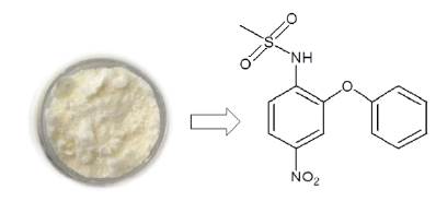
Nimesulide (figure 1) is a non-steroidal anti-inflammatory drug having more affinity to inhibit COX-2 enzyme. It is used to reduce the pain associated with rheumatoid arthritis, and other degenerative joint disorders, dental pain and muscle inflammation. After its oral administration, this drug exhibits a half-life between 1.5 to 4.9 hours, which requires frequent dosing to maintain the ideal plasma concentration for treating inflammatory disorders [18]. In this paper, we study the use of eggshell membrane (ESM), a natural food by-product from egg processing, as an alternative polymeric matrix for drug release systems. He nimesulide was used as a model drug in our release studies. His work shows the incorporation of nimesulide in ESM, the ionic cross-link of this matrix, characterizations of the materials, in vitro drug release studies and application of theoretical kinetic models to the release data.
MATERIALS AND METHODS
Materials
Nimesulide was obtained by extraction from commercial formulations (100 mg tablets) acquired from a local drugstore. Before its use, the extracted nimesulide was purified by column chromatography and characterized by melting point (145-147 °C), 1H NMR and 13C NMR analyses, which proved its purity for analytical tests. He chemical name, molar mass, water solubility and pKa of nimesulide are N-(4-nitro-2-phenoxyphenyl) methane sulfonamide, 308.30 g-mol-1, 10.0 μg-mL-1 and 6.5 [19]. All other reagents used in the experiments were of analytical grade. Aqueous solutions were prepared with deionized water.
Extraction of eggshell membrane
Fresh white chicken eggs (30 units) were treated with 3.0 L of 10.0 % v-v-1 H3CCOOH (acetic acid) for 48 h at room temperature to dissolve the outer layer of the shell and expose the eggshell membrane (ESM). The turgid eggs were ruptured, and the internal content discarded. The ESM were thoroughly washed with deionized water and dried on a Teflon tray for 48 h at room temperature. The dried ESM were ground in a domestic blender using ethanol 95.0 % v-v-1 as solvent, sieved with a sieve size of 18 mesh (1 mm) to remove small particles, and stored in refrigerator until use. This procedure was an adaptation of the eggshell membrane preparation described by Krishnamoorthy et al. (2012) [20] and is based on the acid-base reaction between the CaCO3 (main component of the eggshell) and the acetic acid.
Drug loading
This experiment was adapted from the drug loading method reported by Gonçalves et al. (2005) [21], where the water insoluble biopolymer acts as an adsorbent for the drug. In this procedure, 400 mg of ESM were weighed in an erlenmeyer and immersed in 50.0 mL of 10 g-L-1 nimesulide ethanolic solution at 95.0 % v-v-1 for 48 h at room temperature. The solid samples were filtered, washed with deionized water, dried on a Teflon tray for 48 h at room temperature and stored in a refrigerator until use.
Cross-linking of biopolymer
The cross-linking procedure was carrying out to obtain a biomaterial capable to extend the release of the loaded nimesulide. This experiment was adapted from the ESM mineralization method reported by Wu et al. (1995) [22], which consist of a complexation cross-linking of the biopolymer. This complexation process occurs between the positively charged calcium ions (Ca2+, a Lewis acid) and the negatively charged carboxylic acid groups (R-COO-, a Lewis base), at the ESM surface.
In this procedure, 200 mg of ESM containing nimesulide incorporated were weighed at an erlenmeyer and immersed in 100 mL of 0.01 mol-L-1 HCl containing 10.0 %w-v-1 CaCl2.2H2O. The reaction was allowed to proceed for 24 h at room temperature. The solid sample were filtered, washed with deionized water, dried on a Teflon tray for 48 h at room temperature and stored in the refrigerator until use.
Morphological and functional groups analyses
The solid samples (ESM), with and without nimesulide, and with and without cross-linking (CaCl2), were characterized by Scanning Electron Microscopy (SEM - Vega3 Tescan) and Fourier Transform Infrared Spectroscopy (FTIR - Spectrum Two Perkin Elmer).
Quantification of nimesulide
A standard calibration curve of nimesulide was constructed for the spectrophotometric determination of this drug. Initially, a stock solution of nimesulide 100.0 mg-L-1 was prepared in 50 mL phosphate buffer pH 7.4 (0.10 mol-L-1). Afterwards, nimesulide standard solutions were prepared by appropriate dilutions of the stock solution using the same buffer and containing 0.2 mL of polysorbate 80. The concentration levels of nimesulide ranged from 0.5 to 30.0 mg-L-1. The standard solutions were analyzed with a spectrophotometer (NI-1600 UV Nova Instruments) at 400 nm using the same buffer as blank solution. The equation calculated by linear regression was y = 0.04073x - 0.01771, with a coefficient of determination (R2) of 0.9852.
Evaluation of drug loading
To obtain the amount of nimesulide incorporated in the polymeric matrix this drug was extracted from ESM using exhaustive maceration method at room temperature, and methanol-phosphate buffer (pH 7.4 and 0.10 mol-L-1) 1:1 as solvent. In this procedure, 25 mg of ESM containing nimesulide were weighed in an erlenmeyer and immersed in 8 mL of the extractive solution for 24 h at room temperature. The supernatant solution was carefully collected with a pipette and transferred to a vial keeping the solid inside in the erlenmeyer. This extraction procedure was repeated twice, and the three aliquots of the extractive solutions were transferred to a 25 mL volumetric flask and diluted with deionized water. The concentration of the free drug (in mg-L-1) in the supernatant (extractive solution) was measured with a spectrophotometer at 400 nm using the calibration curve equation. For this quantification, an aliquot of the extractive solution was diluted with the same solution used for the calibration curve construction (phosphate buffer pH 7.4 with polysorbate 80) to ensure that methanol did not compromise the analyses.
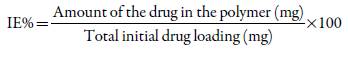
The total amount of nimesulide in the extractive solution (in mg) and the drug content in ESM (in mg-g-1) were calculated. The incorporation efficiency (IE %) of nimesulide in the polymer matrices was calculated, after measurements performed in triplicate, as follows:
In vitro kinetic release studies
Nimesulide release studies from ESM containing this drug, with and without cross-linking (CaCl2), were performed using a proposed spectrophotometric flow system that mimics the USP apparatus 4. A spectrophotometer equipped with a 10 mm "U" quartz flow cell was used as a detector in the flow experiments. A magnetic stirrer (NI 1103-E Nova Instruments), peristaltic pump (DMC-100 MS Tecnopon), pump silicon tube (0.2 mm i.d.) and transparent silicon tubes (0.1 mm i.d.) were also used in the proposed flow system. The inlets of the flask were closed with rubber stoppers to prevent the evaporation of the dissolution medium. In addition, to prevent the entry of suspended solids into the flow system, the inlet of the suction tube was coated with a piece of plastic sponge. Figure 2 shows an illustrative scheme of this system.
Figure 2 Scheme showing the proposed spectrophotometric flow system used in the nimesulide release studies.
The dissolution media used for the release studies in sink condition were: i) 100 mL of hydrochloric acid/potassium chloride buffer (0.10 mol-L-1, pH 2.0 to simulate the gastric fluid, and containing 0.2 mL of polysorbate 80); and ii) 100 mL of phosphate buffer (0.10 mol-L-1, pH 7.4 to simulate the intestinal fluid, and containing 0.2 mL of polysorbate 80). These dissolution media were maintained at a controlled temperature of 37 ± 1 °C (water bath) under magnetic agitation (150 rpm) and a flow rate of 5 mL-min-1. After the thermometer recorded 37 °C, the spectrophotometer was reset at 400 nm with the dissolution medium and 25 mg of the solid sample was inserted into the flask. The absorbance recording was performed from 10 seconds to 60 minutes at predetermined time intervals. The amount of free drug in the dissolution medium was calculated with the calibration curve equation. All drug release tests were performed in triplicate.
Release studies from commercial nimesulide tablets (100 mg) were also carried out using the proposed spectrophotometric flow system. In this procedure, the tablets were previously weighted, and 25 mg of the formulation powder was used in the studies. These formulation samples (25 mg) showed an equivalent mass of nimesulide of 6.07 ± 0.07 mg. The tests were made under the same experimental conditions as the release studies from ESM. These drug release tests were also performed in triplicate.
It is worth emphasizing that the commercial nimesulide tablets were crushed prior to release studies for: 1) to standardize the release procedure for formulation and ESM biopolymer; 2) to approximate the amounts of nimesulide present in such samples; 3) to adapt the sample mass to the flow release system used in tests, which works on a small scale.
Kinetic drug release models
Parameters of nimesulide release kinetics from ESM containing the loaded drug, with and without cross-linking (CaCl2), were determined applying the linearized models and considering the entire time interval of the test (0-60 minutes). The fitting parameters and correlation coefficients (R2) were calculated as follows:
Zero-order model:
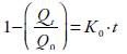
First-order model:
Higuchi release model:

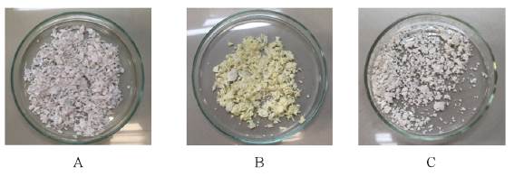
Peppas model:
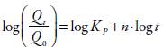
Where Q t is the amount (in mg) of drug present in the dissolution medium at time t, Qp is the total amount (in mg) of drug present in the solid sample, Q t /Q 0 is the drug fraction released at time t, K 0 is the zero-order release rate constant, K x is the first-order release rate constant, K H is the Higuchi constant, K P is the Peppas constant, n is the release exponent (that indicates the mechanism of drug release), and t is the time elapsed in the release study. In the Peppas model, n = 1 corresponds to zero-order release kinetics (case-II transport), 0.5 < n < 1 means a non-Fickian release model (anomalous transport), and n ≤ 0.5 indicates a Fickian diffusion.
RESULTS AND DISCUSSION
Morphological characteristics
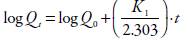
After drying, the ESM obtained from chicken eggs acquired the appearance of white fine flakes. This biomaterial was insoluble in aqueous medium and in other organic solvents. An experimental fact that needs to be reported when working with ESM is the difficulty of transforming its shape. This membrane also cannot be solubilized without compromising its biological structure. For this reason, the ESM was used in this work in its solid form (fine flakes). After the nimesulide incorporation and drying ofthe polymeric matrix, the ESM acquired the appearance of yellow flakes. This color change of the biopolymer is probably due to the natural color of the model drug incorporated (nimesulide).
After drying, the ESM containing nimesulide, and cross-linked with CaCl2 through ionic interactions, acquired again the white color, but the ESM cross-linked with CaCl2 was slightly more rigid than the ESM in nature. Despite membrane bleaching after cross-linking, FTIR analysis and release studies proved that drug still remained incorporated into the ESM. The cross-linking procedure was carrying out in acid media (HCl 0.01 mol-L-1) to avoid the alkalinization of the Ca2+ aqueous solution and the subsequent release of the loaded nimesulide from ESM during this reaction. The figure 3 shows the visible characteristics of these samples.
Physicochemical characterization
The microstructure characterization of the polymeric samples was made by Scanning Electron Microscopy (SEM). As can be seen in the SEM image (figure 4A), the ESM is a biopolymeric fibrous net, with its individual fibers randomly oriented, interwoven, and coalescent. This biostructure is similar for that showed by Baláz (2014) [23].
Figure 4 (A). SEM images of ESM in nature, (B). Nimesulide, (C). ESM containing nimesulide and (D). ESM containing nimesulide and cross-linked with CaCl2 with a magnification of 1500x.
Nimesulide used at this work was also characterized by SEM (figure 4B). The drug, in the solid state, has the aspect of acicular and tabular crystals, a microstructure also described by Sanphui, Sarma e Nangia (2011) [24]. When nimesulide was incorporated into the ESM matrix, the SEM image (figure 4C) shows an apparent thickening of the fibers, a decrease in porosity and the presence of amorphous granules and some crystals, structures which may be the drug adsorbed on the surface. Finally, when the ESM containing nimesulide was cross-linked with CaCl2, the SEM image (figure 4D) shows a microstructure with regions where the fibers appear to agglomerate, which may be the result of the cross-linking process.
The Fourier Transform Infrared Spectroscopy (FTIR) was used to characterize the ESM and nimesulide, individually, and after the incorporation of the model drug in the polymeric matrix, in terms of the presence of some organic functional groups (figure 5).

Figure 5 (A). FTIR spectrum of ESM in nature, (B). Nimesulide, (C). ESM containing nimesulide and (D). ESM containing nimesulide and cross-linked with CaCl2.
The natural ESM FTIR spectrum (figure 5A) shows the presence of peaks (in cm-1) at 3289 (stretching mode of O-H and N-H), 3060, 2932 and 2869 (asymmetric stretching vibrations of the C-H bonds present in =CH and =CH2 groups), 1646 (C=O stretch of amide), 1524 and 1240 (CN stretching/NH bending modes of amide), 1441 (stretching mode of C=C bond), 1066 (stretching mode of C-O bond) and 660 (stretching mode of C-S bond). These signals are in agreement with the ESM FTIR reported by Balaz (2014) [23] and indicate that this biopolymer has various chemical groups such as amine, amide, carboxyl and hydroxyl groups.
The pure nimesulide FTIR spectrum (figure 5B) shows the presence of characteristic peaks (in cm-1) at 3286 (stretching mode of N-H), 1519 and 1342 (asymmetric and symmetric stretching mode of N-O, respectively), 1589 (stretching mode of N-H from the sulfonamide group), 1249 (asymmetric stretching mode of C-O-C from the ether group), 1154 (symmetric stretching mode of S=O) and 976 (stretching mode of S-O-C bond). Several of these FTIR signals are in agreement with those related by Khan et al. (2010-B) [25].
The figure 5C shows the FTIR of the ESM containing nimesulide. In this spectrum some peaks come from the polymer (3289 cm-1 and the shoulder at 1646 cm-1) and from the drug (3286, 1589, 1519, 1342, 1154 and 976 cm-1). The signals from ESM at 1646 cm-1 (C=O of the amide bond) and from nimesulide at 976 cm-1 (S-O of the sulfonamide bond) showed a significant reduction on its intensities, indicating interactions by intermolecular forces, such as hydrogen bonds, between the polymer and the model drug. These results confirm the incorporation of nimesulide in ESM. The broad band at 3433 cm-1 belongs to the bending mode of the hydroxyl group of the adsorbed water.
Finally, the figure 5D shows the FTIR spectrum of the ESM containing nimesulide and cross-linked with CaCl2. In this spectrum also it could be seeing some peaks that come from the polymer (1646 cm-1) and from the drug (shoulder at 3286, 1519, 1342, 1154 and 976 cm-1). These results show that drug remains in the matrix after the cross-linking process. In addition, the broad band at 3433 cm-1 became more intense, indicating adsorbed water. This aspect may be due to the complexation process between the calcium ions (Ca2+) and carboxylic acid groups (at the ESM surface), which may also require some water molecules as Lewis's base.
Drug content in polymer matrices
The amount of nimesulide incorporated in the polymer matrices was determined by spectrophotometry after the extraction process (exhaustive maceration). The nimesulide drug content in ESM (in mg-g-1) showed values of176.83 ± 13.72 mg-g-1 and 122.69 ± 14.86 mg-g-1 for natural ESM and cross-linked with CaCl2, respectively (table 1). The incorporation efficiency (IE) of nimesulide in the studied polymer matrices (natural ESM and cross-linked with CaCl2) was low, showed values of14.14 ± 1.10 % and 9.82 ± 1.19 %, respectively (table 1). In this work, an adsorption method was used for the drug loading since the ESM is an insoluble matrix and, therefore, makes it difficult to obtain a monolithic system with the drug dispersed in the polymer in a more concentrated and homogeneous way. This is the main reason for the low incorporation efficiency (IE) of the drug in this biomaterial. It is inappropriate to compare the IE obtained in this work, with the IE obtained from other studies involving the incorporation of drugs into polymers using the classical emulsion solvent evaporation method, which is the method most widely used.
Table 1 Amount of nimesulide incorporated in the polymer matrices by the adsorption method.
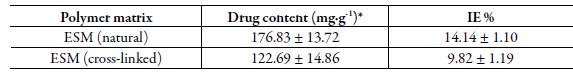
* Average value ± standard deviation (n = 3).
Although the nimesulide incorporation in the ESM matrices have apparently not showed attractive results, it is important to reinforce that this biopolymer has great potential for use in drug delivery systems owing to its biocompatibility and biodegrad-ability. And, because of this, more studies for pharmaceutical applications with this membrane should be stimulated.
In vitro release studies
Figure 6 shows the in vitro release of nimesulide incorporated at the ESM (natural and cross-linked) in sink conditions and at pH 7.4, evaluated using the proposed spectrophotometric flow system method that mimics the USP apparatus 4. The experimental data obtained in these studies were expressed as cumulative concentration and percent release as a function of release time.

Figure 6 Drug release curves of nimesulide from natural ESM and cross-linked with CaCl2 in phosphate buffer at pH 7.4 and 37 ± 1 °C.
Release studies of nimesulide at pH 2.0 (simulated gastric fluid) were also performed for both the matrices (not shown in figure), but in 60 minutes of timespan were not observed significant signals of the drug in dissolution media. These results are explained because nimesulide is a poorly soluble drug in aqueous medium, and at pH 2.0 this drug is not ionized (pH < pKa). These two characteristics do not facilitate the release of this drug from the polymer matrix. The release test with the commercial nimesulide formulation also showed similar results.
Based on the release curves obtained at pH 7.4 (simulated intestinal fluid) and showed in figure 6, it is possible to detect the presence of a moderate burst effect for both studied matrices in the first 10 minutes of experiments, followed by a gradual release phase. According to Freitas and Marchetti (2005) [26], the burst effect is normally attributed to the presence of the drug near the surface of the polymeric particle, which is known to be permeable to water. As time progress, these particles allow the water diffusion into their core and, consequently, the drug diffuses through the polymer matrix.
The release studies in simulated intestinal fluid showed that 26.63 and 19.81 % of the drug were released from natural ESM and cross-linked, respectively, during the first 10 minutes, and that 54.55 and 42.58 % after 60 minutes. These results are expected for a hydrophobic drug such as nimesulide, which tends to remain entrapped in the hydrophobic zones of the polymer. These results also confirmed that cross-linking with CaCl2 extended the drug release from the polymer matrices. This is a promising result considering that cross-linking reagent used is safe for human consumption. In addition, the release studies with the commercial nimesulide formulation showed that 27.57 % and 44.77 % of the drug were released in just 1 and 3 minutes, respectively. In this case, a time greater than 3 minutes of release for the commercial nimesulide formulation was not evaluated because the spectrophotometric signal went beyond the maximum monitoring limit, making the drug quantification unfeasible. This rapid release is a common behavior for immediate release dosage forms. A similar behavior was related by Purcaru et al. (2010) [27]. In their study nimesulide was released from solid formulations in tween 80 aqueous solutions at different concentrations. In 0.5 % tween 80 solution (in combination with phosphate buffer pH 7.4) nimesulide was immediate released, reaching a value of 70.0 % in the first 10 minutes. Based on the Biopharmaceutical Classification System (BCS), nimesulide is considered a BCS 2 drug (poorly soluble and highly permeable) and, therefore, the dissolution process is the limiting step for its release [27].
The release of drugs that are dispersed in (and not covalently bound to) polymer matrices is dependent on the level of drug loading and the solubility of the drug in the matrix [4]. In this context, in pH 7.4, the release of nimesulide from the ESM matrices is favored because of the deprotonation and ionization of sulfonamide group of this drug (pH > pKa). This fact enhances its solubility and favors its dissolution and release process.
In immediate release formulations, such as tablets, all the drugs should be released in less than half an hour, which was not observed in the prepared polymer matrices containing nimesulide (natural and cross-linked ESM) [28]. This behavior makes the ESM a promising biopolymer that can be used as an alternative excipient in extended-release dosage forms.
For comparative purposes, it is valid to demonstrate other studies related to the release of nimesulide from different polymer matrices. In study of Freitas e Marchetti (2005), the authors prepared microspheres of polylactic acid (PLA-Z) containing nimesulide to act as a polymeric carrier for this drug [26]. The results indicated that loading efficiency of nimesulide in PLA-Z microspheres was 70 % using the classical emulsion solvent evaporation method for drug entrapment. The percentage of drug release (in phosphate buffer, pH 7.4) from this matrix was 10.42 % within 60 minutes and 14.22 % after 120 minutes. The obtained results by these researchers showed that developed formulation demonstrated a potential use for sustained release system to vehicle nimesulide.
Raval et al. (2010) prepared nimesulide microspheres using Eudragit RS100 and RL100 together, in a single matrix, but at different combinations of these polymers [29]. The authors obtained a drug entrapment varying between 56.36 and 85.36 % through the emulsion solvent evaporation method. The cumulative release of nimesulide (in phosphate buffer, pH 6.8), in the first hour (60 minutes), varied between 6.71 to 27.04 %. These results showed that drug release from microspheres was highly influenced by the combination of Eudragit polymers, and the release profiles showed that a suitable combination of these polymer materials can produce a new matrix for the sustained release of nimesulide.
In 2017, Verma, Mishra and Nayak prepared microspheres of nimesulide, cross-linked with glutaraldehyde, using the coacervation phase separation technique and three different polymeric carriers (gelatin, pectin and sodium alginate) [30]. The drug content for gelatin, pectin and sodium alginate in each microsphere formulations were 80 %, 50 % and 48 %, respectively. When dissolution studies were carried out, in phosphate buffer pH 7.2, the drug release in 60 minutes was 27.2, 19.54 and 37.8 % for gelatin, pectin and sodium alginate microsphere, respectively. The authors developed a sustained release oral product (microspheres) of nimesulide using hydrophilic carriers, but the cross-linking with glutaraldehyde was necessary to sustain the drug release effectively.
Kinetic drug release models
It is known that drug release from polymeric particles is a complex process inluenced by many aspects such as polymer swelling and/or erosion, drug diffusion and drug dissolution [31]. In this way, the probably release mechanism of nimesulide from the matrices (ESM in nature and cross-linked) was examined based on the magnitude of correlation coefficients (R2) obtained after application of zero-order, first-order, Higu-chi and Peppas models. Table 2 shows the kinetics parameters obtained after the application of these theoretical models.
Table 2 Kinetic parameters obtained after the application of linearized theoretical models on the experimental nimesulide release data.
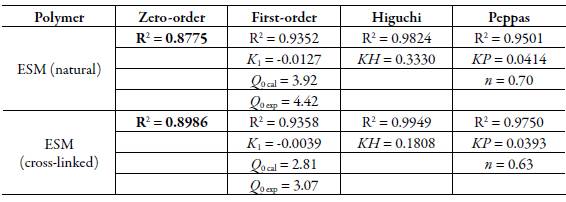
The correlation coefficients (R2) calculated for the polymers tested (natural and cross-linked ESM) (table 2) indicated that nimesulide release was better described by the Higuchi model in both the cases. Results also revealed that polymers tested showed values of n (from the Peppas model) between 0.63 and 0.70, indicating a non-Fickian (anomalous transport) release. These data might indicate that release of nimesulide followed coupled erosion-diffusion mechanism [32]. In other words, both drug diffusion and polymer swelling were important processes for the drug release.
Besides, the kinect parameters of Higuchi and Peppas models indicate that mechanism of nimesulide release from the polymer matrix apparently was unaffected by the cross-linking of ESM with CaCl2.
CONCLUSIONS
The present paper showed the use of eggshell membrane (ESM), a natural food byproduct from egg processing, as a biopolymer in drug release studies. FTIR analysis proved the nimesulide incorporation on ESM. Although the drug loading did not show attractive results at these preliminary studies, it is important to reinforce that this biopolymer has potential use in pharmaceutical formulations owing to its biocompatibility and biodegradability. Furthermore, this paper shows an initial study using ESM as an alternative polymeric material for pharmaceutical purposes. The cross-linking of the ESM containing nimesulide using CaCl2 has been able to extend the release of the hydrophobic drug, showing itself as a safe reagent for human health. The release studies and the kinetic parameters indicated that release of nimesulide from ESM matrices (natural and cross-linked) followed the Higuchi model and a non-Fickian transport. In view of these results, it could be said that ESM has potential to become a water insoluble carrier for extended release of nimesulide. Further studies should be performed in order to achieve an increase in drug release time from ESM, besides checking stability, toxicity, and the correlation of the in vitro-in vivo release profile.

