




 English (pdf)
 Article in xml format
 Article references

 Automatic translation
 Send this article by e-mail
 Cited by SciELO
 Cited by Google
 Similars in
SciELO
 Similars in Google

 Permalink

Introduction
Doxycycline (DOXI) is a semisynthetic wide-spectrum antimicrobial drug tetracycline derivative. Currently, therapeutic applications for DOXI include urinary, intestinal, respiratory, ocular, dental, dermatological, and sexually transmitted infections, but recently DOXI have demonstrated potential application on anti-inflammatory and anticancer treatments. DOXI can exist in hydrochloride and hyclate salts forms with high solubility and high permeability according to the biopharmaceutical classification (BCS) 1,2. Some limitations that can influence the effectiveness of DOXI include instability in biological environment, bacterial resistance, and poor cellular penetration, which hinder drug action in intracellular pathogens 1. The development of drug delivery systems based on carriers like micro and nanoparticles (NP) allow to overcome those limitations, since tetracyclines are easily oxidized and degraded the NP provides greater stability. In addition, nanocarriers with positively charged surfaces, for instance, improve electrostatic interaction with the negatively charged bacterial wall and consequent bactericidal action of antibiotics 1. Besides, the administration of drugs on mucous membranes offers several advantages, including retention at the site of administration, as well as local administration in order to reduce the systemic dose, reducing adverse effects or conversely promoting the absorption of drugs according to the desired effect 3. Mucoadhesive carriers adhere to the mucosa increasing the residence time at the administration site, while in mucus penetration, micro or nanoparticles can spread over the mucosa and penetrate deep regions so that their residence time increases considerably, reaching the epithelium of absorption 4. It has been reported that certain surface properties of nanoparticles influence their mucoadhesive or mucopermeation behavior, especially hydrophilic surfaces such as biopolymers. Among the variety of polymers used for the preparation of carriers is gelatin. Gelatin (GEL) is a biopolymer derived from collagen, which has a positive charge at a pH below its isoelectric point (IP) due to the protonation of amino groups of lysine and arginine residues. It is biocompatible and biodegradable, has high availability and low cost. In addition, the release kinetics can be modified by varying the molecular weight of gelatin, as well as the amount of crosslinking agent during preparation. Gelatin carriers have demonstrated to be suitable for mucus administration, for antibacterial drugs like DOXI. Thus, this works aim to develop and characterize a drug delivery system for Doxycycline based on gelatin, intended to mucus delivery for antibacterial therapy.
Materials and methods
Materials
Gelatin (type A, gel strength ~225 g Bloom), Pluronic F-68 and Doxycycline Hyclate were purchased from Sigma-Aldrich. Glutaraldehyde (GA) grade I (25% solution in water) was obtained from Merck. Regenerate cellulose dialysis bag (MWCS 12-14 kDa) was obtained from Spectra/Por, USA. Ultrapure water was obtained with a Barnstead Nanopure diamond system. All buffer solutions were prepared consistent with the USP30-NF25.
Nanoparticle formulation
Gelatin nanoparticles were prepared using the desolvation method previously reported in the literature with some modifications 5. Type A gelatin (GEL) was dissolved in deionized water at 40.0±1.0 °C with constant magnetic stirring at 250 rpm. Subsequently, the gelatin solution was adjusted to a pH of 3.0±0.1 with HCl 2.0 N and Doxycycline Hyclate (DOXI) was added. On the other hand, Pluronic F-68 (PF68) was dissolved in acetone at a concentration of 3.0% (w/v) at room temperature and constant magnetic stirring. The solution of Pluronic F-68 was added drop by drop to the gelatin solution maintaining stirring with an Ultra Turrax® T18 (IKA). Finally, an aqueous solution of glutaraldehyde (2.0% v/v) was added as a crosslinking agent and kept in constant magnetic stirring for 12 h.
The nanoparticles formulation was optimized in one factor at a time, to obtain a stable dispersion. In this regard, various parameters were investigated for their effects on particle size and PDI. In order to optimize the methodology for obtaining the nanoparticles, the effect of some process parameters were investigated, such as: effect of the stirring speed (1000, 5000, 8000 and 10000 rpm), concentration of Pluronic F-68 (0.5, 1, 2, 3 and 5% w/w), gelatin concentration (10 mg/mL and 20 mg/mL) and volume of the crosslinking agent Glutaraldehyde 2.0 % v/v (100, 300, 500, 1000 and 1500 µL).
Nanoparticle characterization
Particle size and Z potential (ξ)
The hydrodynamic diameter and polydispersity index (PDI) were determined by dynamic light scattering (DLS) technique using a Zetasizer Zen3600 (Malvern Instruments) at 25.0±1.0 °C with a detection angle of 173° (n=3). The Z potential (ξ) was determined with the same equipment by electrophoretic mobility in deionized water (n=3).
Entrapment Efficiency (EE%)
The entrapment efficiency (%) was determined by centrifugation method or indirect method, by UV-VIS spectrophotometric quantification of doxycycline hyclate contained in the clear supernatant after centrifugation (n=3) 6. An aliquot of 1.5 mL of the nanoparticles was placed and centrifuged (Biofuge Primo R) at 14000 rpm for 1.0 h. The absorbance of the supernatant at the maximum absorption wavelength (λmax=343.43 nm) was determined with an S2000 spectrophotometer using a DT1000 deuterium light source, with a SAD500 interface (Ocean Optics, Inc.), using a 10 mm long quartz cell (Prolab). A calibration curve of doxycycline hyclate was prepared in water in a range of 0.5 to 20 μg/mL from a stock solution (n=3) and the linearity of the results was evaluated by an analysis of variance (ANOVA) using the Statgraphics Centurion XVIII® software. The entrapment efficiency was calculated as follows (equation 1):
Scanning electron microscopy (SEM)
The morphology of nanoparticles was determined by scanning electron microscopy (SEM) using a JSM-35 CF (JEOL) microscope at 20.0 kV with gold coating. A drop of freshly prepared NP dispersion was deposited and air-dried at room conditions and coated with gold, finally, the sample was analyzed in vacuum.
Infrared spectroscopy (FT-IR)
The FT-IR spectra were recorded with a Spectrum 400 FTIR/FIR spectrophotometer (Perkin-Elmer). The infrared spectra of the samples were measured in a wavelength range of 4000-400 cm-1.
In vitro profile release
The release profile of the prepared systems was evaluated using the dialysis bag method. An amount of NP dispersion was placed in the regenerated cellulose dialysis bag (MWCS 12-14 kDa, Spectra/Por) previously hydrated in deionized water at room temperature for 12.0 h. The dialysis bag was suspended in the release medium (100.0 mL) of phosphate buffer solution pH 7.0±0.1 at 37.0±1 °C in a closed beaker with constant magnetic stirring (250.0 rpm). At specified time intervals, a volume was extracted from the release medium and analyzed using a spectrophotometric method. The medium was replaced by adding an equal amount of fresh medium, which was preheated to 37.0±1.0 °C, after each sampling to maintain sink conditions. An UV-Vis spectrophotometric quantification was performed at the wavelength of maximum absorbance (λmax=343.43 nm). The analysis was performed on an S2000 spectrometer using a DT1000 deuterium light source, SAD500 port (Ocean Optics, Inc.) with a 10 mm long quartz cell (Prolab).
Mathematical modeling of profile release
There are many theories or kinetic models to describe drug release from controlled or immediate release forms, where the amount of drug released is a function of time. The release data was fitted to two of the most renowned models to describe drug release kinetics and mechanism.
Higuchi model: Higuchi is related to particles of active dispersed in homogeneous matrices submitted to a diffusing medium. The simplified Higuchi model describes drug release as a diffusion process based in the Fick´s law, square root time dependent (equation 2). This model can be used to describe the drug dissolution from several types of modified release pharmaceutical dosage forms, and is the most widely used model to describe drug release from pharmaceutical matrices. Where Mt/M∞ is a fraction of drug releases at time t, k is the release rate constant. The extend model is based on the hypothesis that (i) initial concentration of drug in the system is much higher than the drug solubility, (ii) the edge effect is negligible, which means that diffusion is unidirectional, (iii) drug molecules are much smaller than system thickness, (iv) swelling or dissolution of system is negligible, (v) drug diffusivity is constant, and (vi) in the release environment perfect sink conditions are maintained 7.
Korsmeyer-Peppas model (Power Law): This model is a semi-empirical equation developed by Korsmeyer and Peppas to describe drug release from polymeric systems, it can be seen as a generalization of the observation of the superposition of two apparently independent mechanisms of drug transport, relaxation, and diffusion, usually applied when the release mechanism is not known or when more than one type of phenomenon of drug release is involved (equation 3) 7. Where Mt/M∞ is a fraction of drug releases at time t, k is the release rate constant and n is the release exponent. Depending on the value of n that better adjusts to the release profile, concluding for values of n=0.5 the drug release is governed by Fick diffusion, values of n between 0.5 and 1.0 corresponds to a non-Fickian transport or anomalous transport, and the mechanism of drug release is governed by diffusion and swelling, and n=1.0 corresponds to Case II transport the mechanism driving the drug release is the swelling or relaxation of polymeric chains 7.
All fitting models were performed by non-linear direct fitting and analyzed by using DDSolver program as analysis tool. The examination of the goodness of fit statistics was made by comparison of the coefficient of determination or R-square (R2), the adjusted coefficient of determination (R2adjusted) due to is more meaningful when comparing models with different numbers of parameters.
Results and discussion
In recent years, gelatin nanoparticles have proven to be carriers with advantages for modified drug release. Gelatin nanoparticles have been developed by various methods such as coacervation, solvent emulsion-evaporation, nanoprecipitation and desolvation 8. NP were prepared using the desolvation method, which is a widely used method due to its good reproducibility and ease of preparation. In addition, it allows to modify the characteristics of interest in the NP such as size, Z potential, entrapment efficiency, etc. through the manufacturing conditions 9. It was prepared a solution of gelatin which was adjusted to pH of 3.0±0.1, because in acidic conditions, gelatin type A (Isoelectric point=7.0-9.0), presents a positive charge by the protonation of the amino groups (-NH3+) of lysine residues. During synthesis, the positive charge avoids the uncontrolled agglomeration of gelatin chains, allowing to obtain nanoparticles with better characteristics of size and PDI 10. The desolvation agent (acetone) was used to dehydrate gelatin chains and induce conformational changes in their structure and subsequently induce precipitation of NP 8. Finally, GA was used as a crosslinking agent to stabilize the structure of the NPs in order to prevent their rapid dissolution in aqueous media and a rapid release of the drug at body temperature. GA is a crosslinking agent that has been extensively studied with proteins, as it reacts easily at room temperature with the simultaneous formation of aldimine bonds (-CH=N-) between the amino groups of proteins 11.
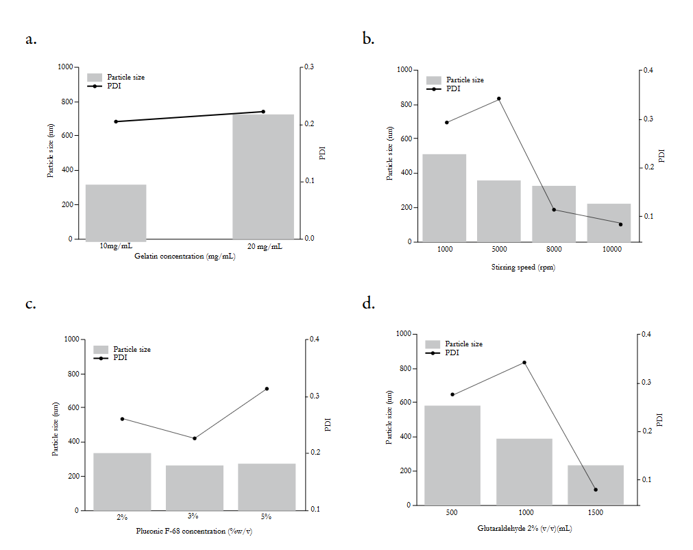
Figure 1 shows the results of formulation studies to obtain NP. The effect of various conditions on manufacturing were investigated, studying one parameter at a time: effect of gelatin concentration (10 mg/mL and 20 mg/mL) (Figure 1a), stirring speed (1000, 5000, 8000 and 10000 rpm) (Figure 1b), Pluronic F-68 concentration (0.5, 1, 2 and 3 w/v %) (Figure 1c), and volume of the crosslinking agent Glutaraldehyde (GA) at 2.0 % v/v (100, 300, 500, 1000 and 1500 µL) (Figure 1d).
Figure 1a showed the results of the concentration of gelatin effect. It was observed that the preparation of NP with a lower concentration of gelatin resulted in a decrease in particle size from 735.3 nm to 341.6 nm, along with a decrease in PDI from 0.224 to 0.208. The low concentration of gelatin allowed the formation of smaller particles with a more homogeneous size distribution. This is due to the lower availability of the biopolymer in the particle formation medium. The gelatin concentration of 10 mg/mL was established and the effect of stirring rate on particle size and PDI was evaluated.
In Figure 1b stirring speed effect was evaluated in particle size (nm) and PDI, in order to obtain a final formulation with particle size ≤ 250 nm and polydispersity index PDI ≤ 0.3 which indicates a narrow size distribution. The use of homogenizers in the development of nanoparticles by desolvation method is rarely reported, however, Jia Xue and Qixin Zhong reported the use of high-speed homogenization for the preparation of thymol hybrid nano dispersions based on gelatin and lecithin 12. It was observed that as the speed of agitation increased, the size decreased from 511.4 nm to 219.8 nm and PDI values were observed from 0.343 to 0.088. The results are similar to other results reported in the literature in that it was observed that increasing the speed of agitation favors the arrangement of chains in small droplets that precipitate allowing the formation of smaller particles. The speed of 10,000 rpm was selected in a high-cut homogenizer (Ultra Turrax®).
Subsequently, in Figure 1c the effect of the surfactant Pluronic F-68 was studied, which allows to stabilize the dispersion of particles and reduce the size according to previous studies reported in the literature 5. At the concentration of Pluronic F-68 from 0.5 and 1%, the formation of NP was not observed but the appearance of large agglomerates which may be due that at low concentrations, there is not enough surfactant to stabilize the formation of NP and there is agglomeration. From 2 to 3% it was observed the NP formation, however, the smaller particles were obtained at the concentration of 3%, since Pluronic F-68 decreases the surface tension between organic and aqueous phase and leads to the formation of smaller solvent droplets 5. However, at higher concentrations of Pluronic F-68, an increase in particle size was observed, which may be due an excess that may interact with gelatin significantly increasing the particle size.
It is known that in gelatin carriers swelling behavior, mechanical and thermal properties depend on the crosslinking degree 13. Glutaraldehyde (GA) is a well-known crosslinking agent, it have an aldehyde group (-CHO) that reacts with the amino group of the lysine residues of proteins leading an aldimine linkage (-CH=N-) generating inter and intra covalent bonds 14. To study the effect of glutaraldehyde as a crosslinking agent with 100, 300, 500, 1000 and 1500 μL aliquots of a 2% v/v aqueous glutaraldehyde solution were added to nanoparticles. After crosslinking reaction, the NP were obtained from ~200 to ~600 nm with PDI from ~0.2 to ~0.3 as shown in Figure 1d. The addition of 100 and 300 μL was studied, however, the formation of a stable dispersion of NP was not observed. It was possible to obtain a stable dispersed system from 500 to 1500 μL. The addition of the crosslinking agent allowed to obtain a stable system with adequate size with a volume of 1500 μL because at lower volumes of Glutaraldehyde the particle size increased which may be due to the agglomeration of the particles.
A decrease in particle size was observed as glutaraldehyde volume increased from 500 to 1500 µL may be related to the intra-particle crosslinking effect. Finally, Glutaraldehyde is consumed during the cross-linking process and with an adequate purification, there is no evidence of in vivo toxicity nor adverse effects have been reported, however minimum concentration of crosslinker was utilized 15,16.
Particle size and Z potential (ξ)
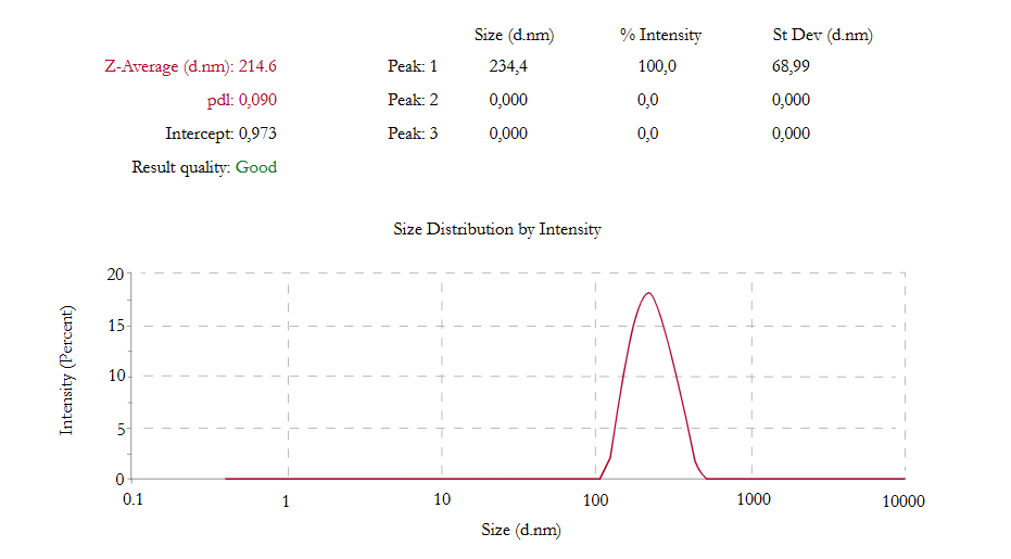
The final formulation presented an average particle size of 214.6 nm with a PDI of 0.090. Figure 2 presents the particle size distribution with the final conditions of the previous formulation studies, where a population with a modal distribution was observed.
Zeta potential is another important physicochemical parameter that influences the stability of colloidal dispersions. High positive or negative zeta potential values (ξ ≥ 30 mV) cause strong repellent forces, whereas repulsion between particles with similar electric charge prevents aggregation of the particles and thus ensures easy redispersion 17. The NP Z potential was 22±1.51 mV, which is similar to other reports. The nanoparticles were positively charged, this result may indicate the presence of gelatin on the particle surface, according to other reports. The zeta potential was relatively low (ξ ≤ 30 mV), however, their surface has a sufficient zeta potential to prevent further agglomeration of the particles observed in good size and PDI measurements 10,18. Also, cationic nanoparticles have demonstrated to improve oral bioavailability of drugs by mucoadhesion 19. Gastrointestinal tract is an attractive place for mucoadhesive delivery systems to modified the release of drugs. The mucosal surface is negatively charged, a positive charge on the polymer like gelatin NP might facilitate the mucoadhesive process. A positively charged polymer may be attracted to the biologic surface by electrostatic attraction, followed by mechanical interlocking of the polymer chains by van der Waals force, hydrogen bond, and other forces 19,20. Also, positively charged colloidal drug carriers may increase the permeability and potential uptake of slightly soluble drugs 19.
Entrapment Efficiency (EE%)
Once the manufacturing conditions for size, PDI and Z potential were determined, the entrapment efficiency of the system was determined with a calibration curve of Doxycycline hyclate from a stock solution, which was linear (R2=0.999) (n=3) in a concentration range of 0.5 to 20 μg/mL. A nanoparticulated formulation is preferred to be with high drug loading capacity to reduce the quantity of the carrier required for administration. The amount of bound drug and the nature of interaction between drug and nanoparticles depend on the chemical structure of the drug as well as the polymer and the conditions of drug loading 13. It is known that hydrophilic drugs may be loaded into gelatin carriers by incubating the drug with aqueous gelatin solution prior to nanoparticle formation. The entrapment efficiency was determined by an indirect method, and the NPs showed a 59.83±1.45% of EE (n=3) which is similar to other reports where DOXI loaded gelatin microspheres were prepared with %EE from 49 to 88% 21.
Scanning electron microscopy (SEM)
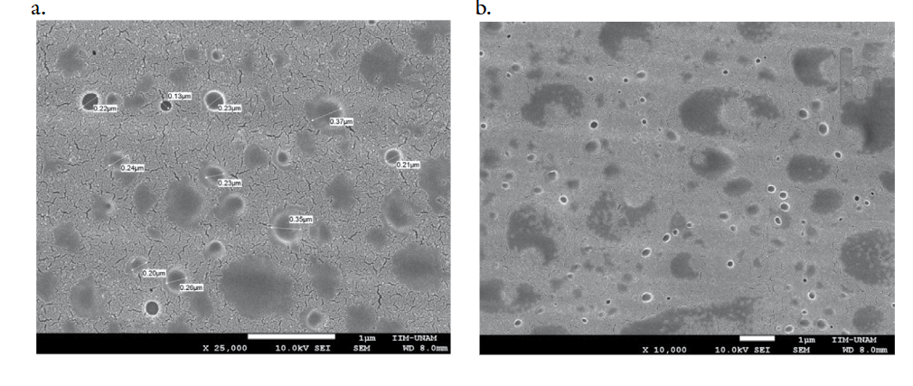
The shape, size and surface morphology of the NPs were determined by SEM. Figure 3 shows the NPs that were obtained with spherical morphology and smooth surface, without cracks or heterogeneities, this morphology is similar to that obtained by other authors for gelatin nanoparticles used in the release of drugs. The NP observed by SEM presented particle sizes similar to the results obtained by the DLS technique (Figure 3a), which is suitable for administration in mucous membranes orally or nasally.
Infrared spectroscopy (FT-IR)
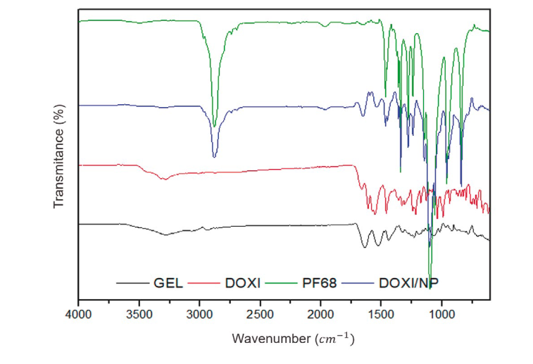
The system obtained together with the individual components (gelatin, Pluronic F68, Doxycycline hyclate) were characterized by FT-IR in the range of 4000 to 400 cm-1, with which the characteristic bands of the functional groups of each component were observed. Figure 4 shows the spectra obtained by FT-IR. Pure Doxycycline (DOXI) has characteristic bands in 3331.85 cm-1 corresponding to the vibrations of the bonds (O-H) and (N-H), in 1663.34 cm-1 corresponding to the primary amine (N-H), 1611.09 cm-1 of the carbonyl group (C=O), 1458.05 cm-1 due to the doubling of the group (CH2), 1329.80 vibrations (C-H), 1216.67 and 1170.63 cm-1 indicative of the vibrations of the link (C-N). In the spectrum of gelatin (GEL) the characteristic bands of 3282.08 cm-1 corresponding to amide A (N-H), 1637.22 cm-1 of amide I (C=O), 1528.97 cm-1 of amide II (N-H) are observed. In the spectrum of Pluronic F68 (PF68) the characteristic bands in 3505.65, 2880.76 and 1099.18 cm-1 corresponding to the vibrations of the bonds (O-H), (C-H) and (C-O) were observed. In the spectrum of NPs loaded with DOXI (DOXI/NP), bands are observed in 3306.22 cm-1 corresponding to amide A (N-H), 1652.15 cm-1 of amide I (C=O), indicative of the presence of gelatin, however there is also the appearance of a new band in 1450.58 cm-1 corresponding to the aldimine bond (R--CH=N--R') which confirms the crosslinking of the amino groups of lysine residues in gelatin with glutaraldehyde (-CHO), the bands indicative of the presence of Pluronic F68 were also observed in 2881.44 and 1102.80 cm-1 corresponding to the bonds (C-H) and (C-O) 8,22.
In vitro release profile
In vitro release studies were performed using the dialysis bag method. Regenerated cellulose bags of pore size of 12-14 kDa (Spectra/Por®) previously hydrated for 12 h were used. Figure 5 presents the release study, which was maintained for 6 hours at 7.0±0.1 at 37.0±1°C it was observed a biphasic patter, since during the first 2 hours the 83.11±1.78 % (n=3) was released. Subsequently, the system was maintained for 5 hours in which up to 91.46 ±1.47 % (n=3) was released. The system presented a profile with a quick release during the first 2 h and then a slow release until 6 h. The profile found may be suitable to achieve effective plasma concentrations quickly and continue with a slow release until the second administration. It has been reported that the profile of this type of system can be modified, by increasing the crosslinking agent. In addition, since the drug has a hydrophilic character, it can be deposited on the surface of the system, which favors a rapid release at the beginning of the study named Burst effect, as other authors have already reported.
An adjustment of the data obtained was made to the mathematical models of Higuchi and Korsmeyer-Peppas, which is useful to explain the speed and mechanism of release. It was observed that, of the selected models, the particulate system showed a better fit to the Korsmeyer-Peppas model (60%), with respect to the Higuchi model, according to the highest values of R2 and R2adj (Table 1). The Korsmeyer-Peppas model allows to determine the parameter n, which is related to different types of release mechanisms, the prepared system presented a value of n of 0.593 which is related to an anomalous transport mechanism (0.5>n>1.0), which is indicative of a combination of mechanisms of Fickian diffusion and swelling. The drug release kinetics from gelatin nanoparticles depends on the rate of water uptake, drug dissolution/diffusion rate and the polymer glass-rubbery transition including matrix erosion/degradation rate 23. Gelatin is a biopolymer capable of providing this type of mechanism since when in contact with water the highly soluble drug can dissolve and diffuse through the channels of the particle, as well as present a certain degree of swelling. Due to its properties like particle size, zeta potential and prolonged release, the prepared system could be a good candidate for administration on mucus membranes. Since its high isoelectric point (IP=7-9), in different types of mucus such as buccal (pH 6.5-7.5), nasal (pH 5.5-6.5), vaginal (pH 3.5-4), etc. the system would remain positive allowing interaction by electrostatic attraction with mucus 4.
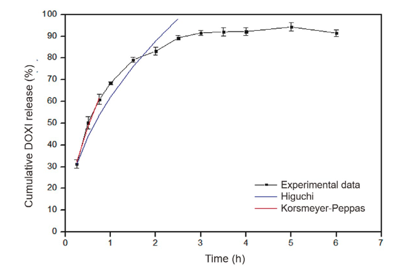
Figure 5. NP profile release (experimental data (•)), Higuchi model (blue line), Korsmeyer-Peppas model (red line)

Conclusions
The process factors and formulation studied successfully lead a doxycycline loaded gelatin nanoparticles with desirable characteristics as low particle size, narrow particle distribution and high drug entrapment efficiency (>50%). The system was characterized using various instrumental techniques and in vitro tests to evaluate its effectiveness. The NP showed prolonged release for 8 hours with an anomalous transport as main mechanism of drug delivery. The system prepared own cationic surface properties to be a possible mucoadhesive system.

