




 English (pdf)
 Article in xml format
 Article references

 Automatic translation
 Send this article by e-mail
 Cited by SciELO
 Cited by Google
 Similars in
SciELO
 Similars in Google

 Permalink


Introduction
Oral microbiota refers to the term used to determine the group of more than 700 species of microorganisms that symbiotically inhabit the oral cavity and that may be beneficial or harmful to the human host. Current evidence demonstrates that the microbiota plays a crucial role in the associations between oral and general systemic health [1].
Streptococci of the mutans group constitute about 70% of the cultivable microorganisms in the oral cavity. They are Gram-positive, ovoid bacteria, organized in pairs or chains that ferment dietary sugars and have acidogenic and adherence capacity. In addition, they initiate and have a strong role in the production, increase and maturation of biofilms [2].
Previously, Streptococcus mutans were thought to be the only cause of dental caries [3]. However, it has recently been shown that Candida albicans also plays a critical role in the development of root caries, causing significant microbial dysbiosis and increased abundance of acidogenic/aciduric S. mutans, which leads to the formation of a more cariogenic polymicrobial biofilm [4].
The microbial biofilm is the polymicrobial grouping involved in a viscous matrix composed of water, exopolysaccharides, proteins, nucleic acids and substances absorbed by the attached microorganisms that bind to the film acquired on the surface of the tooth [5]. This film is formed within 15 minutes after cleaning and polishing the dental enamel and is considered the first step for the fixation of S. mutans and the formation of dental biofilm [6].
Adequate control of the biofilm in the oral cavity results in a lower risk for the development of oral and systemic diseases arising from the oral microbiota [7], and the toothbrush is an important instrument in the promotion of oral health.
Apposite tooth brushing provides partial removal of oral biofilm with reduction of microorganisms related to the etiology of dental caries [8].
However, the brush itself can become an environment for deposition and proliferation of microorganisms in its bristles [9]. In addition, a correlation has been demonstrated between the microbial load found on toothbrushes and the dental status of patients [10].
In this context, the objective of this work was to evaluate, under laboratory conditions, the formation of biofilm on toothbrushes by streptococci of the mutans group and Candida spp. isolated from the oral cavity of students at the State University of Goiás in Brazil.
Material and methods
Obtaining saliva samples
This study was submitted to the Research Ethics Committee of the State University of Goiás (UEG) in accordance with resolutions 196/96 and 466/12 for research involving human beings of the Ministry of Health and the National Health Council through the Brazil Platform with approval number 3.604.978. Consultation and free, prior and informed consent (FPIC) was signed and a copy delivered to the participant before any procedure in this research.
Saliva samples were collected with stimulation by tooth brushing, from 13 students at the Central Campus of the State University of Goiás in the city of Anápolis. Volunteers were included according to the inclusion criteria: being of legal age, agreeing to participate in the research by signing the necessary documentation, having good systemic health, not having eaten in the period between the last brushing and collection, not having brushed their teeth before collection and not having any missing tooth in the oral cavity, except for third molars.
In the act of collecting the material, information was collected and then the volunteers were instructed on brushing with the modified Bass technique [11]. An illustrated insert was presented in order to facilitate brushing and standardize sample collection by the participants, and the brushing time was timed. Toothpaste or dental floss was not used and the tongue was not brushed.
Saliva samples were collected with new, disposable brushes with a size 30 rounded head, 20 tufts, 840 bristles and 14 cm long soft bristles that were previously sterilized in an autoclave at 125 °C for 15 minutes.
Volunteers were taken to the bathroom and requested that, as soon as brushing was completed, the brushes were immediately returned to be placed in Falcon tubes containing 15 mL of 0.9% sodium chloride sterile saline solution for transport, and subsequent processing in the Microbiology Laboratory.
Microbiological processing
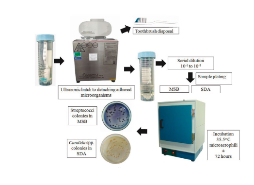
The samples were submitted to sonication to detach the microorganisms adhered to the brushes in an ultrasonic bath at 40 kHz (Ultronique Q 5.9/40A, Indaiatuba, São Paulo) for 5 minutes at room temperature [12]. Then the brushes were discarded and the supernatants were diluted and plated up to 10-8 for the isolation of the collected microorganisms.
Microbial samples were subcultured onto mitis salivarius agar (MSB) (Himedia, Mumbai, India) supplemented with 20% sucrose PA (Neon, São Paulo, Brazil), 0.2 IU bacitracin (Dr. Ehrenstorfer, Augsburg, Germany) and 1% potassium tellurite (Dinâmica, Indaiatuba, Brazil) for the isolation of mutans group streptococci and on Sabouraud dextrose chloramphenicol agar (SDA) (KASVI, São José dos Pinhais, Brazil) for isolation of Candida spp. [13]. The plates were incubated at 35.5 °C in microaerophilia for 72 hours (Figure 1).
After incubation, growth readings were performed in the culture media with a colony counter (MA-6000, Marconi, Piracicaba, Brazil) for the presumptive identification of the microorganisms recovered from each brush.
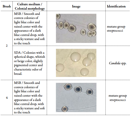
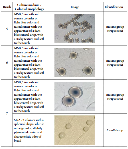
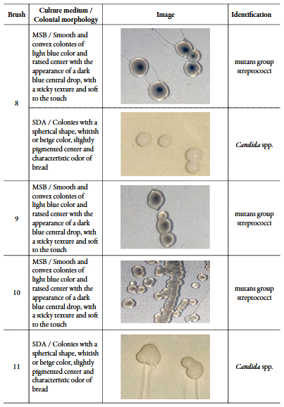
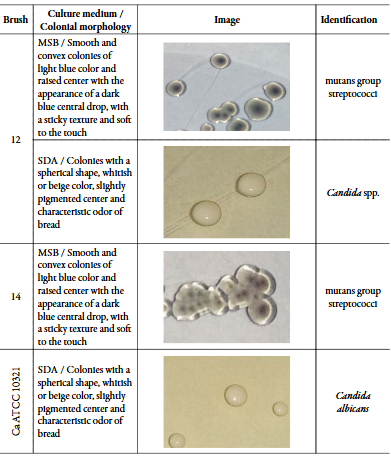
The presumptive identification of the isolated microorganisms was carried out based on the analysis of colonial aspects such as morphology, color, texture and odor after growth in the respective selective culture media. The enriched MSB medium is highly selective for S. mutans, S. sobrinus, S. salivarius and S. mitis. Smooth and convex colonies of light blue color and raised center with the appearance of a dark blue central drop, with a sticky texture and soft to the touch were identified as mutans group streptococci in this study [14], [15].
For the isolation and identification of Candida spp., the criteria used were growth in SDA with chloramphenicol, with formation of whitish, spherical colonies, with a slightly darker center and odor of the plates similar to bread fermentation [16].
After identification, the isolated microorganisms were frozen in aliquots for further studies of biofilm formation [17].
Biofilm formation on brushes
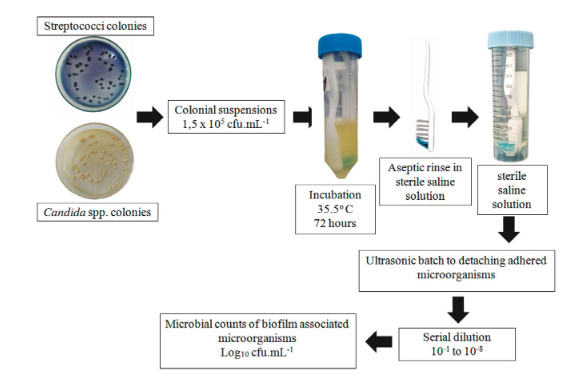
Aliquots of the isolated microorganisms were subcultured in the respective culture media and after incubation at 35.5 °C in microaerophilia for 72 hours, and then the purity of the cultures was checked. Subsequently, three to five typical colonies were dissolved in 0.9% sterile saline solution (0.9% SSS) at approximately 1.5 × 10⁸ cfu·mL-1 with the 0.5 McFarland scale. Then, 200 µL of the suspension was transferred to Falcons tubes with 19,800 µL of Brain Heart Infusion Broth (BHI) (Himedia, Mumbai, India) adjusting and the initial inoculum for the biofilm formation assay at 1.5 × 10⁵ cfu·mL-1.
After preparation of the microbial inocula, sterile toothbrushes were aseptically placed in Falcon tubes and the samples were incubated. Then, the brushes were removed with sterile forceps and then rinsed in a Falcon tube with 20 mL of 0.9% SSS. After this procedure, the brushes were transferred to other tubes with 20 mL of 0.9% SSS and sonicated in an ultrasonic bath at 40 kHz (Ultronique Q 5.9/40A, Indaiatuba, São Paulo) for 5 minutes at room temperature [12] for detachment of attached colonies and subsequent counting in MSB and SDA media, for growth of mutans streptococci and Candida spp., respectively (Figure 2).
All microorganisms were evaluated in three independent replicates. The results of the counts were transformed into log10 and the means and standard deviations were calculated. Sterility controls and the Candida albicans ATCC 10231 were included in the assay.
Results
Isolation and presumptive identification
Of the total of 13 brushes processed, microorganisms were isolated in 12 (92.31%), in 1 (7.69%) brush were not isolated. A toothbrush that showed microbial growth was removed from the study due to the waiving of a volunteer from participating in the investigation.
A total of 15 types of colonies were obtained in the 11 brushes with microbial growth. Of these, 10 colonies were suggestive of mutans group streptococci and 5 colonies of Candida spp. In 6 (54.54%) brushes were isolated only streptococci of the mutans group, in 4 (36.36%) brushes were isolated both microorganisms and in 1 (9.10%) brush only Candida spp. was found.
Table 1 presents the morphological description and presumptive identification of the isolated microorganisms.
Biofilm formation on brushes
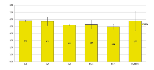
All microbial isolates were able to form biofilms under the tested conditions, with microbial cell counts adhered to the biofilm ranging from 6.75 to 11.00 log10 cfu·mL-1 for mutans streptococci, and 4.94 to 5.79 log10 cfu·mL-1 for Candida spp.
The isolates MS7 and MS14 were the major biofilm formers, with counts of 11.00 ± 2.51 and 10.28 ± 1.70 log10 cfu·mL-1, respectively. The other streptococci remained with more homogeneous counts. The isolated yeast counts were more uniform, as shown in Figures 3 and 4.
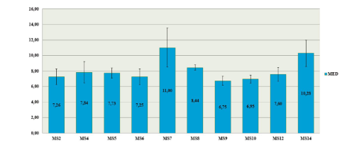
Figure 3 Mutans group streptococci cell count (log10 cfu·mL-1) recovered from the microbial films adhered in toothbrushes. MS: mutans group streptococci; cfu: colony forming unit.
Discussion
The oral cavity is colonized by several types of microorganisms, with the establishment of a transient microbiota, generally controlled by microbial antagonism with the resident microbiota and by routinely employed hygiene [1]. In the present study, brushes were used to isolate mutans group streptococci and Candida spp. of the oral cavity of volunteers. Subsequently, we verified the ability of the isolates to form biofilms on sterile brushes. In the first stage of the study, microorganisms were isolated in 92.31% of the brushes used to obtain the microbial samples. Of the 15 microorganisms isolated, 10 were mutans group streptococci and 5 were Candida spp.
Tooth brushing is essential for oral hygiene, however the use of a toothbrush requires care such as proper cleaning and frequent replacement to minimize contamination, because during brushing, part of the oral microbiota can be transferred to the brush and the toothbrush, and from then on, the microbial load tends to increase and diversify with each use [18], [19].
The mutans group streptococci are present in 90% of the world population, while C. albicans are isolated in 30-50% [20]. S. mutans are related to the installation and progression of dental caries due to their ability to primarily adhere to the smooth surface of the tooth and allow the subsequent aggregation of other acidogenic microorganisms, with increased sucrose metabolism, lactic acid release and maturation of polymicrobial biofilm [21].
Yeasts of the Candida genus are part of the oral microbiota in situations of normality of the host, but they can behave as pathogens when local and systemic factors are triggered [22]. In addition, C. albicans has the ability to colonize the oral mucosa and denture surfaces, and is reported as a frequent cause of oropharyngeal infections and has cariogenic potential, being found in 10% of decayed teeth without S. mutans[23].
By itself, C. albicans has no cariogenic capacity, but in the presence of sucrose, aggregation with S. mutans occurs, increasing the amount of aciduric acid-tolerant microorganisms and the production of extracellular matrix. Therefore, there is a relationship between the levels of caries, the amount of microorganisms and their ability to form biofilms. In this way, the mixed biofilm of these microorganisms can intensify the carious processes [24], [25]. Furthermore, Candida is an opportunistic fungus, and oral pathological manifestations are directly related to the immune status of the host [26] since the occurrence of microbial biofilm in this anatomical site is related to the oral health of the patient.
The data presented in figures 3 and 4 indicate that all the isolates were able to form biofilms on the brushes, with high counts of microorganisms associated with the biofilms. These results are similar to the results obtained by other authors, although using different substrates and methodologies. In a study [27] on S. mutans biofilm formation on bovine tooth enamel, the counts obtained in the control group ranged from 5.00 to 10.00 log10 cfu·mL-1. The results found in the viability controls of studies that evaluated the inhibition of biofilm formation by S. mutans were 6.5 and 10.00 log10 cfu·mL-1, respectively [28], [29]. Regarding the cell counts associated with Candida biofilm, other authors found counts ranging from 4.02 to 9.50 Log10 cfu·mL-1 cells [30]-[33]. Biofilms constitute a form of existence of microorganisms encapsulated in an extracellular matrix that holds cells together and forms a three-dimensional structure resistant to extrinsic adversities [34]. In the oral cavity, the biofilm is adhered to the surfaces of the mouth in general and tends to establish itself and increase if there is no chemical or mechanical interference [35]. Thus, microbial control methods are used to reduce microbial biofilm and maintain oral health, the use of mouthwashes and tooth brushing are common methods used for this purpose.
Conclusion
In summary, in this work we evaluated the biofilm formation on toothbrushes by mutans group streptococci and Candida spp. isolated from the mouths of students at the State University of Goiás. The sample collection methodology proved to be effective, as microorganisms were not recovered in just one brush collected. In the second part of the study, it was detected that all isolates were able to form biofilms on the brushes, and that the isolates showed considerable counts of microorganisms associated with the biofilm, with emphasis on the counts of streptococci of the mutans group. In agreement with other authors [36], [37], the results obtained in this study highlight the importance of taking care with the decontamination of toothbrushes in the promotion of oral health.

