




 English (pdf)
 Article in xml format
 Article references

 Automatic translation
 Send this article by e-mail
 Cited by SciELO
 Cited by Google
 Similars in
SciELO
 Similars in Google

 Permalink


Introduction
The study of medicinal plants began a long time ago, going through generations until nowadays, due to its easy access, low cost and compatibility with popular traditions. The commercialization of medicines of plant origin has increased due to scientific advances in the chemical, pharmacological and toxicological areas [1]. Given this context, copaiba oils are widely used due to their many properties, such as medicinal, cosmetic and industrial [2].
The copaiba plants belong to the family Leguminosae, subfamily Caesalpinioideae, genus Copaifera [3, 4]. They are common to Latin America and West Africa, and usually live about 400 years, reaching heights between 25 and 40 meters [5, 6]. In Brazil, they are in the Southeast, Central-West and Northern regions, mainly covering the states of Amazonas, Pará and Ceará [7-9]. There are 72 described species of Copaifera, more than 20 of them are in Brazil, among which 17 are endemic. The main species are: Copaifera officinalis L., Copaifera reticulata Ducke, Copaifera multijuga Hayne, Copaifera confertiflora, Copaifera langsdorffii, Copaifera cariacea and Copaifera cearensis Huber ex Ducke [10, 11].
The oil from these trees — copaiba oil or balm — varies in yellow gold to brown coloring, depending on the species. The oil is extracted by making an incision in the trunk of the tree, it is composed of a solid, non-volatile resinous part formed by diterpene acids, corresponding to 55 to 60% of it, diluted in the other part, an essential oil, composed of sesquiterpenes [12-15]. This oil composition varies depending on the concentration and nature of the diterpenes and sesquiterpenes present, according to species variation, biological factors and abiotic factors [13-15]. Some of the active components in these oils are β-bisabolol and β-caryophyllene, both with anti-inflammatory action, in addition to bactericidal action in the second [16].
Indigenous populations of Brazil's Northern and Northeast regions have used the oil to treat various diseases since the 16th century [17]. Nowadays, it is popularly used as a wound healer, anti-inflammatory, antiseptic in wounds, antitumoral, eczema, psoriasis and urticaria, anti-inflammatory of the urinary tract, in pulmonary affections (coughs, pneumonia, bronchitis, antiasthmatic, colds and flu), sinusitis, scarring of small irritations of the scalp, dysentery, urinary incontinence, cystitis, leucorrhoea, analgesic and antitetanic [18-22].
Despite being used to treat various diseases, there are no studies on the effects of copaiba oil on gliomas. Gliomas are part of the group of neuroepithelial tumors that correspond to 31% of primary tumors (neuroepithelial, meninges, cranial and paraspinal nerves, germinative, sellar region and hematopoietic) and 80% of malignant tumors on the central nervous system (CNS) [23]. Among 1% of all neoplasms, it can be seen that this type of tumor in adults is responsible for about 2% of deaths attributed to malignant gliomas. In glioblastoma multiforme, the more aggressive type, the average survival time of patients is approximately one year after diagnosis, regardless of the strategy used in the treatment [24].
This study analyzed and characterized 3 types of oil from different species of copaiba tree from different regions of Brazil through gas chromatography with mass spectrometry detector (GC-MS) and easy ambient sonic-spray ionization with mass spectrometry detection mass spectrometry (ESI-MS). Evaluating cell viability and oil cytotoxicity in human glioma cells (U251).
Materials and Methods
We studied 3 different species of copaiba, one from the region of Pernambuco (1), and two others from the region of Manaus (2 and 3), all from Brazil. The Research Ethics Committee of the State University of Campinas (UNICAMP) approved this study through protocol number 2414-1.
Chemical Analysis
We used two methods to perform the GC-MS and ESI-MS.
Esi-ms
For this study, the spectra were obtained in the negative mode, using a mass spectrometer with Orbitrap mass analyzer, Q Exactive™ Hybrid Quadrupole-Orbitrap equipped with an ESI source (Thermo Scientific, San Jose, USA). The conditions of analysis were resolution (140,000); spray voltage (3.5 kV); 3 micro-scans per spectrum; capillary temperature (275 °C). The Xcalibur software was used to process the mass spectra (version 2.0, Service Release 2, Thermo Electron Corporation).
CG-MS
GC-MS analyzes were performed using a gas chromatograph (HP6890) with a mass spectrometry detector (HP5975) equipped with a HP5-MS fused silica capillary column (30 m × 0.25 mm × 0.25 μm). The carrier gas was helium with flow rate of 1 mL per minute. The injector and detector temperatures were 220 and 290 °C, respectively. The injection volume was 1 μL and the samples were at a concentration of 15 mg/mL. The compounds were identified from their retention index in the NIST library database, using a homologous series of n-alkanes (C8-C22).
Cytotoxicity and Cell Viability Assay
We performed two tests to determine the cytotoxicity and cell viability of the 3 samples of copaiba oils in the glioma cell line (U251), the MTT assay and Neutral Red. The experiments were performed six times and the results are presented as mean ± standard deviation. The positive control drug was doxorubicin hydrochloride.
Cultivation of the Cell Line, U251
The U251 human cell line (glioma) was kindly given by the CPQBA - UNICAMP. We cultured the samples of this strain in cell culture flasks (75 cm3) containing RPMI 1640 (R6504, Sigma) supplemented with 10% fetal bovine serum (16000-044, Gibco), 2% Glutamax (35050-061, Gibco), 1% sodium pyruvate (S8636, Sigma) and 0.2% Pen Strep (15070-073, Gibco). The cells were incubated at an initial concentration of 3-10×105 cells/mL and maintained at 37 °C in a humidified atmosphere of 5% CO2. When they reached 70-80% confluency, the cells were used for cytotoxicity and cell viability assays. The cell lines were stored in N2.
MTT
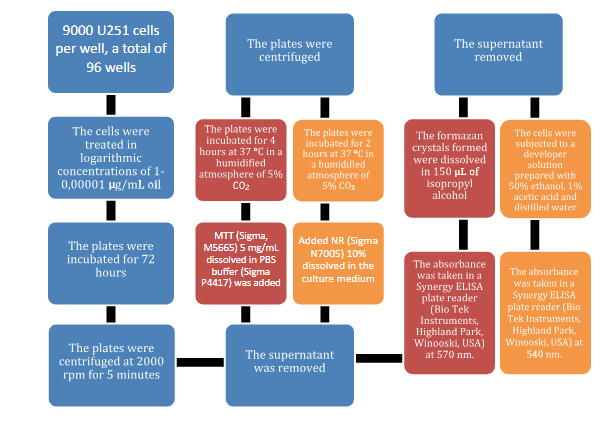
Protocol [24]. The U251 cells (9×10⁵ cells/well) were distributed in 96-well plates. The cell lines were treated with copaiba oil using 10-5 μg/mL concentrations. The cells were incubated for 72 hours with copaiba oil, then the plates were centrifuged at 2000 rpm for 5 minutes. The supernatant was removed, and MTT (Sigma, M5665) 5 mg/mL dissolved in PBS buffer (Sigma P4417) was added. The plates were incubated for 4 hours at 37 °C in a humidified atmosphere of 5% CO2. The plates were centrifuged again after this period, the supernatant was removed, and the formazan crystals were dissolved in 150 μL of isopropyl alcohol. The absorbance values were obtained using a Synergy ELISA plate reader (Bio Tek Instruments, Highland Park, Winooski, USA) at 570 nm. We expected 100% cell viability in the negative control.
Neutral Red
Protocol [25]. The U251 cells were distributed in 96-well plates as described on the MTT protocol. The cells were incubated for 72 hours with copaiba oil, the plates were also centrifuged at 2000 rpm for 5 minutes. The supernatant was removed, and the 10% NR (Sigma, N7005) dissolved in culture medium was added. The plates were incubated for 2 hours at 37 °C in a humidified atmosphere of 5% CO2. After this procedure, the plates were centrifuged again, the supernatant was removed, and the cells were subjected to a developing solution prepared with 50% ethanol, 1% acetic acid and distilled water. The absorbance values were obtained using a Synergy ELISA plate reader (Bio Tek Instruments, Highland Park, Winooski, USA) at 540 nm. We expected 100% cell viability in the negative control.
Figure 1 shows both assays described above for a better understanding:
Results
Chemical analysis
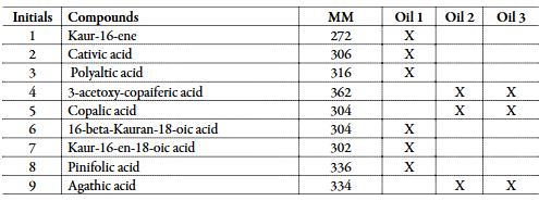
The ESI-MS spectra below present the chemical fingerprint of all 3 oils in the negative mode (Figure 2).
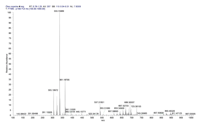
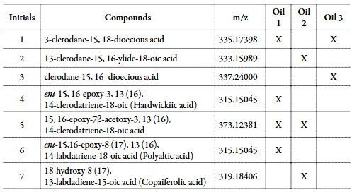
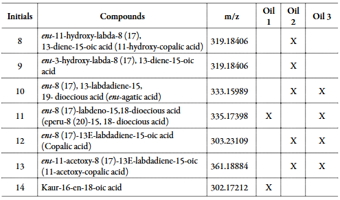
Our analysis of the spectra was based on a review study and tables 1 and 2 show the compounds identified in the oils through ESI-MS and GC-MS methods [21]:
Cytotoxicity and Cell Viability
The mean and standard deviation were calculated with the values found in the experiment, and from them we obtained the cell viability graphic and the IC50 value, which represents the required concentration of each oil to cause the death of 50% of the treated cells, as shown in the Figures 3 and 4 and Table 3.
MTT
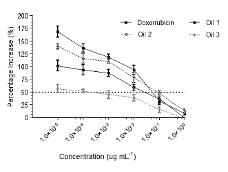
Figure 3 Graph of cell viability after 72 hours of treatment using Copaifera spp oil in a human glioma cell line (U251) for the MTT assay.
Neutral Red
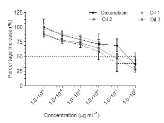
Figure 4 Graph of cell viability after 72 hours of treatment using Copaifera spp oil in a human glioma cell line (U251) for the NR test.

Correlation between MTT and Neutral Red Test
There was a significant correlation between the MMT and Neutral Red assays (Figure 5). We used the Pearson correlation coefficient to establish the correlation (Table 4).
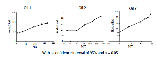

With a confidence interval of 95% and α = 0.05
The results show α = 0.05 and r values greater than 0.9, which indicates a strong correlation between both methods used. Despite being used to assess the viability/cytotoxicity of cells, each method we used needs different processes to measure this characteristic.
Photomicrographs
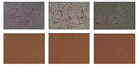
The photomicrographs below show the effects of all oils on the U251 cells at a concentration of 10-1 μg.mL-1 after 72 hours of treatment (Figure 6).
We can observe that the treated cells suffered intense nuclear pleomorphism, show little cohesiveness, nuclear pyknosis, and sometimes scant cytoplasm, dense chromatin, and nuclear atrophy. Cell death can also be observed in photomicrographs.
Discussion
We found a total of 23 compounds through the chromatography methods used, 14 through ESI-MS and 9 through GC-MS. 3 compounds are common to both techniques, Polylactic acid, Copalic acid, and kaur-16-en-18-oic acid. Gas chromatography identifies gases of lower molar mass, the sesquiterpenes, while ESI-MS identifies those of greater molar mass, the diterpenes. The number of compounds found in Oil 1 was 10, also 10 in Oil 2 and 8 in Oil 3.
The MTT assay consists of reducing yellow-colored tetrazolium salt (3-(4,5-dimethylthiazol-2-yl)-2,5-diphenyltetrazolium bromide — Sigma M2128) through the succinic dehydrogenase of the enzyme present in the mitochondria of the tumor cell, which acquires a purplish color, is measured spectrophotometrically at 570 nm [25].
The Neutral Red test is based on the ability of viable cells to incorporate the dye in the lysosomes; thus, the red staining is evaluated by spectrophotometry at 540 nm.
Oil 3 presented the lowest IC50 value in the MTT assay, which was lower than the positive control, followed by Oil 1 and Oil 2, however, both presented greater values than doxorubicin.
For the Neutral Red test, all 3 oils presented IC50 values lower than the positive control, Oil 2 presented the best value.
Conclusion
This study allowed us to observe some characteristics of copaiba oil and some of its compounds and its effects on tumor cells. Despite having different compositions, all 3 oils affected the cell line U251 positively. The results from the cell viability tests show slight differences regarding the IC50 values of Neutral Red versus MTT assays for the 3 types of tested oils, which suggests that the oil interferes at the concentrations studied with cellular metabolic activity and integrity of the liposome membrane.

