


 text in
  English (pdf)
 Article in xml format
 Article references
 Send this article by e-mail
 Cited by SciELO
  Cited by Google
  Similars in
SciELO
Similars in
SciELO  Similars in Google
 Permalink
INTRODUCTION
Placenta accreta is the abnormal insertion of the placenta to the uterine wall beyond the fibrin layer or Nitabuch layer1, secondary to total or partial decidual absence2, with trophoblast villi penetration into the myometrium and even into the serosa or adjacent organs3,4. Three variants of placenta accreta have been proposed according to the degree of trophoblast invasion: accreta, increta and percreta5. Placenta accreta impairs or hinders normal placental detachment6,7, constituting a relevant problem, because although it occurs in 0.1-0.3% of pregnancies2, up to 80% of patients develop massive bleeding due to abnormal placental detachment8, creating a considerable risk of maternal morbidity and mortality9.
Women with uterine scars due to surgical procedures such as cesarean section (c-section) or myomectomy have a higher risk of placenta accreta, and this explains why the incidence has increased ten-fold over the past three decades, clearly in association with the growing number of cesarean sections10-13. Another risk factor is placenta praevia or low-insertion placenta (placental tissue that extends over or close to the internal os), which may occur in up to 9.3% of pregnancies3, 1-3% of which are accompanied by placenta accreta14,15.
Given the threat posed by placenta accreta, the best approach is prenatal diagnosis3,10-12,15 in order to ensure timely referral to centres with the human and technical resources for adequate delivery care and response to potential complications such as massive bleeding, thus reducing the risk of maternal death9,16-20. In patients with risk factors, it is advisable to assess placental implantation after 12 weeks in the office using 2D ultrasound and Doppler or nuclear magnetic resonance in order to look for signs that may suggest placenta accreta21,22. Magnetic resonance imaging (MRI) is used to assess posterior placenta accreta or when invasion of adjacent organs is suspected. However, it has high cost, not readily available, and some patients may feel claustrophobic23,24. Moreover, no significant improvement in outcomes has been found with the use of this imaging modality in placenta accreta pregnancies25.
The sensitivity of 2D ultrasound described in the literature ranges between 86% and 100%, while specificity is around 66% to 98%26-29.
These variations may be partly explained by the population studied, by the fact that it is operator-dependent30 and by the ultrasonographic criteria used. In Colombia, there are three reports related to ultrasound diagnosis of placenta accreta, all of them with limitations in terms of patient number and selection and the type of information provided31-33. On the other hand, there are reports that describe good accuracy of Doppler ultrasound in placenta accreta34.
Given the variability of accuracy indicators of 2D ultrasound for the diagnosis of placenta accreta, the potential usefulness of adding Doppler to the prenatal study, and the scarcity of research conducted in this country, the objective of this study was to assess the diagnostic performance of combining obstetric 2D ultrasound plus Doppler for the diagnosis of this condition in high-risk women in a Level III perinatal care centre in the city of Bucaramanga, Colombia.
MATERIALS AND METHODS
Design and population. Diagnostic test accuracy assessment study in pregnant women seen for prenatal care and who ended their pregnancies between 2014 and 2016 at Hospital Universitario de Santander (Santander University Hospital - HUS). This institution is the referral centre for the public hospital network in the northeastern region of Colombia and provides services mainly to low-income patients affiliated to the contributive insurance regime of the Colombian social security system. All pregnant women over 18 years of age receiving care during the three years of the study and who met the following criteria were included: a) 12 weeks or more of gestational age; and b) a positive history of uterine surgery (cesarean section, myomectomy or curettage) or placenta in anterior location, low or praevia (total or partial). Patients in whom ultrasound imaging could not be performed were excluded. Convenience sampling was used. The goal was to include all the pregnant women who met the inclusion criteria.
Procedure. Candidates for entering the study were identified by any of the six specialists in maternal-foetal medicine working at the Santander University Hospital when called in for assessment by the obstetric emergency service or when patients were referred from another institution to the perinatology clinic. When candidates were identified and met the inclusion criteria, they were asked to sign a written informed consent. Baseline data were captured in a Microsoft Excel® data sheet administered by the first author. One of the six specialists in maternal-foetal medicine performed the transabdominal and transvaginal 2D ultrasound on a full bladder together with trans-abdominal Doppler using a General Electric Voluson E8® ultrasound machine, looking for the following signs of placenta accreta on 2D ultrasound: thinning of the uterine wall < 1 mm, absence of retroplacental space, disruption of the hyperechoic line between the uterine serosa and the bladder, abnormal placental venous lakes, placental bulging, and focal exophytic masses. Doppler determinations included the presence of diffuse lacunar flow throughout the placenta extending to the myometrium, turbulent lacunar flow with peak velocities > 15 cm/sec, hypervascularity of the placenta-bladder interface, and presence of subplacental venous complexes2,4,21,22. Patients were defined as having a high probability of placenta accreta depending on whether imaging showed at least two criteria on 2D ultrasound and one more on Doppler35, in accordance with the recommendations by Finberg and Williams(4) and Chou et al.(21) Diagnosis was made by one of the authors (LAGS) based on the stored images, before the end of pregnancy.
All patients in whom image analysis showed a high probability of placenta accreta were explained what their clinical condition meant, the need for cesarean delivery, and the risk of needing a hysterectomy if no placental separation was achieved, reason why the procedure was performed by an obstetrician assisted by a specialist in oncogynaecology in the event placental separation proved impossible. Patients with a low suspicion of placenta accreta were allowed to go into trial labour, and some of them ended up requiring cesarean section due to other obstetric reasons. Specialists in the gynaecology and obstetric emergency service provided care to patients who had a vaginal delivery, following the institutional protocols in cases of placental retention.
Measured variables. The following variables were described: maternal age, gestational age, timing of ultrasound assessment, history of uterine surgery and presence of placenta praevia, high and low probability of placenta accreta, type of imaging suggesting placenta accreta, route of delivery, ease of placental detachment, visual or clinical findings of placenta accreta. The diagnosis of placenta accrete was established using a complex gold standard. The criterion used in patients who underwent vaginal delivery was lack of placental detachment after 30 minutes of delivery, controlled cord traction and active use of uterotonics. Negative diagnosis after vaginal delivery was applied in patients in whom the absence of placental detachment was not due to cervical contraction or structural uterine abnormalities and with complete placenta on examination after birth, and the absence of abnormal vaginal bleeding before discharge4,6,7,36,37. Diagnosis in patients taken to cesarean section was made on the basis of in situ evidence of placenta accreta, defined by the attending expert obstetrician-gynaecologist, and in patients taken to hysterectomy during the cesarean intervention or in the immediate postpartum period in accordance with the clinical criteria of placenta accreta. The surgical specimen was studied by the pathology service of the Santander University Hospital.
Statistical analysis. Sensitivity, specificity, positive and negative predictive values, positive and negative likelihood ratios (LR) and 95% confidence intervals (CI) were estimated when comparing prenatal ultrasound diagnosis plus Doppler rated as high or low probability of placenta accreta with the diagnosis obtained using the gold standard.
Ethical considerations. The research protocol was approved by the Ethics and Scientific Research Committee of Universidad Industrial de Santander and the Santander University Hospital. All the patients gave their written informed consent to participate in the research. The information obtained was stored electronically and adequate confidentiality was preserved making patients anonymous at the time of database analysis by each individual author.
RESULTS
Overall, 73 patients who met the inclusion criteria were identified. They had all been referred to HUS due to different obstetric reasons, but only three due to suspected placenta accreta. Of them, 22 patients (30%) were excluded because of the inability of the maternal-foetal specialists to perform imaging studies for administrative reasons. Consequently, 51 patients were included.
The median age of the patients studied was 29 years (inter-quartile range [IQR] 15-35). Median gestational age at the time of ultrasound assessment was 36.3 weeks (IQR 34.1 - 38.0 weeks); 46 (90.2%) patients were assessed during the third trimester of gestation. The surgical risk for placenta accrete found most frequently was a prior cesarean section, followed by curettage and myomectomy (Table 1).
Table 1 Risk history and findings on 2D ultrasound and Doppler suggesting placenta accreta in women at a high risk for this condition in Bucaramanga, Colombia, 2014-2016
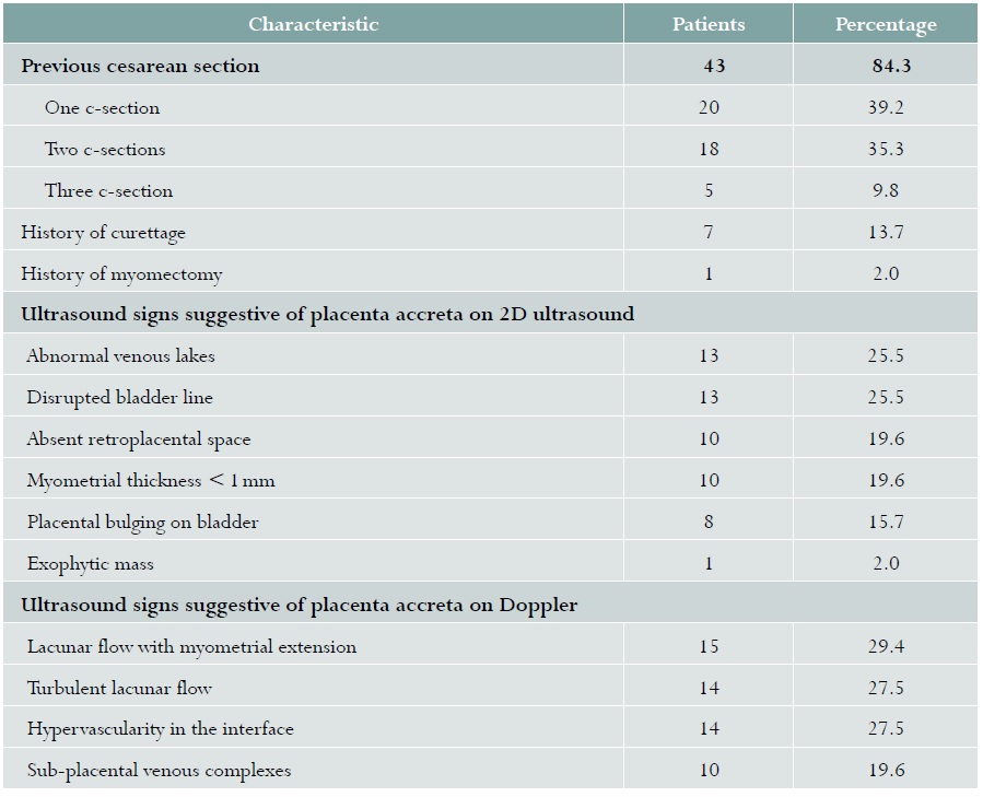
Note: Given a patient could have more than one ultrasound and Doppler image, the per cent sum may be greater than 100%.
Of 51 patients, 21 (41.2%) had anterior implantation placenta and 30 (58.8%) low implantation placenta (11 [36.7%] without occlusion of the internal os, 66 [20.0%] with partial occlusion, and 13 [43.3%] with total occlusion).
A total of 16 (31.4%) patients had a high probability of placenta accreta according to the imaging studies, and 35 (68.6%) had a low probability. The most frequent signs on 2D ultrasound were interruption of the hyperechoic line between the uterine serosa and the bladder and the presence of abnormal placental lakes, while on Doppler the most frequent was the presence of turbulent sonolucent vascular lakes (Table 1). High imaging probability of placenta accreta was declared in one patient assessed during the first trimester of pregnancy, in two of the four who were assessed during the second trimester, and in 13 of the remaining 46 who were assessed during the third trimester.
Pregnancies ended in cesarean delivery in 35 (68.6%) patients: in 16 with the high imaging probability of placenta accreta, and in 19 in the low-risk group due to different obstetric reasons.
Hysterectomy was performed in the 16 patients with the high probability of placenta accreta, due to placental detachment impossibility as a result of placenta accreta documented in situ by the gynaecology specialist. On the other hand, one of the 16 patients who had a vaginal delivery had difficulty with placental detachment, leading to hysterectomy during the management of the retained placenta; in situ assessment of the surgical specimen confirmed the diagnosis of focal placenta accreta.
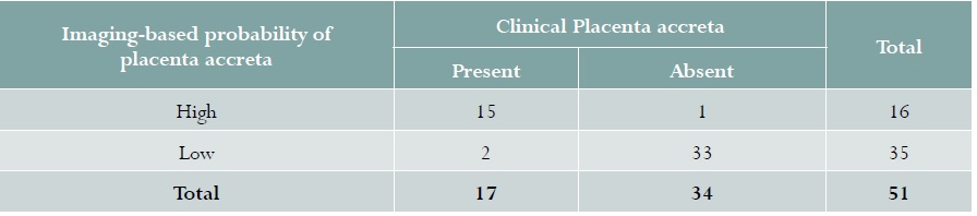
Therefore, clinical placenta accreta was found in 17 patients, representing 33.3% of the study population. Of them, 15 had been declared before delivery based on high imaging probability, while one of the remaining 34 who did not present clinical signs of placenta accreta had been declared as having a high probability (Table 2). Two of the patients with low probability had placenta accreta. The sensitivity and specificity of 2D ultrasound plus Doppler for the diagnosis of a high probability of placenta accreta were 88.2 % (95% CI 70.0-100) and 97.1% (95% CI 89.9-100), respectively, with a positive LR of 30.0 (95% CI 4.3-208.5) and a negative LR of 0.12 (95% CI 0.03-0.45).
DISCUSSION
This study shows that the high-probability rating for the presence of one criterion in 2D ultrasound and an additional criterion on Doppler in pregnant women at a high risk of placenta accreta had a sensitivity of 88.2% and a specificity of 97.1%, with positive and negative likelihood ratios of 30.0 and 0.12, respectively. These figures are similar to those reported in 2013 by D’Antonio et al. in their meta-analysis35, where ultrasound had a sensitivity of 90.7% (95% CI: 87.2-93.6), a specificity of 96.9% (95% CI: 96.3-97.5), a positive LR of 11.01 (95% CI: 6.1-20.0) and a negative LR of 0.16 (95% CI: 0.11-0.23) for the detection of placenta accreta before delivery. However, this meta-analysis is not the best benchmark for our results because among the many reasons that make comparison difficult is the fact that it included only publications in English that used the four ultrasound criteria considered as the most frequently associated with the presence of invasive placenta. Moreover, Doppler was not used in the study which contributed 54% of the patients assessed27.
When comparing our study with international research, we observe that Wong et al.28 report a sensitivity of 89%, specificity of 98% with a positive LR of 44.5 and a negative LR of 0.11, in a retrospective study, while ours was a prospective design; however, placental ultrasound and Doppler were assessed. Calì et al.26 found a 90% sensitivity and 100% specificity with diagnostic criteria similar to the ones used in our study. Comstock et al.27 in their 2004 report used ultrasound as the only diagnostic test, but their results are very different from ours given that Doppler was not used as a diagnostic test and the equipment was also different from ours, considering a time period of more than 10 years between the two studies. The studies mentioned above26-30 were carried out during the second and the third trimesters. In this study we assessed 1 case as a high probability of placenta accreta in the first trimester of pregnancy, and 2 out 4 cases in the second trimester, which were then correlated with the clinical findings at the time of birth; however, the majority of our cases were assessed during the third trimester.
In Colombia, three studies have been described, all of them prospective31-33 and conducted by highly qualified researchers with experience in the diagnosis of placenta accreta. However, Parra et al. used placental Doppler and magnetic imaging and reported a sensitivity of 90% with a limited number of patients and a high selection bias. Studies like those of Ferreira et al.(31) and Vargas et al.33 have a similar design to ours but the frequency with which the condition occurred was low, preventing estimation of positive or negative likelihood ratios.
Prospective patient follow-up, state-of-the art assessment technology and highly trained personnel in maternal-foetal medicine are strengths of this study.
Weaknesses include not having used histopathology as the gold standard method for diagnosis in all patients38. However, performing it in patients who do not undergo hysterectomy is not feasible, making clinical diagnosis an option in cases in which the uterus is not excised39. On the other hand, there is a risk of differential verification bias, considering that the gold standard in patients undergoing c-section was different from the one used in patients who had a vaginal delivery.
An attempt was made to control this bias using a clinical definition of placenta accreta at the time of birth and during the postpartum period based on manifestations such as placental retention during birth, bleeding during hospitalisation in patients with vaginal delivery or abundant bleeding in the implantation site following placenta removal during the cesarean section40. On the other hand, the inability to carry out studies in 30% of candidate women increases the risk of selection bias, considering that it was not possible to determine whether these patients had focal placenta accreta that might affect the sensitivity or specificity of 2D ultrasound and placental Doppler.

