Introduction
Various psychological and neuropsychological studies have contributed to the detailed description of the clinical picture presented by children with attention deficit hyperactivity disorder (ADHD), not only regarding the effect of the alteration 1-5, but also, other psychological processes that may be involved 6-8. These findings do not necessarily establish a relationship between what is observed at clinical behavioral level, and the functional or developmental stage of the central nervous system.
Some authors have tried to correlate the clinical picture of ADHD with possible alterations in the central nervous system structures 9-10; most imaging studies such as positron emission tomography (PET) and functional magnetic resonance coincide in pointing out that subjects suffering from this syndrome reflect an underlying deficit in the frontal lobes and the right hemisphere 11,12. However, these studies have focused on studying the central nervous system without considering the cognitive and behavioral aspects of children with attention deficit disorder.
It should be noted that the ADHD syndrome is diganosed with the help of the questions listed in the DSM-IV TR 13 and DSM-V manuals 14, and after applying the questionnaire, the behavioral level and the structural or functional aspects of the central nervous system are individually assessed. Although neuropsychological, neurological and psychological evaluation data have been analyzed 15,16, the findings have not been correlated with each other, with particular neuropsychological difficulties nor with specific electroencephalographic data to establish typical patterns of brain electrical activity in children with ADHD at various stages in preschool and elementary school children.
Previous studies have analyzed the execution of neuropsychological evaluation tasks through EEG data in Mexican preschool children diagnosed with ADHD by neuropediatrics 17,18. These studies showed the typical difficulties from the neuropsychological viewpoint, whose origin cannot be interpreted only as frontal or cortical. Among the encountered difficulties, not only severe problems with regulation, control and motor sequential organization were found, but also with spatial analysis and synthesis, therefore, rejecting the hypothesis of a single alteration of predominantly cortical frontal systems 19-21.
In subsequent studies with preschool children diagnosed with ADHD, an insufficient level of non-specific general cerebral activation was established, which was corroborated with data obtained from EEG 17. In this work, the visual qualitative analysis of the examination revealed a dysfunctional state in the deep structures of the brain stem based on the appearance of particular patterns of electrical brain activity; these patterns are related to the outbreaks of synchronized waves in the central and posterior regions of both hemispheres 17,22.
After analyzing a broader preschool population of Mexican and Russian children, it was observed that in those diagnosed with ADHD using the DSM-IV TR 13 questionnaire, no pattern correlated with the immature functional status of the cerebral cortex was identified in the EEG 18,23-25; however, the presence of unfavorable EEG patterns was observed at the subcortical level, specifically at the upper brain stem (fronto-thalamic) and lower brain stem (diencephalic) in all cases evaluated. 16 to 50 preschool children diagnosed with ADHD were evaluated in this study.
In the case of the fronto-thalamic level, patterns synchronized as slow waves are recorded in the anterior sectors of both hemispheres, whereas in the diencephalic case, slow sharp waves are recorded in the caudal sectors simultaneously in both hemispheres. In both cases the record was taken using monopolar assembly, which is not usual for the commonly used visual analysis 22,26,27. On the contrary, these two patterns have been presented separately in the children's control group -those without developmental and learning problems- of the same age. The results of this study allowed rethinking the functional role of cortical-frontal structures in the ADHD syndrome in preschool children, confirming the involvement of subcortical regulatory systems at fronto-thalamic and diencephalic levels with higher probabilities than those presented at the frontal cortex level.
Studies involving the population of children in the first three grades of elementary school showed that, unlike preschool age, there is no single profile of neuropsychological difficulties and functional status of cortical and subcortical brain structures 28.
This work aims to continue the line of research that links EEG patterns with neuropsychological performance to correlate the data obtained through neuropsychological assessment with EEG records in Mexican children attending the last three years of elementary school and who were diagnosed with ADHD.
Materials and methods
Subjects
14 right-handed children (nine boys and five girls), diagnosed with ADHD and 10 children with no learning disability (five boys and five girls) were included. All children were regular students in urban public elementary schools in Puebla, Mexico, aged between 9 and 12, and were enrolled in fourth, fifth and sixth grades. The ADHD diagnosis was established and confirmed in writing by a specialist -paidopsychiatrist or neurologist- from outside the research team. Children in the control group showed no signs of hyperactivity, neurological or psychiatric diseases or psycho-pedagogical problems.
All children participated in accordance with the Declaration of Helsinki, with understanding and informed consent of each parent and school teachers; this study was also approved by the local ethics committee.
Procedure
All children were administered the qualitative neuropsychological assessment in a single 60-minute session outside school hours within its facilities. Then each child underwent EEG recording while awake at the clinical headquarters of Universidad Autónoma de Puebla. The results of both assessments were analyzed and contrasted with the aim of establishing the agreement from the neuropsychological and electroencephalographic point of view, in order to indicate the state of maturity and functionality of cortical and subcortical brain structures.
Neuropsychological assessment
Children Brief Neuropsychological Assessment 29 was used for neuropsychological assessment; this instrument qualitatively assessed the functional brain systems based on psychological and neuropsychological theories by Vygotsky 30 and Luria 31,32, and also assessed neuropsychological factors such as functional brain mechanisms 33. Particularly, the functional status of regulation and control, sequential motor organization, phonemic integration, kinesthetic integration, spatial integration and overall activation tone of the brain (arousal) were analyzed for the visual, motor and verbal tasks.
The analysis of the results is based on a qualitative assessment by experts of the presence of specific types of errors or the lack of them, which point the weak functional state of cortical and subcortical brain mechanisms 34-37.
Electroencephalogram
Participants were recorded with NicView System (Nicolet Biomedical Inc.) EEG equipment, with sampling rates of 250 Hz, according to the international 10-20 system in the following cortical regions: F3, F4, F7, F8, T3, T5, T4, T6, P3, P4, O1 and O2. The registration was carried out under wakefulness conditions performing the following operations: a) opening and closing the eyes, b) photostimulation (8-12 Hz) and c) hyperventilation (90 sec).
Monopolar and bipolar montages were used for each record. The monopolar montage allowed assessing the functionality status of different levels of the brain stem through the identification of synchronous deflected patterns that can be seen in the visual analysis; these patterns, depending on their location, expressiveness and representation, indicate the involvement of various subcortical levels. Bipolar montage allowed assessing the functionality state of various caudal areas of the central nervous system 22,27.
EEG visual qualitative analysis
The assessment of the functional status and degree of brain maturation was performed through EEG visual qualitative analysis, and was based on the correlation of the lines identified in the EEG record with the functional status of the brain and, in particular, with its structures 38. The qualitative analysis of EEG was performed by assessing four blocks of parameters identified based on the systemic organization of the brain 22,29; these parameters are used to assess the overall functional state of the cerebral cortex and its correspondence with age norms, general brain effects, functional status of cortical areas particularly related to each hemisphere, and subcortical regulatory systems status 18,22,27. The same qualitative analysis allows to differentiate artifacts from the particular patterns of EEG.
The visual clinical analysis was established by two experts on the subject and those patterns corresponding to the above parameters were selected. In each subject, an average of 20 minutes of electroencephalogram records were analyzed to obtain a homogeneous analysis of each study. In addition, the Kendall correlation coefficient test was used to assess the level of agreement among experts in the visual analysis of the electroencephalogram.
It is important to remember that these types of analysis are necessary to obtain a quantitative average of the opinions or prejudices about a phenomenon or fact.
Results
Neuropsychological assessment data analysis allowed establishing the particular characteristics of the performance of control children and children with ADHD. Similarly, observing the differences in the performance of the assessment tasks by children was possible based on the presence and absence of particular errors.
Children had no difficulties in phonemic and tactile kinesthetic value integration tests, so no differences were found. By contrast, differences were observed in 1) tasks that assess the regulation and control of the activity; 2) the motor sequential organization and dynamic praxis and 3) the spatial analysis and synthesis. In all these cases, errors were committed by children diagnosed with ADHD (Figure 1).
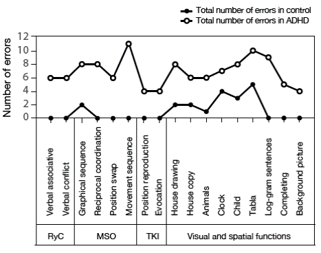
Figure 1 Number of errors for each task in the protocol. R&C: regulation and control; MSO: Motor sequential organization; ICT: tactile kinesthetic integration. Source: Own elaboration based on the data obtained in the study.
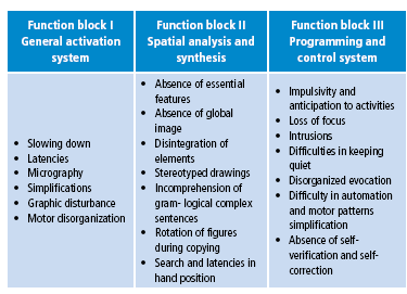
To specify the type of commitment and to systematize the sample, a qualitative analysis of the types of errors under the concept of the three brain function blocks proposed by Luria (33) was performed. Syndromic analysis set specific profiles for each subject reflecting the functional status of the different brain mechanisms (Table 1).
Table 1 Types of errors observed in children with attention deficit hyperactive disorder related to brain function blocks.
Source: Own elaboration based on the data obtained in the study.
The qualitative analysis based on the systematization of error types (Table 1) allowed finding three predominant clinical profiles: 1) regulation and control difficulties (11 cases), 2) lack of activation in the cortical tone work (2 cases), and 3) severe difficulties in spatial functions (1 case). The patterns identified along with the characterization of EEG data according to the three established profiles are presented below.
Profile 1. Functional regulation and control deficit
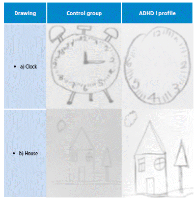
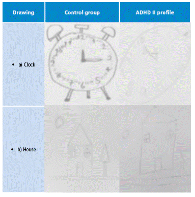
Children with this profile presented impulsivity errors, rotation of elements, perseverance and lack of planning, which are not observed in children of the control group. Table 2 shows the graphic performance while drawing a clock and a copy of a house made by profile children and by children of the control group.
Table 2 Examples of graphic performance: one children when drawing a clock and copying a house.
Source: Own elaboration based on the data obtained in the study.
Simplifications, impulsivity and loss of focus were observed in associative conflict and sequential organization motor tests in dynamic praxis in hands and fingers.
Regarding EEG qualitative visual analysis, in this group of subjects two variants were found: one pointing unfavorable functional status of the superior brain stem, mainly at the fronto-thalamic level (eight children), which is reflected as slow synchronized waves in the frontal records that appear in both hemispheres, and another which presents features of the diencephalic level, which is characterized by bilateral synchronized patterns in the form of slow waves in subsequent records (three children).
An example of the diencephalic and fronto-thalamic levels is shown in Figure 2. EEG patterns were bilateral and synchronous, allowing the verification of their subcortical origin, according to the visual clinical model of interpretation.
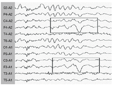
Figure 2 Functional changes in brain electrical activity in the form of synchronous and bilateral waves on the theta band in front and central sectors (monopolar records) groups. Source: Own elaboration based on the data obtained in the study.
Through the Kendall correlation coefficient, coincidence among experts in the analysis of EEG was determined. The test showed a level of agreement between subjects of 0.974 (p<0.05).
Profile 2. Difficulties in visual and spatial functions
Children with this profile presented errors such as lack of proportion and integration in the task of copying a clock and a house (Table 3).
Table 3 Examples of graphic performance when drawing a clock and copying a house corresponding to profile 2.
Source: Own elaboration based on the data obtained in the study.
The visual qualitative analysis of EEG of this child shows deviant patterns of electrical activity in caudal sectors -occipital and parietal- (Figure 3). The data is consistent with results of neuropsychological assessment, which allows confirming expressive difficulties in spatial functions.
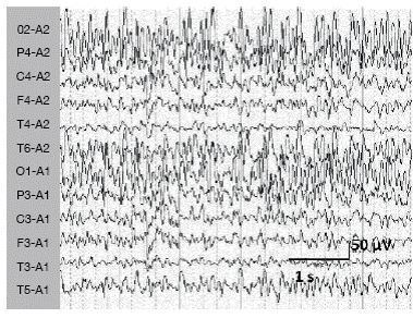
Figure 3 Synchronic and bilateral functional changes in the form of hypersyn-chronic alpha rhythm or groups of sharp waves in the caudal sectors (monopolar record). Source: Own elaboration based on the data obtained in the study.
The EEG visual qualitative analysis of this child shows deviant patterns of deep origin in the brain stem, including paroxysmal discharges in parietal cortical areas.
Through Kendall correlation coefficient, coincidence among experts in the analysis of EEG was determined. The test showed a level of agreement between subjects of 0.861 (p<0.06).
Profile 3. Lack of activation in the cortical tone work
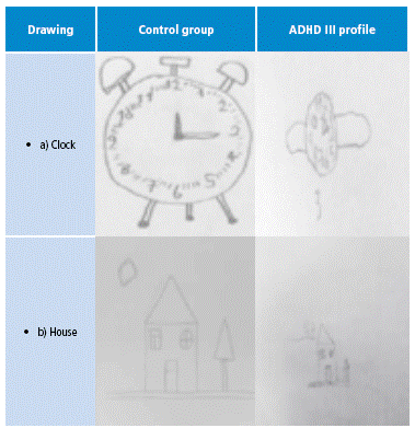
For this group of children, all graphic tasks show traits of instability demonstrated in specific errors such as micro and macro letters, omissions, slowing, problems with tilt, spaces between the perceptual elements and loss of baseline (Table 4).
Table 4 Examples of graphic performance when drawing a clock and copying a house corresponding to profile 3.
Source: Own elaboration based on the data obtained in the study.
Regarding non -graphical tasks, slight kinesthetic integration difficulties, traits of impulsivity, slowing and praxical motor inaccuracies, low capacity to perform all tasks of evaluation, requests of breaks and support by adults were observed (Figure 4).
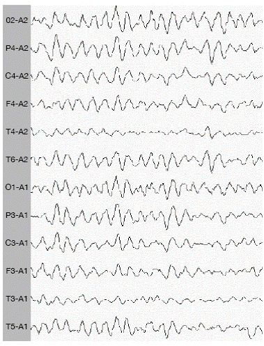
Figure 4 Functional changes in synchronous and general activity in the form of slow alpha of hypothalamic origin. Source: Own elaboration based on the data obtained in the study.
The visual qualitative analysis of EEG identified a non-optimal functional status in the lower brain stem. The same functional status of EEG was observed in three profile 1 children with prevalence of difficulties in regulation and control.
Through Kendall correlation coefficient, coincidence among experts in the analysis of EEG was determined. The test showed a level of agreement between subjects of 0.885 (p<0.05).
Discussion
Several disciplines have been involved in the study of attention deficit disorder in preschool and school- age children since children with this condition are the main source of clinical diagnosis of these disorders. Questionnaires such as DSM-IV TR and the DSM V, where the individual is categorized within a large list of possibilities through a series of questions aimed at identifying behavioral traits, are widely used evaluation tools.
In many cases, children can only be classified into diseases such as ADHD or combined diseases when the focus is solely on observable symptoms or those that are noticed by a primary caregiver. Thus, the child is no longer perceived as an individual with a unique set of difficulties and problems 37. At the same time, the presence or absence of these behavioral symptoms does not establish a clear relationship with the level or type of cortical or subcortical brain structures.
In some previous studies, identifying different characteristics of inadequate functional status of various neuropsychological mechanisms, including regulation and control, motor sequential organization 20, spatial analysis and synthesis 19,20 and cortical tone regulation, was possible 18,21. These studies have found that preschool children are often diagnosed with attention deficit in the presence of all these problems simultaneously, which implies that in children who are diagnosed with attention deficit in preschool, specifically between ages 5 and 6, it is common to see difficulties with regulation and control, lack of integration and spatial orientation, and constant fluctuations in the general state of activation.
In contrast, this work has found that the difficulties associated with different brain mechanisms may be presented separately in children of school age, in particular in the three final grades of the elementary school. As noted by the results, the data allow appreciating at least three different neuropsychological profiles, which were not observed in the case of preschool children with the same diagnosis.
In cases of EEG visual data qualitative analysis, patterns different from those observed in preschoolers were found. It is worth mentioning that neuropsychological difficulties profiles may be clinically related to electroencephalographic profiles.
Previous studies with preschool children have shown that those diagnosed with attention deficit and difficulties characterized by functional deficit in regulation and control, spatial integration and insufficient activation of arousal brain activation -based on a visually qualitative analysis- present patterns of immaturity in the fronto-thalamic regulatory system -superior stem- and dysfunctionality in non-specific regulation related to ascending effects from the reticular formation at diencephalic level 17,18,30. These results were obtained in various populations of Mexican and Russian children 17,18,22,27. In this paper, the visual qualitative analysis of the electroencephalogram allowed to establish three types of electroencephalographic patterns.
In the cases with difficulties in the regulation and control mechanism, EEG analysis revealed diverted bioelectric patterns from fronto-thalamic source that signal the state of immaturity of the frontal cortex and the different thalamic nuclei 25,27,29,39. Such patterns are expressed as theta-synchronized slow waves in the anterior sectors. There is no data in the literature in favor of the non-optimal participation state of this regulatory system in groups of children between 6 and 8 years old diagnosed with ADHD 29,30,40. Neuropsychological assessment in these children reveals typical errors of simplification, impulsiveness, loss of focus and inability to perform tests with dynamic praxis; a correlation between standard errors is also evident in neuropsychological tests in the presence of immature states at anterior brain stem structures level and, at the same time, it was established that the level of the cerebral cortex corresponds to the age norm in all these cases.
As to the cases of alterations in general activation of brain work, the analysis showed changes in the electrical activity originated in the brain stem and the midbrain. In other studies, the authors noted that the presence of bilateral slow waves in the caudal sectors is related to the low level of cortical excitability 41 and correlates with the low level of activation from the lower stem and the reticular formation 22,27,42. These patterns of brain electrical activity were found in children with neuropsychological profile 2; based on the neuropsychological evaluation, these children require constant support, are exhausted before performing tasks and demonstrate instability traits in all graphic and motor tasks.
In cases of regulation and control difficulty and non -optimal status of tone activation of brain work, a positive overall maturity level of the brain was observed. Deviant electroencephalographic patterns are related to the weak state of subcortical operation, which was demonstrated in the synchronized nature of deviant patterns.
Finally, for the only case of this sample with severe difficulties with space obtained in the neuropsychological evaluation, EEG analysis revealed an unfavorable functional state in the parieto-occipital cortical structures along with a state of immaturity of the lower brain stem. Classical neuropsychological literature exposes that spatial difficulties are related to functional deficits in posterior cerebral areas 16,28, so it is interesting to note that patterns of synchronized high amplitude sharp waves are recorded in the parieto-occipital sectors.
Thus, the combination of syndromic neuropsychological qualitative analysis with qualitative visual analysis of EEG allowed establishing a relationship between the functional status of neuropsychological mechanisms with the non-optimal functional status of the various subcortical levels. Such interdisciplinary combination of neuropsychological assessment, accompanied by EEG visual qualitative analysis showed a high level of correspondence between the level of maturity or functional state of different cortico-subcortical structures with clinical manifestations observed.
The data from this research does not indicate that these variants may be the only ones that can be identified. Thus, some studies have found cases suggesting the involvement of the right hemisphere and other combinations of difficulties 30,43,44.
One limitation of this study is undoubtedly the small sample size, which is related to the need for having a homogeneous population of children in the last three grades of regular elementary school who have the same diagnosis issued by a third specialist, and the possibility to participate in the study of neuropsychological evaluation and EEG recording.
Despite this limitation, this study found that school-age children who are diagnosed with ADHD are not a homogenous group and are qualitatively different. Both neuropsychological and electroencephalographic analyzes can set several variants. These data cast doubt on the clinical efficacy of a single diagnosis with a generalizing name such as attention deficit, as it becomes clear that children cannot be differentiated from traditional questionnaires answered by caregivers in relation to purely behavioral traits. These diagnostics have the potential to clarify the relationship between different levels of analysis which lack adequate predictive level.
Conclusions
The qualitative neuropsychological assessment allows addressing various clinical pictures that reject the idea of a single diagnosis of attention deficit disorder.
Qualitative visual analysis of EEG confirms the absence of a single clinical picture, so it is possible to identify various patterns diverted through visual qualitative analysis of the electroencephalogram.
The search for correspondence between the electrophysiological correlation of EEG and neuropsychological assessment can become an important means for the accuracy of functional mechanisms underlying the difficulties of learning and development.


