


 English (pdf)
 Article in xml format
 Article references
 Send this article by e-mail
 Cited by SciELO
  Cited by Google
  Similars in
SciELO
Similars in
SciELO  Similars in Google
 Permalink
Introduction
Esophageal cancer is one of the worst prognosis neoplasms: it is the eighth most common and holds the sixth place as a cause of death worldwide among other types of cancer 1. Its incidence varies; on the one hand, it is higher in Asian countries where rates range from 50 to 130 cases per 100 000 inhabitants 2 and, on the other hand, in countries like Colombia, the rate is much lower according to Globocan 3, since it is 1.2 cases per 100 000 inhabitants, with a mortality rate of 1.1 cases per 100 000 inhabitants.
The incidence of esophageal cancer has increased dramatically over the past three decades due to increased adenocarcinoma of the distal esophagus. The incidence of the disease increases with age -the average age of diagnosis is 68 years- and occurs more frequently in men (4:1). Squamous cell carcinoma is the most common esophageal malignant tumor -more common in the proximal and medial third- and its incidence has remained stable in the past years; however, the incidence of distal esophageal adenocarcinoma and gastroesophageal junction has increased substantially 4.
Most cases of this type of cancer are diagnosed in advanced stages, which is why the prognosis is poor. Clinical manifestations are dysphagia, weight loss, chest pain, malnutrition and anemia. All symptomatic patients should have a diagnostic evaluation to determine those who are candidates for a multimodal management, that is, who has the option of having the tumors resected with or without neoadjuvant management. Taking into account the stage of the disease, a large percentage of patients who are diagnosed undergo a merely palliative management; only 15% of patients with symptomatic esophageal tumors have a possibility of resection 5.
The placement of auto expandable esophageal prosthesis (AEP) before neoadjuvant therapy is a procedure that is used as a method to improve dysphagia, and to maximize nutritional supplementation and the response to treatment prior to surgical resection. Within the palliative treatment of patients with unresectable symptomatic esophageal tumors, AEP are highly effective for improving dysphagia by allowing swallowing food orally and improving the nutritional status and quality of life 6. The priority in the palliative treatment of unresectable esophageal and advanced gastroesophageal region tumors is to improve the quality of life rather than to prolong the survival period 7.
The management of this disease requires adequate staging, as this is a predictor of patient survival and allows selection of treatment 8. Although the variables that are part of the system of clinical and pathological staging can be considered predictors of survival, there are other variables related to tumor size and nutritional status that have not been sufficiently evaluated as predictors of mortality.
The aim of this paper is to show the experience with endoscopic insertion of the AEP for the management of patients with tumors in the esophagus and in the gastroesophageal region attended at a high complexity oncological management institution, and also to describe a group of socio-demographic and clinical variables, to set the frequency of mortality and to assess its association with the aforementioned variables.
Materials and methods
After a review of digital medical records, patients that had an AEP placed at Instituto Nacional de Cancerología in Bogotá between January 2010 and March 2012 were identified. Thus, 135 consecutive patients were found with AEP as a secondary treatment of malignant obstruction of the esophagus, gastroesophageal or chest junction that extrinsically occluded these structures. Since such treatment is directed at symptomatic management, cases of patients in advanced stage of the disease and patients with prolonged survival expectancy were included in the sample. These patients formed a cohort in which the follow-up start date was the date of AEP placement. The information was evaluated retrospectively, using the information provided by the institutional clinical records; the quality of the records of the variables considered in the study was supervised by an institutional monitoring team.
Each patient was monitored from the time of insertion of the prosthesis until death, until discharge from hospital or at maximum two years after the insertion of the AEP; the latter two events were censured for purposes of survival analysis, all cases were right censored.
The placement of the AEP was conducted by experienced endoscopists and the selection of the type of prosthesis was determined by the specialist in charge of each case. The prosthesis had to be 4cm longer than the tumor stenosis, ensuring that the 2cm proximal and distal segment would allow anchoring and impede their movement or migration.
For the analysis, socio-demographic variables (sex and age), clinical variables -main symptom, histological diagnosis, history of previous expansions, location and length of the stenosis- and variables related to the procedure of inserting the prosthesis -length and type of prostheses, technical and clinical success of the procedure, and immediate complications- were included. Technical success was defined as insertion or placement of the prosthesis in proper position with passage of the contrast medium. Clinical success was defined as the ability to swallow food after placement of the prosthesis, with improvement of the dysphagia score -Grade 0: normal ability to swallow; Grade 1: ability to swallow a semi- solid diet; Grade 2: ability to swallow a soft diet; Grade 3: ability to swallow a liquid diet; Grade 4: dysphagia-. The measurement of these variables was made based on what was written in the clinical records of the institution.
Regarding statistical analysis, continuous variables were summarized with means or medians, standard deviation (σ) and ranges according to the symmetry of the distributions. Descriptive statistics for categorical variables were reported as percentages. The difference between means was assessed using t-tests. To establish the frequency of the mortality rate, the survival function was estimated with the Kaplan-Meier method and incidence densities were calculated. Moreover, the difference between survival functions was assessed using the log-rank method and survival functions for variables sex, age (dichotomous at 65 years), length of the prosthesis (dichotomous at 12cm) and pre-dilations were compared.
On the other hand, the effect of the above variables was determined, including albumin levels over the risk of death during follow-up, estimating the hazard ratio with confidence interval at 95%; for this, Cox proportional hazards models were used. The model assumptions were evaluated for each of the independent variables using graphical tools (parallel cumulative risk) and a global test based on Schoenfeld residuals. Statistical analyzes were performed with the R version 3.1.1 program and two-tailed tests and significance levels of 5% were used.
The research that supports the results of this paper was approved by the ethics and research committee of Instituto Nacional de Cancerología.
Results
Out of 135 patients who underwent AEP insertion, 122 (90.4%) had esophageal tumors and esophagogastric junction, and 13 (9.6%) had mediastinal tumors causing extrinsic esophageal compression. 79.3% of patients of the cohort were male (n=107). The age of the patients had a mean of 63.7 years (σ=12.7), ranging between age 22 to 90; the average age for women was 64.3 (σ=3.4) and for men 63.5 (σ=1.1); the age difference between men and women was not significant.
Among non-mediastinal tumors (n=122), the most common histopathological diagnosis was adenocarcinoma in 84 patients (68.9%), which was located in the distal esophagus (n=8) and in the gastroesophageal region (n=6). Squamous cell carcinoma was reported in 38 patients (31.1%), and 15 of these tumors were located in the distal third of the esophagus, 16 in the middle third and 7 in the upper third.
The most common symptoms were dysphagia (n=91, 67.4%), vomiting (n=18, 13.3%) and aphagia (n=5, 3.7%). The serum albumin levels before insertion had an average of 3 (σ=0.7). The length of the stricture had a median of 6cm -range between 2 and 13cm- and the prostheses length of 12cm -range between 8 and 20 cm-. 60% of patients (n=81) required dilations before insertion; they were done ensuring no more than three dilators of ascending diameter per dilation session. The endoscopic insertion was performed without fluoroscopy to all patients.
Tecnostent prostheses were used in 126 patients, Micro-Tech in 8 and Boston in 1. The insertion of the prosthesis was performed in 78 hospitalized patients (57.7%) and 57 ambulatory patients (42.3%). Ambulatory patients received clear liquid diet within the first six hours after insertion, which was sustained for 24 hours in order to give time to the full radial expansion of the prosthesis to allow the intake of a semi-soft diet. Hospitalized patients received clear liquid diet between 6 hours and 24 hours after insertion of the prosthesis. Feeding was enteral for patients who received the prosthesis in surgery, through advanced probe, as some of them were in the intensive care unit with ventilatory support.
The technical and clinical success for insertion of the prosthesis was 85.9% (n=116). There were 13 complications in total, which corresponds to less than 10%. Major complications included chest pain in eight patients (5.9%), bleeding in four patients (3%) and perforation in one patient who died later (0.74%).
Each of the patients in the cohort provided between 1 and 757 days of follow-up (25.2 months) and all patients provided a total of 556 months. The median survival rate was 146 days (4.9 months). In the follow-up period, 76 deaths occurred (56.3%), and the estimated mortality rate was 13.7 deaths per 100 patients/ month (95%CI: 10.9-17.1). The survival function estimated by the Kaplan-Meier method is presented in Figure 1. It shows that the probability of survival presents a sustained reduction at 10 months and that, after that period, it tends to be more stable. When comparing survival functions according to the strata of variables such as sex (male-female), age (under or over 65) and previous dilations (yes or not), no significant differences were found (log-rank test, p>0.05).
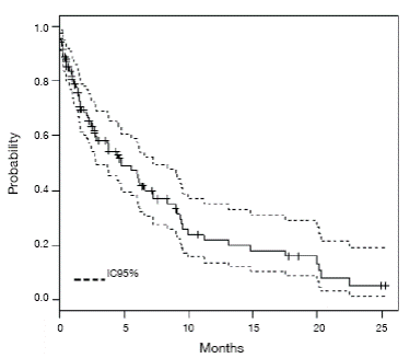
Figure 1 Kaplan Meier survival function with 95%CI. Source: Own elaboration based on the data obtained in the study.
For the variable "prosthesis length" -longer or shorter than 12cm-, a significant difference in survival functions was found -log-rank test, X2 1gl=5.5; p=0.019- (Figure 2). This variable was used as a substitute for the length of stenosis as only 79 patients were recorded and their incorporation into the models affected the accuracy of the estimates. The hazards ratio for serum albumin levels estimated using a Cox model, was 0.64 (95%CI: 0.41-0.90). Table 1 shows the raw hazards ratios, and it is possible to see that those patients with higher albumin levels had a lower risk of death during follow-up; it was also observed that the risk of dying during follow-up is almost double in patients whose prosthesis were longer than 12cm.
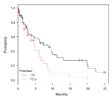
Figure 2 Survival functions depending on the size of the prosthesis. Source: Own elaboration based on the data obtained in the study.
Table 1 Raw ratios for mortality risk in patients with esophageal insertion of self-expandable prosthesis.
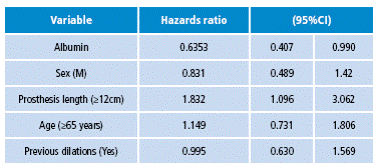
Source: Own elaboration based on the data obtained in the study.
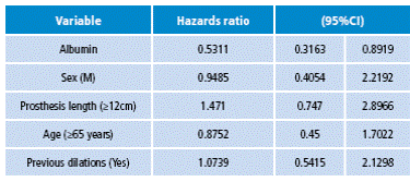
When incorporating the above variables into a proportional hazards model and estimating the adjusted hazards ratios, it can be seen that the effect of the size of the prosthesis vanishes but the significant effect of serum albumin levels (Table 2) is maintained. As mentioned before, the proportional hazards assumption is verified using graphical tools.
Discussion
The most common tumors of the esophagus are squamous cell carcinoma and adenocarcinoma. While the treatment of these two histologic types is often the same, epidemiology is completely different 5. When the patient attends consultation due to dysphagia, the disease is locally advanced and more than 50% of patients present distant metastases 9.
Most patients are treated for palliation with surgery, chemotherapy, radiotherapy and/or endoscopic therapy. Given the high morbidity (13-22%) and mortality (36-71%) with palliative surgical procedures, surgery in these patients is not the best alternative, if we consider that these patients have a median survival rate longer than six months.
A greater effort has been put into less invasive palliation methods, particularly during the insertion of AEP 9,10.
It is clear that the palliation of malignant esophageal obstruction should be based on the safety of the procedure, the restoration of swallowing, decreased hospital stay and reasonable costs 10.
Placing an AEP before surgery is a concept proposed and evaluated to improve swallowing, allowing the application of neoadjuvant therapy and maximizing the nutritional status 7.
In general terms, AEP are reserved for patients with unresectable esophageal cancers 7.
Different studies have evaluated mortality in patients with this disease and its management. For two years, Gray 11 studied 53 patients, out of which 60.4% of cases corresponded to adenocarcinomas, 37.7% to squamous cell carcinoma and one a patient with undifferentiated lesion. He also found mortality at 30 days in 6 patients (11.3%), complications classified as major and minor -bleeding, perforation, pain, and aspiration pneumonia- in 23 (43.4%) and a median survival rate of 84 days (3 months) 11. Specifically, regarding patients treated with AEP, Eroglu et al.12 reported 170 between 2000 and 2008, with a median survival rate of 177 days, being chest pain the most common complication in 31.7%. Stewart et al.8 reported 138 between 1999 and 2009, with a median survival rate of three months, and also chest pain was the most frequent complication in 36% of cases. Dobrucali & Caglar 13 reported 90 cases between 2000 and 2009, with a median survival rate of 134 days. Finally, Castaño et al.14 reported 99 cases between 1999 and 2004, with an average survival rate of 20 weeks, and technical and clinical success of 97%. The most common complication was chest pain in 12% of patients.
The findings of this study are consistent with the published series, where two thirds correspond to adenocarcinomas, with average survival rates of 53 to 139 days and mortality rates of 7.1% to 17% 11,15,16. Literature shows no difference in survival in relation to histological type 9: the reported estimates of mortality (11.3% mortality at 1 month and median survival rates between 53 and 177 days) are similar to those found here.
All patients were fitted with the prosthesis under direct endoscopic vision and without fluoroscopic support. The Wilkes series 16 confirms the results published by other groups and shows that the placement of the prosthesis under these circumstances, in the hands of experienced groups, can be safe and is comparable to the results published by institutions using fluoroscopic control. The question is whether fluoroscopy without endoscopy is better than endoscopy without fluoroscopy. There is no doubt that experienced groups with high volumes of patients may use endoscopy alone or fluoroscopy alone. In the clinical practice, management should be adapted to each case. Since fitting a prosthesis is not an innocuous procedure with enough complications, safety must come from experience. Endoscopy with fluoroscopy has advantages, for example, in the proximal esophagus. The use of both methods is recommended outside centers with extensive experience 17.
90.37% of the patients studied did not show complications with the insertion of the prosthesis. Chest pain within the first 48 hours was one of the most common issues 19).
Other complications described in the literature are tumor growth above the upper edge of the prosthesis, food impactions by obstruction, vomiting, migration -the most common phenomenon in closed prosthesis- aspiration of gastric contents -especially in prostheses that exceed the gastroesophageal region- incomplete expansion, bleeding and perforation, which is very rare. Usually none of the complications is associated with the type of prosthesis or insertion method 19.
It is important to note that there are different brands and different types of prosthesis, which can be coated or partially coated, and which differ in their structure and strength of radial expansion 19.
Na Kyu et al. evaluated the efficacy and safety of the prosthesis in patients with malignant esophageal strictures by reviewing 645 patients with different types of prostheses, classified from the first to the seventh generation, for 22 years. These authors found that the prostheses made from nitinol achieve greater adaptability and flexibility compared to those made with stainless steel, and that patients with nitinol prosthetics had significantly less chest pain compared with those with prostheses made with steel (4.1 vs .15.8; p<0.001) 20. Polyurethane-coated prosthesis showed increased tumor growth rate by degradation of the membrane compared to polytetrafluoroethylene-coated prosthesis. For these authors, the technical success rate was 99.4% and clinical success 95.5% 20.
The current staging system for the AJCC (American Joint Committee on Cancer) does not use the length of the tumor stenosis. Since 1987, the decision to use the deep invasion of the esophageal wall was taken, that is, the "T" more than the length of stenosis 8,21. In this paper, the length of the prosthesis is included as a substitute for the length of the stenosis because the latter was not found in all cases.
In literature, the length of the tumor is a significant predictor of survival, so it was measured at three and five years against an increase in the length of the tumor, with an interval of 1cm, showing a sharp drop in survival when surpassing 3cm. Patients with tumors <3cm had a median survival rate of 79.7% at three years and of 68.4% at five years compared to when the injury was >3cm, with a median survival rate of 23.3% at three years and of 10.6 % at five years (p<0.0001) 8,22. Similar results were seen in patients when the length of stenosis was associated with nodal spread N1 or N0, being the best survival rate in patients with lesions smaller than 3 cm and N0 8.
When variables such as age, sex, histology, tumor location and type of surgery are reviewed, literature does not find a statistical association with survival. Eloubeidi 23 found that tumor length is significant in patients with localized disease, but not in patients with advanced disease.
Wang et al.24 evaluated serum levels of C-reactive protein (CRP) and other variables like the number of white blood cells, alkaline phosphatase, transaminases, body mass index, platelet and albumin levels in patients with esophageal cancer taken to radiation therapy, A multivariate analysis of prognostic factors with statistical significance showed that only CRP and albumin were predictors of overall survival. The survival rates at two years, with and without, hypoalbuminemia were 58.5% and 0%, respectively (p<0.001) 24.
Some limitations of this study include the failure to analyze other variables related to mortality (clinical status, comorbidity), lack of measures of all clinically relevant outcomes -patient's perception, clinical response, quality of life, evolution of the nutritional state- and the failure to incorporate the size of the stenosis into the models -prosthesis length was used instead- due to the lack of registration in medical records. The latter is a common limitation of retrospective studies, especially those based on clinical records.
In conclusion, the results of this study suggest that AEP offer an alternative for improvement of symptoms in patients with esophageal tumors in advanced stages. This is a technique with few complications and probabilities of technical and clinical success at around 90%. The patient's nutritional status and the length of stenosis caused by the tumor are variables to be assessed before each procedure as they seem to be related to mortality.

