


 English (pdf)
 Article in xml format
 Article references
 Send this article by e-mail
 Cited by SciELO
  Cited by Google
  Similars in
SciELO
Similars in
SciELO  Similars in Google
 Permalink
Introduction
Lesions of the nail and the fingertips are common in children and occur, mainly, due to home accidents with doors -for example, when closing doors or during games at school-. Crush traumas, puncture wounds and partial or total amputation of fingertip segments may occur with involvement of the nail, the soft tissue of the finger and the distal phalanx 1-6.
These lesions correspond to 2% of all accidents received in a pediatric emergency department 1. A good initial treatment prevents deformities that can affect positively function and aesthetics of the patient.
The objectives of this work are to analyze the results obtained in different cases related to injury to the nail and the fingertip and to review the current status of treatment.
Materials and methods
This is an observational and descriptive study of clinical cases of children assessed because of injuries to the nail and the fingertip for six months in a pediatric orthopedic service. Fingertip injuries included patients with trauma from the distal phalange to the distal interphalangeal joint.
Patient demographics, type of injury, the anatomical site affected and treatments were analyzed. Finally, literature was reviewed and recommendations were given for the treatment of nail injuries. Data were stored and analyzed using Microsoft Excel 2010. This was a minimal risk job, based on findings from patients, with informed consent and approved by the hospital ethics committee.
Results
66 children, with an average age of 4.6 (age ranged from 9 months to 17 years), who suffered from fingertip injury were studied. 60.3% were male, 55% of the cases occurred in the left hand and 29% in the third finger. 88% of cases were related to traumas caused by crushing (68% corresponded to closing doors), thus becoming the most frequent injury (Table 1).
The nail was compromised in 98% of cases, the soft tissue of the finger in 40%, and fractures of the distal phalanx were found in 55% of the cases, out of which 12 cases required osteosynthesis with 1.0 mm Kirschner Wire since there was a displaced fracture of the distal phalanx. The sterile matrix of 64% of patients was compromised and the germinal matrix, in 34%.
The most common surgical procedures were suture of the nail matrix (60.6%) and suture of the soft tissue (25.7%), with replacement of the same nail or using some substitute, all performed under general plus local anesthesia due to the type of population. Advancement flaps, such as VY, were required in 12 patients (18%).
Although no immediate complications occurred, we were unable to determine long term complications since many patients could not be monitored.
Discussion
The fingers of children are exposed to trauma, especially in the fingertips, which can be injured or amputated; the most vulnerable site is the nail and all its parts. The fingertip injury in children is described as the "door-smashed finger", since closing doors is the most common cause of these traumas 1-8, which is confirmed in this research after observing 45 cases (68%) with this type of injury.
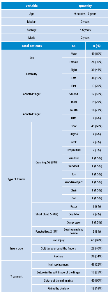
Nails play several important roles in hand function: they protect the dorsal surface of the distal phalanges, increase the sensitivity of the fingertip, facilitate grabbing small objects and have an important aesthetic role. In order to plan appropriate treatment of traumatic lesions of the nails, thorough knowledge of their anatomy and physiology is required. Nails are composed of a nail plate structure, 0.5mm thick, and surrounding soft tissues 6,7,9-11; it is divided into the paronychium, the soft tissues on the sides of the nail, the eponychium -which is the soft tissue surrounding fingernails-, and the hyponychium, the area between the nail bed and the fingertip below the free edge of the nail, which functions as a waterproof seal that protects against infections 6,7,10 (Figure 1).
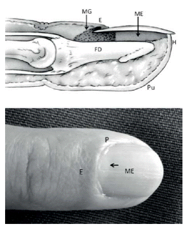
Figure 1 Normal anatomy of the nail and the fingertip. E: eponychium; H: hyponychium; P: paronychium; Pu: soft tissue; FD: distal phalanx; MG: germinal matrix; ME: sterile matrix; Black Arrow: lunula. Source: Own elaboration based on the data obtained in the study.
The nail bed is the place where the nail sticks and is divided into the proximal (germinal matrix), and the distal (sterile matrix); their union is known as lunula. The germinal matrix produces nail keratin and the sterile matrix provides adhesion. The nail bed is nurtured by a wide network of blood and lymph vessels, where many anastomoses occur, which allow using flaps in the bed or the matrix in any reconstruction surgery.
Nail growth depends on factors such as sex, age and habits; the growth rate is approximately 0.1 mm/day (0.5 mm/week) 1,6,11. Most nail injuries are caused by crushing trauma 2-4.11; in approximately 50% of cases, the lesions are associated with phalangeal fractures or damage of the soft tissue of the fingers. After a trauma, the nail stops growing for about three weeks, then an increase in the growth rate is seen over the next 50 days and, finally, a slow growth is observed for 30 days more; growth returns to normal 100 days after the trauma 1,6. During this period, a cross thickening of the nail represents signs of previous trauma (Beau's line).
Adequate primary care is of great importance to achieve a smooth nail, without scars. The wound should be sutured precisely to avoid secondary deformities: a scar on the dorsal roof (eponychium) leaves a line or stripe on the surface of the nail; a scar in the germinal matrix produces a slit or may prevent the growth of the nail, and a scar on the sterile matrix can cause a division of the nail or a distal detachment to the lesion (Figures 2 and 3) . Only one year after the trauma and its treatment, the final outcome of the nail can be observed 2,11-14.
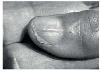
Figure 2 Outcome seen in the nail plate due to eponychium damage. Source: Own elaboration based on the data obtained in the study.
Figure 3 Hook nail tilted toward the palm due to the damage of soft tissues and the loss of the distal phalanx. Source: Document obtained during the course of the study.
Subungual hematoma
The subungual hematoma is a typical injury that occurs after a hammer blow, and its consequence is a subungual hematoma without exit, instead of losing the nail. Many of these hematomas do not increase in size and are confined under the nail, but in some cases, they grow and pain increases, behaving like a small compartment syndrome that requires prompt treatment.
Treatment of subungual hematomas depends on size and behavior; when they are small and the pain is minimal, no intervention is necessary since they incorporate into the nail and move towards the free edge as the nail grows. When they are small but cause pain or occupy up to 50% of the nail bed, it must be drained. An easy draining method implies drilling one or two holes on the surface of the nail with a hypodermic needle, in circular movements (Figure 4); hematoma pressure allows blood to flow out easily, quickly improving the patient's pain and allowing the nail to stick to its bed progressively 5-7,9,15.
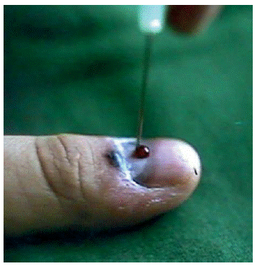
Figure 4 Maneuver with a hypodermic needle to remove a subungual hematoma. Source: Own elaboration based on the data obtained in the study.
In hematomas bigger than 50%, looking for an association of a fracture of the distal phalanx is mandatory, so lifting the nail and exploring the nail bed is recommended to verify the existence of an injury in the sterile or germinal matrix that requires suture 9,11,12.
For the scanning procedure of the nail bed, the nail is lifted with mosquito forceps or a small spatula, starting from the free edge, which is carefully separated from the bed and, depending on the type of lesion, completely removed with small rotating movements; it can also be left as a pedicle in some portion of the paronychia or eponychium 6,16. The suture matrix must be done with separate points of a resorbable monofilament 6/0 (Figure 5).
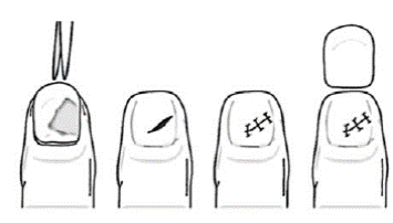
Figure 5 Removal of the nail to explore and suturing the sterile matrix. Source: Own elaboration based on the data obtained in the study.
The nail should be kept to be relocated, and can be used as a biological dressing during repair. In this way, it will fulfill the functions of shaping the nail bed, avoiding adhesions in the bottom of the nail between the eponychium and nail bed and, in the case of associated fractures in the distal phalanx, providing support as a splint and improving postoperative comfort.
When relocating the nail, making perforations is advisable in order to facilitate the process of draining the accumulated blood. The relocated nail must remain fixed in his bed and in the proximal bottom by a suture, preferably in the form of x, avoiding stitches through the nail bed 5,6,16-18.The new nail grows pushing the nail-ferrule, which is replaced in a period of 1 to 3 months.
Wounds and lacerations of the nail matrix and surrounding tissues
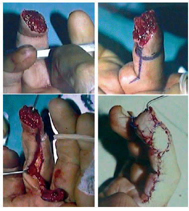
When avulsion or dislocation occur, the nail must be lifted to be replaced at the end of the procedure; if only sterile or germinal matrix are wounded, the nail must be sutured as indicated above (Figure 6 and 7).
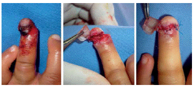
Figure 6 Crush injury of the tip of the fourth finger. The nail is lifted and an injury in the sterile matrix and paronychium is observed. The matrix is sutured with resorbable 6/0. Source: Own elaboration based on the data obtained in the study.
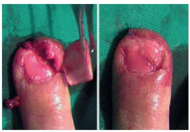
Figure 7 Crush injury. When the nail is lifted, hyponychium and sterile and germinal matrix damage is observed. It is approached with an initial suture in the hyponychium and then in the nail bed. Source: Own elaboration based on the data obtained in the study.
The loss of the nail bed may occasionally occur, which is solved through a flap in the matrix of the same finger, if necessary. Grafting from the nail matrix of the big toe or adipose flap rotation must be used when large losses occur 6,7,19-21.
The wounds that compromise the paronychium, the hyponychium or the eponychium should be sutured carefully. Sometimes, it is impossible to recover the nail, which makes a temporary replacement necessary for about two to three weeks, as it will prevent adhesions at the bottom of the eponychium and throughout the matrix during the healing process.
Soft and hard synthetic elements, such as pieces of x-ray film or the envelope of the suture, have been used in healing procedures, as well as non-adherent gauze, but these are easily contaminated and are hard to place on the nail bed. The most practical method used is a small flexible sheet removed from the sterile bag of the saline solution, which allows easy cutting and settles to the nail bed and the bottom of the nail 6,16,22-25.
In the case of deeper injuries associated with fracture of the distal phalanx, a fracture osteosynthesis with a nail, or alternatively a hypodermic needle, is recommendable.
Bandages are left in the nail or fingertip and must be replaced every three to five days for a period of 20 days, moment when the sheet that protects the nail bed is removed; then the area must be lubricated with petroleum jelly or cream. If the patient has his own nail-ferrule, it is allowed until new nail growth and falls by itself.
Wounds and lacerations of the nail with soft tissue damage
Besides the treatment described for lesions of the nail matrix and surrounding tissue of the nail, and according to the injury in the soft tissue, a simple suture should be done using local advancement flaps, such as VY, or a bit more complex flaps such as homodigital or heterodigital neurovascular island flaps 20,26-38 (Figure 8 and 9). This must be done by a specialist, but while the type of procedure is determined, the finger should be washed with a local anesthetic and covered with lubricated plasters.
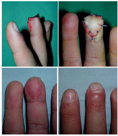
Figure 8 Loss of a fingertip that has been solved with VY advancement flap. Source: Own elaboration based on the data obtained in the study.
Conclusions
Nail lesions are the result of a contusion or compression against the underlying distal phalanx. The type of injury depends heavily on the energy and direction of the trauma. Different injuries can be observed as subungual hematomas, simple lesions of the nail and the nail bed, and more complex lesions with soft tissue or nail loss and associated fractures.
A proper anatomical knowledge and a good analysis of the type of injury and the compromised structures allow selecting an appropriate treatment, which, in turn, prevents secondary deformities with decreased secondary reconstructions of the nail that are more complicated and have unpredictable results.
Finally, promoting accident prevention programs at home and in schools, aimed at parents and teachers, is recommended.

