


 English (pdf)
 Article in xml format
 Article references
 Send this article by e-mail
 Cited by SciELO
  Cited by Google
  Similars in
SciELO
Similars in
SciELO  Similars in Google
 Permalink
Introduction
Learning anatomy is an art. Centuries of curiosity, dedication and intelligence allowed our ancestors to learn, through anatomical dissection, about what today are the basics of medicine. 1 The traditional method of teaching and learning anatomy includes on-campus classes given by a professor with experience and knowledge on the subject. In addition, students receive classes in dissecting rooms using anatomical specimens, where they can perform identification and differentiation of structures, dissection and injection, relying on anatomy texts such as atlases that offer images in two dimensions (2D). 2
Based on the international nomenclature, human anatomy has a great variety of structures, and three main methods used for their study are regional anatomy, systems anatomy and clinical anatomy. 3 Anatomical illustrations are fundamental for medicine learning. In the past, they were obtained by dissecting animals, given that the inspection and dissection of a human corpse was illegal and the person who engaged in this practice could be sentenced to death. The founder of modern anatomy, Andreas Vesalio, began to make illustrations based on direct observations and dissections on human cadavers in the 16th century, turning dissection into a fundamental part of the class for his students. The anatomist found errors in the information previously described by Galen regarding the anatomy and function of the human body 4, and although Leonardo da Vinci dissected human corpses before Vesalio, his findings were not disclosed because it was illegal. Perspective can be found in da Vinci's drawings, and this contributed to a three-dimensional approach to the body. 5
Currently, in order to perform dissections and explorations of structures 6, texts such as anatomy atlas are used, as they offer 2D illustrations, although they do not allow perceiving their depth and do not provide enough perspective or detail to grasp correctly the structures. Learning should focus on personal development because it is an empirical process, making practices in the dissection hall fundamental; even so, difficulties in understanding continue to occur due to insufficient cadavers, deterioration of structures and ethical limitations. 7
In this context, the most important historical achievement of anatomy has been the improvement of graphic tools. Several historical figures have concentrated their efforts on the development of new tools or methods that allow achieving greater realism in the illustrations; however, the lack of technology in certain places has limited the ability to progress significantly, reducing the learning options to the use of literary tools and cue cards. With the incorporation of virtual media in society, it is essential to continue with the efforts and create tools that increase the similarity of the anatomical graphic resources with reality.
Two-dimensional illustrations and the ability of the draftsman were the only tools available in the past; today three-dimensional resources allow for spatial understanding and the analysis of a complete structure with only one graphic resource, compared with the illustrations that need many images to analyze only one structure without preserving spatial comprehension. Digital tools, along with traditional learning methods, optimize the understanding of cardiac anatomy. The main evolutionary objective of the study of anatomy is, then, to ensure that multimedia tools bring the student closer to reality. 8
In this regard, multiple strategies have been developed with new software technologies that have proven to be very useful for increasing educational performance, particularly in specialties related to health sciences, because computational tools have the ability to create very similar scenarios that complement the learning process, although they can never replace direct experience with reality. 9
This study was formulated from the perspective of a student when realizing that the hours in the dissection hall were not enough to study and review the structures, because that is the only place where teaching with a three-dimensional approach can occur, achieving greater spatial location with respect to orientation, relationship and nomenclature. In this context, the following questions arose: if the basis of the anatomy begins with dissection, why not using software with the structures found? Can traditional teaching in cardiac anatomy be optimized?
Interactivity and multimedia tools in education are of great importance. Studies show a marked increase in the interpretative capacity of students when they are involved in an educational model equipped with interactive multimedia tools. 10 One of these tools is called Anatomage, which consists of a digital table for virtual 3D dissection. Its implementation has shown favorable outcomes in 63% of students, who claim to have increased their knowledge; in addition, 78% stated that the interactivity of the tool has more impact. 11
Technological alternatives with greater acceptance include 3D software, which, compared with anatomy atlases, have an unlimited number of perspectives that facilitate perception thanks to the interaction with the rendered model. 12 Another study reported greater facility to understand two-dimensional images after studying with 3D web tools. 13
One of the greatest challenges of teaching consists in identifying the methods preferred by the students, since it is important to develop the appropriate didactic approach to involve students with the information taught. This is important in subjects that require great memory effort, for which generating dynamics that are pleasant enough to prevent information from being forgotten is also recommended. After making a 3D model of the larynx, a satisfaction survey found that 93% of the students preferred the technological tool and 87.4% recognized greater facility to understand its anatomy. 14,15
Taking this into account, the objective of this research was to evaluate the impact of using 3D rendering as a digital tool on cardiac anatomy learning.
Materials and methods
This is an experimental study that included third-semester students of a medical program as study population. The population was divided into two groups consisting of 40 students each: the 3HA group, which corresponded to the study or intervention group, and the 3HB or control group. At the time of the study, participants were taking the subject Anatomy II, in the specific context of cardiac anatomy learning.
The selection of students for each group was random, for a total population of 80 students. Those who failed the subject and were enrolled in a program other than Medicine were excluded. Academic performance analysis was not performed according to sex or age.
Both groups attended lectures and practices in the dissection hall, that is, they used the traditional methodology. The 3HB group only followed the traditional methodology, which corresponds to 6-hours lectures and classes in the dissection hall for 4 hours, using texts of anatomy atlases. The 3HA group received traditional instruction and also 2 hours of class in which digital pedagogical strategies applying the 3D Heart-tomy program in the computer room were used, in order to support the theoretical and practical components. The 3D Heart-tomy file used was run using the Blender v2.78 software, which is open source. The file was prepared by the company Botero Mechatronics SAS based on real hearts obtained in the dissection hall.
The digital didactic instrument consisted of an interactive three-dimensional projection, as shown in Figure 1. This interactive learning tool frames and splits each of the structures to understand their relationship and the composition of the heart.
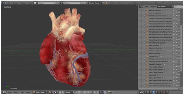
The 3D Heart-tomy allows identifying the structure with its name, moving it, separating it and analyzing its unions and direction. Students can also see all the structures through real-time manipulation. This program has the option of orbiting the 3D model, allowing any perspective as desired, has a list of the nomenclature of each structure, and highlights the name. In the same way, it is possible to hide some sections to visualize the inner part of the heart and there is an Rx option to visualize its interior without hiding the exterior. Finally, a file contains a self-qualifying test that allows students to analyze what structures are being forgotten.
Classes were taught by the same professor and the heart related topics addressed during the virtual based classes were as follows: external configuration and large vessels, internal configuration, epicardium, myocardium, endocardium, electrical system, coronary circulation, innervation and images.
The objective of the theoretical tests in the classroom and the practical test in the dissection hall was to measure the conceptualization of the topic related to cardiac anatomy, with an approval score of at least twelve correct questions (60%), equivalent to a passing grade of 3.0.
Five independent evaluators and experts in the field reviewed these tests and validated the relevance of the questions in relation to the educational level of the groups under study and the requirements on heart knowledge for medical students of the subject Anatomy II.
To avoid bias while grading, the students were identified through their student identification number and the group to which they belonged was only known when the grades were entered into the system, after the tests had been graded.
In addition, an opinion poll was conducted to evaluate the students' opinion of the impact of 3D Heart-tomy as an implementation of new technologies for teaching and learning about cardiac anatomy.
Information was obtained strictly based on the criteria defined for carrying out a medical-legal autopsies contained in Decree 786 of 1990. 16 In addition, all the principles of the Declaration of Helsinki 2013 for medical research were followed 17, thus ensuring respectful treatment of the pieces obtained during the study and maintaining the confidentiality, dignity and integrity of the deceased person. According to the parameters established by the Resolution 8430 of 1993 of the Ministry of Health of Colombia 18, this investigation was presented before the Ethics Committee on Human Research of the Faculty of Health Sciences of Universidad de Ciencias Aplicadas y Ambientales (UDCA), which granted its approval through Minutes 20160401, issued in April 2016. Informed consent was obtained from all participants.
Results
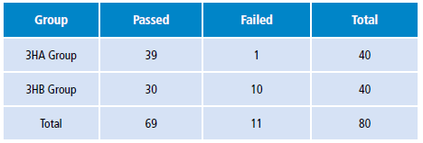
The results obtained were compared after being typed in a table. The amount of grades <3.0, the amount of grades between 3.0 and 3.9 and the amount of grades ≥4.0, both in the theoretical test and the practical test in the dissection hall, were retrieved (Table 1).
Table 1 Statistical results of the theoretical and practical tests of the study group and the control group.

Source: Own elaboration.
The results show a difference between both groups. The average grade of the 3HA group was 4.3 compared to the 3HB group, which had an average grade of 3.6. In this case, a favorable result can be seen in the group that used the 3D tool, which is 14% higher than the result of the group that did not use it.
Regarding the proportion of students who passed the test, the 3HA group obtained a percentage of 97.5%, while the 3HB group obtained 75%. This showed that 25% of the control group failed and only 2.5% of the group that used the digital tool failed the subject.
There were also positive results for the 3HA group in terms of the number of students who achieved the highest score, 22.5%, while only 7.5% obtained it in the control group, which shows results that favor by 15% the group that used the technological tool.
After analyzing the last result, most grades greater than or equal to 4.0 were obtained by the 3HA group and 70% of the students in this group are in the mentioned range; meanwhile only 40% of the students in the control group obtained a 4.0 or higher grade. A 30% difference in favor of the members of the 3HA group.
Data of the theoretical test were organized according to the distribution to find the value of X2, which sought to prove the hypothesis that the traditional method of learning and the use of technology strengthen learning of cardiac anatomy, as described in Table 2.
The results were obtained through OpenEpi. The value was compared with the table of critical values of the distribution X2 having 0.005 as margin of error. The value of X2 in the table for this margin is 8.5, which is greater than the calculated value and allows concluding that the hypothesis for the theoretical test can be accepted.
The odds ratio (OR) is 13 (lower limit: 1 576, upper limit: 107.2), which means that students who used the software are 13 times more likely to pass the theoretical test. Although the interval is wide, the confidence interval is greater than 1.
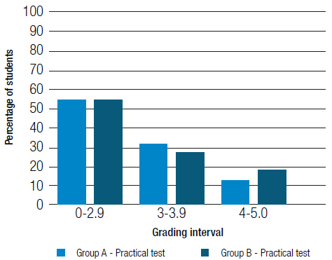
The differences in the practical tests were not significant between the groups, being 2.75 the average grade in the intervention group and 2.5 in the control group. It should be noted that the result was low for both groups, but a little higher for the group that used the 3D tool.
The results of the practical test were organized to determine the corresponding value of X2, which was 0 and can be seen in Table 3.

The alternative hypothesis cannot be accepted for the practical test, because the OR is 1, so there is no statistical significance in passing the practical test using the software.
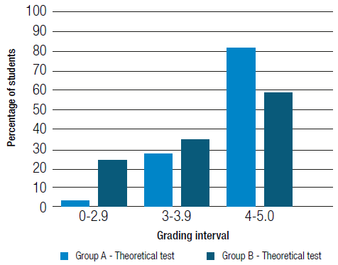
Each grade of the theoretical test obtained by a specific student was typed in a Microsoft Excel sheet, by means of which Figure 2 was generated; it is possible to see that the results of the 3HA group are notoriously higher than those of the 3HB group.
The results of the practical test did not differ much in both groups; however, students in the control group obtained grades below the minimum grades obtained by those in the intervention group, which evidences a better performance in the latter, as it can be seen in Figure 3.
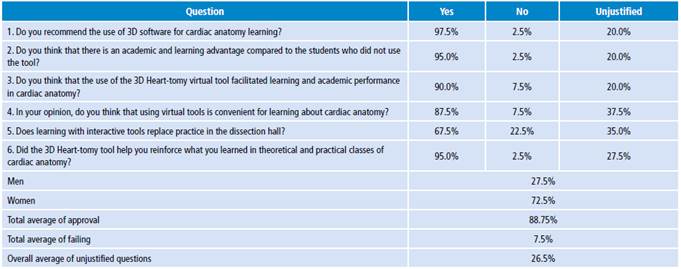
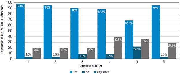
The survey yielded satisfactory results; the percentages of passing together with the percentage of unjustified questions can be seen in and failing grades obtained by students for each question asked, Table 4 and Figure 4.
Discussion
A new field of medical education was born with the advent of new technologies. In recent years, the several benefits that digital media can provide and how much they could improve the learning process have been explored. For the specific case of anatomy, this research has similar results to other studies conducted on the use of 3D tools, which confirm that they are very useful as an additional means of teaching subjects on anatomy.
When analyzing the data and Figure 2, it can be seen that the trend line in the 3HA group is constantly above the trend line observed in the control group. The performance of students who use digital tools has been higher in multiple studies than in students who use non-technological means; computer-based instruction allows the student to be more actively involved, facilitating the achievement of effective learning.
There are few comparative studies; however this study is similar to those performed by Lim et al.14 and Hopkins et al.19, who took 3 groups of people made up of 74 students who were enrolled in the pre-test, laboratory session and post-test (pro sections n=26, 3D model n=23, hybrid n=25) in 17 separate group learning sessions, where each participant attended a single lab session. Students showed a significant improvement of knowledge in the post-test. Although the difference in the results was not that high, there was a better performance in those who used the 3D tool, which correlates with what is reported here. The students then responded to a questionnaire in which 82% preferred to have the usual study tools together with the 3D tool. Observations from the Lim et al. study 14 ensure that 3D tools do not replace any other study method and may work better if used as a complement, which is consistent with the results obtained here.
A similar study 20 compares whether a three-dimensional presentation is more effective for teaching than a presentation supported by a traditional book or not. This research involved 46 students who were divided into two groups, one that participated in the interactive computer-based learning module with 3D images and another that received their lessons in computers using non-interactive 2D images. After each teaching module, students completed a satisfaction survey and nine points of anatomical knowledge after the test. The group that used the 3D tool scored higher in the post-test than the group that used the 2D tool, with an average score of 74% and 64%, respectively; these results were similar to those obtained in the present study 20.
In the post-test satisfaction survey, the group that used the 3D tool expressed higher satisfaction and showed significantly higher figures compared to the students in the control group that used the 2D tool. After verifying the pre-test and post-test analysis, although a higher score was found, it was not statistically significant. Like other authors, in this study, the satisfaction survey showed that students preferred using three-dimensional tools for learning.
In the opinion poll, six questions were asked to students who used 3D software in order to evaluate the impact of the implementation of a digital didactic tool for teaching cardiac anatomy on learning in third-semester students. The result in terms of student satisfaction with the software was quite positive and in line with other studies that found great satisfaction in the use of digital tools for the study of anatomy.
Use of render, as a cognitive tool, facilitates the learning of human cardiac anatomy. 21 In most studies, as found here, the use of digital environments proves to be efficient for learning cardiac anatomy. 22
Conclusions
Participating students agreed that the 3D rendering tool facilitates the learning of cardiac anatomy and improves theoretical academic performance due to the interactivity they are given by it. This opinion resembles statements from other studies and shows an increase in learning capacity thanks to the implementation of new digital technologies.
Didactic and digital 3D media facilitate the understanding of the location of the structure and works as a pedagogical tool; however, it does not replace practices in the dissection hall.

