


 English (pdf)
 Article in xml format
 Article references
 Send this article by e-mail
 Cited by SciELO
  Cited by Google
  Similars in
SciELO
Similars in
SciELO  Similars in Google
 Permalink
Introduction
The human eye is an organ constituted by an assembly of lenses with the capacity to capture light and convert it into electrical impulses, which are transformed into images by the brain.1 These impulses are transmitted to the visual cortex, located at the back of the head, by a set of nerve fibers called optical pathways.2 The movements of the eyeball in the orbital cavity occur thanks to the extrinsic muscles, which are innervated by the common ocular motor, trochlear, abducens, and trigeminal nerves, cranial pairs that relay sensitive information.1
Eyes are the organ that makes possible the sense of sight and, therefore, they are necessary to interact properly with the environment. For this reason, health professionals (optometrists, ophthalmologists, and general practitioners) involved in their care must learn in a practical and correct way their anatomy during their training.
There is currently controversy among anatomists as to whether dissection is the best method for teaching this scientific discipline.3 In recent decades, this method has gained detractors because of the limitations that professionals and students face when they need to perform dissection procedures, such as the difficult access to cadavers and legal restrictions on their use.4,5 Nevertheless, corpse dissection continues to be the preferred method for teaching and learning anatomy.6
With this in mind, the present research revisits this classic method and proposes an innovative modification of the Caldwell-Luc procedure, a surgical intervention indicated for treating chronic sinusitis and odontogenic maxillary sinusitis,7,8 to facilitate the learning of the structures surrounding the eye among health professionals. To this end, this study sought to modify the Caldwell-Luc procedure with a simultaneous superior approach that allows access to all the extrinsic structures of the eye contained in the orbital cavity and, in this way, facilitate its study in situ.
Materials and methods
Descriptive study. First, pig eyes were dissected so the students could gain the necessary experience to carry out the dissection in a human corpse. Then, the orbital cavity contents were dissected by planes in a human cadaver based on an exhaustive review of the literature in the ClinicalKey, PubMed, LILACS, and SciELO databases, as well as in textbooks of human anatomy and embryology.
The procedures were performed in the Human Anatomy Laboratory of the Faculty of Health Sciences at the Universidad de Ciencias Aplicadas y Ambientales U.D.C.A by the research group using the necessary biosafety tools. The following materials were used: dissection instruments; scalpel blades # 10, 11, 15, 18, 23 and 25; two pig heads (preserved with formalin and glycerin in the laboratory); Stryker® autopsy saw (model 810 -BD001) for cutting bones; visual protection equipment and a Nikon® D-5500 camera with 17mm lens.
Before performing the anatomical dissection of the human orbital cavity, students practiced the procedure on two pig heads, which were purchased in the Quirigua neighborhood marketplace; this is to make clear that no animal sacrifices were performed. It should be noted that pigs were used because of the anatomical similarities between its eyes and human eyes.3 A cut was made in the maxillary bone of one of the heads, while the eyeball was dissected keeping this bone intact in the other one. The purpose was to determine which of the two procedures allowed visualizing a larger amount of structures.
Then, the modified Caldwell-Luc technique was utilized in the human orbital cavity, as described in 1972 by Walter.9 Two incisions were made, one on the face midline from the forehead, going through the nasal dorsum and the nasolabial fold until the upper lip, and the other from the medial region to the lateral region, starting from the cut made on the midline until the right auricle. Then, the frontal belly of the occipitofrontal muscle and the orbital and palpebral portions of the orbicularis oculi muscle were exposed. Subsequently, the subcutaneous and muscle tissues were removed from the surface of the zygomatic and maxillary bones to expose these bone structures and, therefore, visualize the infraorbital artery, the infraorbital vein, and the infraorbital nerve.
Afterwards, using the autopsy saw, a 2cm square cut was made on each side and the bone block was removed to see the infraorbital artery, the infraorbital vein, and the infraorbital nerve. Then, the subcutaneous tissue from the orbit was dissected and removed to improve the visualization of the extrinsic structures contained in this cavity.
In addition, a cut was made beginning at the base of the skull, crossing the minor wing of the sphenoid bone, continuing through the anterior cranial fossa to the frontal bone at the base of the skull, and then continuing around the anterior cranial fossa with a lateral cut up to its limit with the middle cranial fossa; finally, an anterior cut was made until the initial point of the first cut was reached. A bony piece of the skull was left free to approach the structures contained in the orbital cavity from an upper view, so these structures were cleaned and separated for proper identification.
The original Caldwell-Luc procedure was modified by performing a surgical resection of the maxillary bone and the medial edge of the zygomatic bone, that is, where this bone articulates with the maxillary bone. Another cut was made at the base of the skull on the limit between the orbital cavity and the superciliary arch. Once the common tendinous ring was dissected, the ophthalmic artery was identified.
Ethical and legal considerations
This study was conducted following the ethical principles set forth in the Declaration of Helsinki10 and the ethical standards established in Resolution 485 of 2002 issued by Instituto de Medicina Legal y Ciencias Forenses, which regulates the procedures for the destination of corpses and anatomical components obtained from them for teaching and research purposes.5 Likewise, the provisions of Decree 1546 of 1998, which establish that scientific institutions and hospitals may use unclaimed cadavers, as well as organs or anatomical components obtained from them for teaching and research purposes, were taken into account.11
All guidelines and parameters that ensure due respect for human corpses and their use to support and expand medical, surgical and scientific knowledge established in articles 47 and 48 and the principles for biomedical research on animals set forth in article 87 of Resolution 8430 of 1993, issued by the Colombian Ministry of Health,12 were followed. Also, according to this resolution, since the study was conducted on a corpse in the dissection hall, it is considered to be free of risk.12
Results
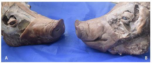
The structures contained in the orbital cavity of both pig heads were exposed (Figure 1). This practice allowed the students to obtain the necessary skills to carry out the dissection in the human cadaver.
Source: Document obtained during the study.
Figure 1: Dissection performed on pig heads. A: pig's head with resection of the maxillary bone; B: pig's head with complete maxillary bone.
During the procedure, it was determined that cutting the maxillary bone facilitates the approach to the orbital cavity since it allows identifying the structures contained in this cavity and dissecting them in a more practical way.
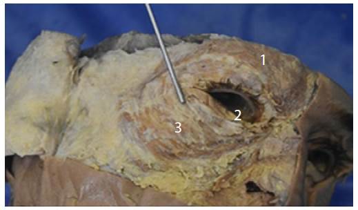
By dissecting the human corpse, it was possible to observe the frontal belly of the occipitofrontal muscle and the orbital and palpebral portions of the orbicularis oculi muscle, as well as the infraorbital artery, the infraorbital vein, and the infraorbital nerve (Figure 2).
Source: Document obtained during the study.
Figure 2 Cuts made in the skin on the sagittal midline of the face and transversally up to 2 cm below the right auricle. 1: frontal belly of the occipitofrontal muscle; 2: palpebral portion of the orbicularis oculi muscle with complete detachment of the skin covering it; 3: orbital portion of the orbicularis oculi muscle.
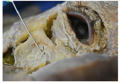
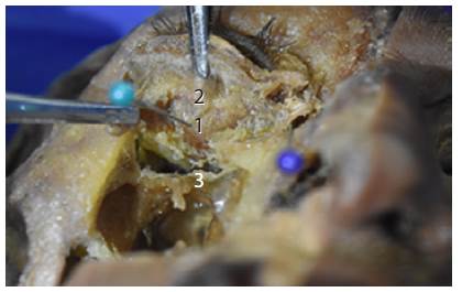
The square cut made in the middle third of the zygomatic bone, that is, where this bone articulates with the maxillary bone (Figures 3 and 4), and in both lateral portions of the right half of the maxillary bone, that is, in the articular proximity with the zygomatic bone, allowed visualizing the infraorbital artery, vein and nerve, and the occipitofrontal and orbicular muscles of the eye. This cut is one of the modifications made in the present study to the original procedure proposed by Caldwell-Luc in 19729 and to other modified procedures that preserved the original approach.
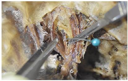
Another modification to the original technique and its variants in the present study was a cross section made at the base of the skull to allow the observation of the infraorbital artery, infraorbital vein and infraorbital nerve, and the common tendinous ring, which reaches the orbital cavity from the cranial cavity. After this cut was made, the following structures were visible: the eyeball; the lacrimal gland; the lacrimal vein; the frontal nerve; the frontal sinus; the olfactory bulb; the pituitary gland and pituitary stalk; the levator palpebrae superioris muscle; the superior, oblique, medial and lateral rectus muscles; the oculomotor (III cranial pair), trochlear (IV cranial pair) and abducens (VI cranial pair) nerves; and the lacrimal, frontal and nasociliary branches of the ophthalmic nerve (V1) of the trigeminal nerve (V cranial pair) (Figure 5).
Source: Document obtained during the study.
Figure 3 Square cut made in the zygomatic bone. 1: infraorbital nerve passing through the infraorbital foramen after the dissection of the anatomical structures.
Source: Document obtained during the study.
Figure 4 Cutting and resection of the square block of the zygomatic bone with anteroinferior view of the orbital cavity. 1: inferior rectus muscle; 2: inferior oblique muscle; 3: infraorbital nerve.
Discussion
Currently, the Caldwell-Luc procedure is not being used for the purpose for which it was initially intended since minimally invasive procedures are preferred. With this technique, indicated for treating chronic sinusitis, the sinuses are approached through the oral cavity.13,14 Multiple modifications have been made to the original version, so it has become a dissection technique that preserves the morphology of the orbital cavity structures and provides a real and accurate perspective of their location.
In 2003, Gonül et al.15 proposed an anatomical model for the study of the morphology of the orbital floor that encouraged the creation of other models based on the Caldwell-Luc procedure. At the University of Costa Rica, Rodríguez-Vargas & Guevara-Arroyo16 proposed an anatomical model to visualize, in a more precise manner, the vascular and nervous structures of the orbital floor, in which the musculature is lifted until finding the bony structures. Then, a part of the zygomatic bone and the maxillary bone are removed precisely under the orbital cavity, and the vascular and nervous structures of the anterior segment of the orbital roof are dissected.
In contrast with the original Caldwell-Luc procedure and the modifications proposed by Gonül et al.15 and Rodríguez-Vargas & Guevara-Arroyo,16 in the present study, a variation in the way of approaching the anatomical structures of the face and a cross section at the base of the skull in the superciliary arch were performed. The cross section was added to remove the anterior cranial fossa from the base of the skull to approach the structures of the orbital cavity from an upper view in an axial plane, as seen in Figure 5.
The proposed modification is based on a complete dissection of all the structures attached to the orbital cavity while advancing in depth, that is, skin, subcutaneous cellular tissue, muscles, bones, nerves and blood vessels, favoring learning of the cranial morphology and the extrinsic structures of the eye. Therefore, the modification proposed in this research is a new technique and a model for studying the anatomy of the orbital cavity and its contents.
The authors do not think that there were study limitations; however, they agree with Mora-Villate et al.3 that the study of the orbital cavity morphology can be difficult since it is limited to virtual models and illustrations that do not offer a real impression of this structure and its contents. This shortcoming is solved by our proposal, as it has been proven that the use of dissection techniques improves the morphological learning of any anatomical structure.3 Table 1 compares the original Caldwell-Luc procedure and previous modifications, and describes the relevance of the findings of this study.
Table 1 Comparison of the Caldwell-Luc procedure and related modified dissection techniques.
| Technique (year) | Approach | Objective | Access | Target dissection structures | Modification and innovation | Additional structures found |
|---|---|---|---|---|---|---|
| Original technique9 | Surgical | Paranasal sinuses | Oral cavity, canine fossa | Maxillary sinuses | Original surgical procedure | Infraorbital nerve. Face and gums preserved |
| Gönül et al.15 (2003) | Anatomical study | Maxillary sinuses | Transmaxillary approach | Maxillary sinuses | Modification | Face and nerves preserved |
| Rodríguez-Vargas & Guevara-Arroyo16 (2013) | Anatomical study | Orbital floor | Transmaxillary approach | Orbital floor structures | Modification and innovation: resection of the maxillary and zygomatic bones | Rectus and inferior oblique muscles, infraorbital nerve |
| Muñoz- Castaño et al. (2018) * | Anatomical study for the teachinglearning process | Extrinsic structures of the eye contained in the orbital cavity | Bone resection, dissection with transmaxillary and transfrontal approach | Rectus muscles of the eye, levator palpebrae superioris, gland and oblique muscles, nerve structures | Modification and innovation: resection of the maxillary and zygomatic bones, cross section at the base of the skull | All extrinsic structures of the human eye, vascular and nerve structures of the face and base of the skull |
* Technique proposed by the authors of this study.

