


Services on Demand
Journal
Article
 Article in xml format
 Article references
 Send this article by e-mail
Indicators
-
 Cited by SciELO
 -
 Access statistics
Related links
-
 Cited by Google
 -
 Similars in
SciELO
Similars in
SciELO -
 Similars in Google
Share

 Permalink
Revista Colombiana de Ciencias Pecuarias
Print version ISSN 0120-0690On-line version ISSN 2256-2958
Rev Colom Cienc Pecua vol.23 no.3 Medellín July/Sept. 2010
Detection of bovine herpesvirus 5 (BoHV-5) in formalin-fixed, paraffin-embedded bovine brain by nested PCR in Colombian cattle¤
Detección de herpesvirus bovino 5 (BoHV-5) en cerebro incluido en parafina y fijado en formalina, por PCR anidada, en bovinos colombianos
Detecção de herpesvírus bovino 5 (BoHV-5) em cérebro incluído em parafina e fixado em formalina, pela PCR, em bovinos colombianos
Francisco J Pedraza1*, MV, MSc, Becario de Doctorado del CNPq-Brasil; Antonio C Alessi2, MV, PhD; Edel F Barbosa-Stancioli3, MV, PhD.
1Departamento de Salud Animal, Universidad de Caldas, Manizales Colombia. Doutorando FCAV-UNESP, Jaboticabal, SP, Brasil. 2Departamento de Patologia Veterinária, Universidade de Estadual Paulista (FCAV-UNESP) Jaboticabal, São Paulo, Brasil. 3Departamento de Microbiologia, Universidade Federal de Minas Gerais, Belo Horizonte, MG, Brasil.
(Recibido: 2 noviembre, 2009; aceptado: 15 junio, 2010)
Summary
Fifteen cases of viral meningoencephalitis in Colombian cattle were tested by nested PCR analysis for the detection of bovine herpesvirus 5 (BoHV-5). All fatal cases had shown severe neurological signs and had occurred following natural outbreaks of the disease. The neurological infection was histologically characterized by mild to moderate inflammatory changes in the brain and cerebellum, including meningitis, mononuclear perivascular cuffing, gliosis, haemorrhage, and the presence of Gitter cells (macrophages) accompanying large areas of malacia. No intranuclear inclusion bodies were seen in any of the cases. Results from BoHV-5 molecular extraction analyses showed there were five positive cases thus confirming the presence of the virus in Colombia.
Key words: bovine herpesvirus, polioencephalomalacia, viral encephalitis.
Resumen
Quince casos de meningoencefalitis viral en ganado Colombiano fueron analizados por reacción en cadena de la polimerasa (PCR anidada) para la detección del herpesvirus bovino 5 (BoHV-5). Todos los casos mostraron severos signos neurológicos antes de morir y ocurrieron como brotes naturales de la enfermedad. La infección neurológica fue caracterizada histológicamente por leves a moderados cambios inflamatorios en el cerebro y cerebelo, incluyendo meningitis, manguitos perivasculares, gliosis, hemorragia y la presencia de células Gitter (macrófagos) acompañando grandes áreas de malacia. No se encontraron cuerpos de inclusión intranucleares en ninguno de los casos. Los resultados del análisis de la extracción molecular de BoHV-5 revelaron la existencia de cinco casos positivos, lo cual confirma la presencia del virus en Colombia.
Palabras clave: encefalitis viral, herpesvirus bovino, polioencefalomalacia.
Resumo
Quinze casos de meningoencefalite viral em gado bovino Colombiano foi analisado pela reação em cadeia da polimerasa (PCR) para a detecção do herpesvírus bovino 5 (BoHV-5). Todos os casos mostraram graves sinais neurológicos antes de morrer y aconteceram como surtos naturais da doença. A infecção neurológica foi caracterizada histologicamente por leves a moderados câmbios inflamatórios no cérebro e cerebelo, incluindo meningite, manguitos perivasculares, gliose, hemorragia e a presencia de células gitter (macrófagos) acompanhando grandes áreas de malacia. Não se encontraram corpos de inclusão intranucleares em nenhum dos casos. Os resultados da análise da extração molecular do BoHV-5 por PCR revelaram a existência de cinco casos positivos, os quais confirmam a presença do vírus na Colômbia.
Palavras chave: encefalite viral, herpesvirus bovino, polioencefalomalacia.
¤ Para citar este artículo: Pedraza FJ, Alessi AC, Barbosa-Stancioli EF. Detection of bovine herpesvirus 5 (BoHV-5) in formalin-fixed, paraffin-embedded bovine brain by nested PCR in colombian cattle. Rev Colomb Cienc Pecu 2010; 23:292-298.
* Autor para correspondencia: Francisco J Pedraza. Departamento de Salud Animal, Manizales, Colombia. E-mail: fpedraza@udecaldas.edu.co
Introduction
Bovine encephalitis caused by Bovine Herpesvirus 5 (BoHV-5) was initially described in 1962 when the virus was isolated following an outbreak of the disease that killed several calves in Australia (Lemos et al., 2002). Initially, the virus was considered identical to the one causing Infectious Bovine Rhinotracheitis (IBR). However, some outbreaks of the disease causing exclusively neurological signs led to suspect there was a variant of such agent exhibiting neurological disease properties (Moretti et al., 1964; Watt et al., 1981; Weiblen et al., 1989). In 1986, through molecular techniques discrimination, such agent was designated 1.3 bovine herpesvirus (Studdert, 1989). In 1992, the International Viral Taxonomy Committee named it BoHV-5 (Roizman, 1992).
Clinically, the condition may be misdiagnosed as rabies, pseudorabies, polioencephalomalacia resulting from thiamine deficiency, lead or salt intoxication, among other conditions (Sanches et al., 2000; Lemos et al., 2002; Spilki et al., 2003).
BoHV-5 has been reported in Europe (Barenfus et al., 1963; Bartha et al., 1969), Canada (Beck, 1975), the United States (d`Offay et al., 1993), and South America (Carrillo et al., 1983; Colodel et al., 2002; Pérez et al., 2002). For some authors, the occurrence of the disease in the south hemisphere (Australia, Argentina, and Brazil) is more important (Riet-Correa et al., 1989; Lemos et al., 2002; Halfen et al., 2000). This is a sporadic disease involving calves. Morbidity may reach 50%. Neurological signs include depression, anorexia, isolation from herd, ocular and nasal serum discharge, slight sialorrhea, muscular tremor -which is particularly evident on the head and the neck- and hyperesthesia to both touch and noise, followed by loss of sensorial refl;exes (particularly visual refl;exes although involvement of auditive and dermal refl;exes has also been described) (Vasconcelos et al., 1993; Salvador et al., 1998; Lemos et al., 2002).
Circle wandering, ataxia, obstacle crashing, trismus, tongue tone decrease, difficulty for grabbing food and drinking water, nystagmus, teeth grinding (bruxism), decubitus prolong position with difficulty to return to normality, catatonia, and the finally ventral and lateral decubitus prostration, circling movements and death (Beltrao et al., 2000; Colodel et al., 2002; Pérez et al., 2002). The encephalitis by herpes virus involves in a very frequent way the gray substance of the cerebral cortex, though it could also affect the white substance, being a wide neural necrosis (Jubb et al., 1993; Colodel et al., 2002; Lemos et al., 2002). One can observe macroscopically fl;attening of the cerebral convolution and the cortical malacia (Colodel et al., 2002; Lemos et al., 2002; Pérez et al., 2002).
The lesions correspond microscopically to an acute necrosis non-supurative meningoencephalitis, widely distributed, which can vary from mild to severe degree. The lesion is characterized for neural necrosis, gliosis, rupture of the neuropil and monocular perivascular cuff mainly of lymphocytes, macrophages named Gitter cells and occasionally neutrophils in the gray substance of the encephalon (Jones et al., 1997; Salvador et al., 1998; Silva et al., 1999; Pedraza and Alessi, 2006). A leptomeningitis has also been reported lymphocytic histiocytic with perivascular muff and focal gliosis or diffuse. The lesions are conclusive by the severe citonecrotic changes, which are particularly prominent in the cerebral hemisphere. The encephalitis shows its specificity for the intranuclear eosinophilic inclusion bodies that are presents in the astrocytes and cell core and they can be correlative with different degrees of infection according to the quantity (Salvador et al., 1998; Colodel et al., 2002; Lemos et al., 2002).
The purpose of the present investigation was to detect genetic evident through the molecular extraction of the DNA of the BoHV-5 from nervous tissue of bovines that suspiciously died of herpetic encephalitis and in this way determine the existence of this virus as a possible cause for cows mortality for first time in Colombia.
Materials and methods
Virus and Cell
Sample used as reference strain was BoHV-5 Brazilian isolate EVI-88 - titre of 106,25 TCID50/50 ml - provided by Dr. Paulo M. Roehe (FEPAGRO Saúde Animal / Instituto de Pesquisas Veterinárias Desidério Finamor, Rio Grande do Sul State, Brazil). The virus was replicated in the Madin Darby Bovine Kidney (MDBK) cell line. MDBK cells were grown with minimal Essential Media-Eagle (MEM) supplemented with 8% bovine fetal serum, penicillin (10.000 IU/L), streptomycin (0.2 g/L) and fungizone (2.5 mg/L).
Paraffin-Embedded Brain Tissue
A retrospective study was conducted using 15 formalin-fixed, paraffin embedded brain samples of Colombian cattle with non-suppurative encephalitis. Paraffin blocks came from the National Veterinary Diagnostic Laboratory of the Instituto Colombiano Agropecuario (ICA) in Bogotá, Colombia. We reviewed the histopathological results of the file from 2000 to 2004.
Fifteen samples were selected from two hundred negative cases to the rabies disease (by indirect immunofl;uorescence test) using a histopathological criteria of compatibility with BoHV-5 infection such as perivascular cuffing, mononuclear cell infiltration, areas of malacia and Gitter cells (macrophages). None of the cases showed inclusion bodies.
The cases analyzed were ten males and five females of the Zebu or Zebu crossed breed, aged between six months and five years, with clinical symptoms of neurological disease. Non-suppurative encephalitis post-mortem diagnosis was possible with the microscopic examination.
Extraction of Viral DNA
The paraffin-embedded samples were cut (5 μm) and mounted on microscope slides. The DNA was extracted according to Ben-Ezra et al. (1991) and Greer et al. (1991), with the following modifications: the excess paraffin was removed from the slide with a fl;ame, and the resulting material scraped into a 1.5 ml test tube using a needle. The remaining paraffin was removed by soaking in 900 μl of Xilol at room temperature, with vigorous shaking for thirty minutes. The sample was then centrifuged at 1.400 rpm for 20 minutes, the supernatant discarded, and the sediment washed with 100% ethyl alcohol and centrifuged at 13.000 g for 15 minutes. The supernatant was discarded and the sediment dried by vacuum for 15 minutes at room temperature and resuspended in 100 μl digestion buffer (Tris 50 mM pH 8.5; EDTA 1 mM; Tween 20 - 0.5%), and Proteinase K (0.2 mg/ml) was added. The samples were incubated at 37 ºC for 3 days; each day 3 μl of proteinase K (20 mg/ml) was added. Afterwards, it was centrifuged at 1.400 rpm for 15 minutes and the supernatant was transferred to a new test tube and heated to 95 ºC for 8 minutes, followed by a phenol-chloroform DNA extraction (Invitrogen Corp.). DNA of EVI-88 reference sample was extracted from cell supernatant using phenolchloroform.
PCR assay
A nested PCR based on coding region of glycoprotein G, US4 gene of BoHV-5 was used (Gomes et al., 2003). The PCR consisted of 35 cycles in total; in the first round each cycle consisted of 60 seconds at 95 ºC, 60 seconds at 61 ºC followed by 60 seconds at 72 ºC. An initial time of 1 min a 98 ºC was included before the first cycle and a final extension of 6 min. at 72 ºC was included at the end of the last cycle. For the nested PCR, 1 ml of this product was used with internal primers, using the same program above, except for annealing step (60 seconds at 57 ºC). The PCR products were analyzed in 1.2% agarose gel (89 mM Tris-borate, 2 mM EDTA, pH 8.2) and stained with ethidium bromide (0.5 μl/mL).
Results
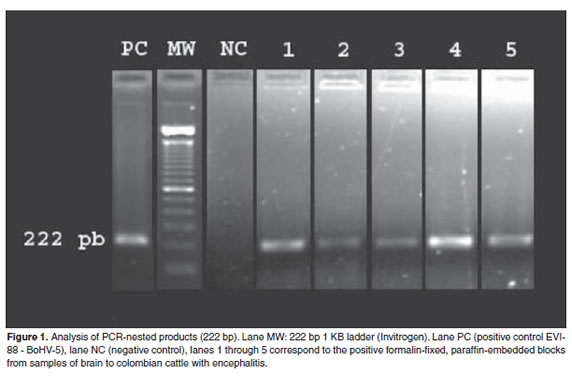
The nested-PCR assay employed in this study detected the presence of BoHV-5 as the casual agent in five of affected cases evaluated (33.3%) among the 15 paraffin-embedded samples tested. The external PCR generated a product of 592 bp and the internal PCR a product of 222 bp (sense: TACGGACTGCCGGATTAA, antisense: GTCACCACTACCACCGCCGCCAAC) (Figure 1).
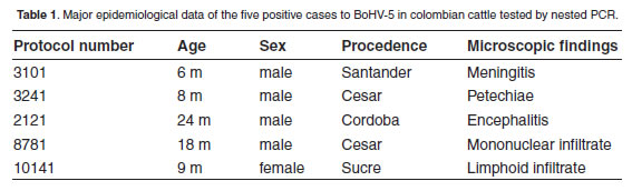
All five cases involved patients with previous history of neurological disease. These cases involved both young and adult animals. In all the cases the veterinarians responsible for the submissions requested diagnosis of rabies. In all cases histological changes were observed in the examined brain sections and showed histological changes typical of herpesvirusinduced meningoencephalitis, notably necrotizing encephalitis (Table 1).
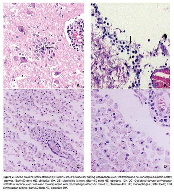
The histological lesions were classifi ed mainly as non-suppurative encephalitis with neuronal degeneration, neuronophagia, and without acidophilic intranuclear inclusions in neurons and glial cells could be observed (Figure 2).
Discussion
Several PCR-based methods have been developed for rapid detection of BoHV-5 in fresh tissue, in spite of the detection of BoHV5 from fixed tissues be largely unexplored, particularly for routinely processed bovine autopsy specimens. Recovering nucleic acid from archived formalin-fixed, paraffin embedded blocks would significantly expand the opportunity for understanding the BoHV-5 epidemiology obtained from negative samples for rabies infection and bovine spongiform encephalopathy disorder (Ferrari et al., 2007).
The use of PCR for diagnosis of bovine herpesvirus 5 is a useful tool in the identification of the virus using nervous tissue samples kept in formaldehyde (Silva et al., 2007). Up to this moment it had not been possible in Colombia to establish with certainty the presence of the virus. Although it is known that it is a virus spread worldwide, the lack of reports made difficult the research on this type of encephalitis because health authorities did not consider cautious to speculate about the presence of a disease with a big economic impact in other countries. Previous studies performed in herds reporting with nervous symptoms that sometimes recovered (although showing some sequels), reported sera positive to BoHV-5, however, the possibility of a crossed reaction between this virus and BoHV-1 (already identified in the country) did not allow clarity about the presence of the agent causing herpetic encephalitis. One aspect hampering the analysis is the difficulty to differentiate between antibodies generated after infection and those generated by the commonly used vaccination against infectious bovine rhinotracheitis.
Some brain tissue samples with histological diagnosis compatible with herpetic encephalitis were tested by immunohistochemistry using the technique standardized by Prof. Dr. Eduardo Flores in the Federal University of Santa María (Rio Grande do Sul, Brazil) showing slight positivism in three cases (data not shown) confirmed by PCR in this study, and explaining the difficulty in antigenic recuperation due to the long time interval between fixation and processing (up to three months in some cases). That time interval did not allow the correct staining using monoclonal antibodies. Finally, molecular extraction of BoHV5 DNA from brain tissue of colombian cattle, and the positive PCR results using primers registered in Gene Bank leaves no doubt about the presence of this virus in this South American country, suggesting its inclusion as a differential diagnosis. This study will allow also the development of research to establish the economic impact of the disease, and the possibility of establishing measures to control a disease apparently present in the country from several years ago.
Acknowledgement To Dr. Darío Mogollón and to the Instituto Colombiano Agropecuario (ICA) for providing the cases thereof and the Dra. Ona Jurksaitis for her valuable help in the translation of text. References 1. Barenfus M, Della-Quadri CA, Mcintyre R, Schroeder RJ. Isolation of infectious bovine rhinotracheitis virus from calves with meningoencephalitis. J Am Vet Med Assoc 1963; 143:725-728. [ Links ] 2. Bartha A, Hajdu G, Aldassy P, Paczolay G. Occurrence of encephalomyelitis caused by infectious bovine rhinotracheitis virus in calves in Hungary. Acta Vet Acad Sci Hung 1969;19:145-151. [ Links ] 3. Beck BE. Infectious bovine rhinotracheitis encephalomyelitis in cattle and its differential diagnosis. Can Vet J 1975; 16:269-271. [ Links ] 4. Beltrão N, Flores EF, Weiblen R, Silva AM, Roehe PM. Infecção e enfermidade neurológica pelo herpesvírus bovino tipo 5 (BHV-5): coelhos como modelo experimental. Pesq Vet Bras 2000; 20:144-150. [ Links ] 5. Ben-Ezra J, Johnson DA, Rossi J, Cook N, Wu A. Effect of fixation on the amplification of nucleic acids paraffin-embedded material by polymerase chain reaction. J Histochem Cytochem 1991; 39: 351-354. [ Links ] 6. Carrillo BJ, Ambrogí A, Schudel A, Vázquez M, Dahme A. Meningoencephalitis caused by IBR Virus in Calves in Argentina. Zbl Vet Méd B 1983; 30:327-332. [ Links ] 7. Colodel, E.M., Nakazato, L., Weiblen, R., Mello, R.M., Da Silva, R.R.P. Meningoencefalite necrosante em bovinos causada por herpesvirus bovino no Estado de Mato Grosso, Brazil. Ciência Rural 2002; 32:293-298. [ Links ] 8. d'Offay JM, Mock RE, Fulton RW. Isolation and characterization of encephalitic bovine herpesvirus type 1 isolates from cattle in North America. Am J Vet Res 1993; 54:534-539. [ Links ] 9. Ferrari HF, Luvizotto MCR, Rahal P, Cardoso TC. Detection of bovine herpesvirus type 5 in formalin-fixed, paraffin-embedded bovine brain by PCR: a useful adjunct to conventional tissuebased diagnostic test of bovine encephalitis. J Virol Method 2007; 146:335-340. [ Links ] 10. Gomes LI, Rocha, MA, Souza JG, Costa EA, Barbosa-Stancioli EF. Bovine herpesvirus 5 (BoHV-5) in bull semen: amplification and sequence analysis of the US4 gene. Vet Res Commun 2003; 27:495-504. [ Links ] 11. Greer CE, Peterson SL, Kiviat NB, Manos MM. PCR amplification from paraffin-embedded tissues. Effects o fixative and fixation time. Am J Clin Pathol 1991; 95:117-24. [ Links ] 12. Halfen DC, Vidor T, Braga FM, Van Der Laan CW. Imunogenididade do Herpesvírus bovino tipo 5 (BHV-5) em vacinas inativadas de diferentes formulações. Ciência Rural 2000; 30:851-856. [ Links ] 13. Jones TC, Hunt RD, King, NW. Veterinary Pathology, 6ed, Philadelphia, Williams and Wilkins. 1997. [ Links ] 14. Jubb KVF, Kennedy PC, Palmer N. Pathology of Domestic Animals, Fourth Ed. San Diego, Academic Press Inc. 1993. [ Links ] 15. Lemos RAA, Barros N, Brum KB. Enfermidades de interesse econômico em bovinos de corte. Perguntas e respostas. Campo Grande MS, Editora UFMS, 2002. [ Links ] 16. Moretti B, Orfei Z, Mondino G, Persechino A. Infectious bovine rinotracheitis, clinical observations and isolation of virus. Vet Ital 1964; 15:676. [ Links ] 17. Pedraza FJ, Alessi AC. Encefalitis bovina por herpesvirus bovino tipo 5 (HVB-5). Una revisión. Rev Colomb Cienc Pecu 2004; 17:2:148-155. [ Links ] 18. Pérez SE, Bretschneider G, Leunda MR, Osorio EA, Flores EF. Primary infection, latency, and reactivation of bovine herpesvirus type 5 in the bovine nervous system. Vet Pathol 2002; 39:437-444. [ Links ] 19. Riet-Correa F, Vidor T, Schild AL, Méndez MC. Meningoencefalite e necrose da córtex cerebral em bovinos causados por herpesvirus bovino 1. Pesq Vet Bras 1989; 9:13-16. [ Links ] 20. Roizman B. The family Herpesviridae: An update. Arch Virol 1992; 123:432-445. [ Links ] 21. Salvador SC, Lemos RAA, Riet-Correa F, Roehe PM, Osorio AL. Meningoencefalite em bovinos causada por herpesvírus bovino tipo 5 (BHV-5). Pesq Vet Bras 1998; 18:76-83. [ Links ] 22. Sanches AWD, Langohr IM, Stigger AL. Doenças do sistema nervoso central em bovinos no Sul do Brasil. Pesq Vet Bras 2000; 20:113-118. [ Links ] 23. Silva AM, Weiblen R, Irigoyen LF, Roehe PM, Sur HJ. Experimental infection of sheep whit bovine herpesvirus type5 (BHV-5): acute and latent infection. Vet Microbiol. 1999; 66:89-99. [ Links ] 24. Silva MS, Brum MCS, Loreto ELS, Weiblen R, Flores EF. Molecular and antigenic characterization of Brazilian bovine herpesvirus type 1 isolates recovered from the brain of cattle with neurological disease. Virus Res 2007; 129:191-199. [ Links ] 25. Spilki, F.R., Franco, A.C., Esteves, P.A., Teixeira, M.B., Esteves, P.A. Bovine herpesvirus type 5 (BHV-5) in a calf with rabies. Pesq Vet Bras 2003; 23:1-4. [ Links ] 26. Studdert MJ. Bovine encephalitis herpesvirus. Vet Rec 1989; 125:584. [ Links ] 27. Vasconcelos RM, Varaschin MS, Wouters F. Meningoencefalite bovina por herpesvírus. Anais Encontro Nacional de Patología Veterinária, Santa Maria-RS Brazil. 1993. [ Links ] 28. Watt JA, Jonston WS, Mac Leod NS, Barlow RM. Infectious bovine rhinotracheitis and encephalitis. Vet Rec 1981; 108:63. [ Links ] 29. Weiblen R, Barros CSL, Canabarro TF. Bovine Meningoencephalitis from IBR vírus. Vet Rec 1989; 124:666-667. [ Links ]

