


Services on Demand
Journal
Article
 English (pdf)
 Article in xml format
 Article references
 Send this article by e-mail
Indicators
-
 Cited by SciELO
 -
 Access statistics
Related links
-
 Cited by Google
 -
 Similars in
SciELO
Similars in
SciELO -
 Similars in Google
Share

 Permalink
Revista Colombiana de Ciencias Pecuarias
Print version ISSN 0120-0690
Rev Colom Cienc Pecua vol.25 no.4 Medellín Oct./Dec. 2012
CLINICAL CASES
Feline aortic thromboembolism: first case reported in Colombia¤
Tromboembolismo aórtico felino: primer caso reportado en Colombia
Tromboembolismo aórtico felino: relato do primeiro caso na Colômbia
Víctor M Molina1, MV, MSc; Juliana Estrada G1, MVZ; Sergio A Salas1, MV, cMSc; María S González1*, MV, MSc.
1 INCA-CES Research Group, Faculty of Veterinary Medicine and Zootechny, CES University, Calle 10 A # 22- 04, Tel. 4440555 Ext 1550, Medellín, Colombia.
* Corresponding author: Maria S Gonzalez, Faculty of Veterinary Medicine and Zootechny, CES University, Calle 10 A # 22- 04, Tel. 4440555 Ext 1550, Medellín, Colombia. Email: mgonzalez@ces.edu.co
(Received:19 april, 2012;accepted:10 september, 2012)
Summary
Anamnesis and treatment approach: a cross-breed tomcat, 5 years old, with bilateral pelvic limb paresis was treated with saline solution hydration (20 drops/in), tramadol (2 mg/kg IV every 8 h), methylprednisolone succinate (18 mg, every 6 h), dimethyl sulphoxyde (0.36 mg diluted in sodium chloride, twice a day, IV) and ketoprofen (2 mg/kg, every 24 h) with no response to this treatment schedule. Clinical and laboratory findings: according to results of thorax and abdomen radiological tests, coldness in pelvic limbs and lack of bleeding after a deep nail cutting, clinical diagnosis of feline aortic thromboembolism was established and the cat was subjected to euthanasia after informed consent. A hypertrophic cardiomyopathy and thrombus in the lumbar abdominal aorta were found at necropsy. Conclusion: the first case of feline aortic thromboembolism in Colombia is reported and the most relevant findings and treatment schedule are discussed.
Keywords: aorta, hypertrophic cardiomyopathy.
Resumen
Anamnesis y aproximación terapéutica: un felino mestizo de 5 años de edad, con cuadro neurológico fue tratado con solución salina (20 gotas/min), tramadol (2 mg/kg i.v. cada 8 horas), succinato de metilprednisolona (18 mg totales cada 6 horas), dimetilsulfoxido (0.36 mg diluidos en cloruro de sodio cada 12 horas e.v.) y ketoprofeno (2 mg/kg cada 24 horas), sin responder al tratamiento. Hallazgos clinicos y de laboratorio: ante los resultados de las radiografìas de tórax y el abdomen, las extremidades pelvianas frías y el no sangrado ante corte profundo de uña, se estableció el diagnóstico de tromboembolismo aórtico felino. El gato fue sometido a eutanasia previo consentimiento informado. A la necropsia fue hallada una cardiomiopatía hipertrófica y un coágulo en la porción lumbar de la aorta abdominal. Conclusiones: este es el primer reporte de un caso de tromboembolismo felino en Colombia, se discuten los hallazgos clínicos y el esquema de tratamiento más relevante.
Palabras claves: arteria aorta, cardiopatía hipertrófica.
Resumo
Anamnese e abordagem de tratamento: um felino mestiço de 5 anos de idade com quadro neurológico foi tratado com solução salina (20 gotas / min), tramadol (2 mg / kg iv c / 8 horas), succinato de metilprednisolona (18 mg c total / 6 h), sulfóxido de dimetilo (0.36 mg de cloreto de sódio diluído em q12h ev) e cetoprofeno (2 mg / kg C/24 h), não respondendo ao tratamento. Achados clínicos e laboratoriais: com os resultados do exame radiológico de tórax e abdôme, membros pelvianas frios e falta de sangramento ao corte profundo da unha, foi diagnosticado com tromboembolismo aórtico felino. O gato foi eutanasiado prévio consentimento informado. Na necropsia foi encontrada cardiomiopatia hipertrófica e um coágulo na porção lombar da aorta abdominal. Conclusões: Este é o primeiro reporte de tromboembolismo felino na Colômbia, neste artigo se discutem os achados clínicos e o cronograma de tratamento mais relevante.
Palavras-chave: aorta, cardiomiopatia hipertrófica.
Introduction
Arterial thromboembolism (ATE), or feline systemic arterial thromboembolism, is a disease generally related to a subjacent pathology of the heart, such as hypertrophic cardiomyopathy (HCM), dilated cardiomyopathy (DCM), restrictive cardiomyopathy (CMR), and non-classified ischemic cardiomyopathy (August, 2006; Baty, 2004; Bonagura, 2009; Kirk et al., 2002). Due to its similarity with carcinogenic embolism in humans (Sodikoff, 1988), ATE was proposed as the name for carcinogenic arterial thromboembolism (August 2006). Thrombus formation is the consequence of vascular stasis preceding clot formation (Bonagura, 2009). Thromboembolism is the consequence of vascular obstruction caused by an intracardiac fibrin clot (August, 2006; Ettinger, 2007). ATE has a prevalence rate of 12% in the United States (Ettinger, 2007). Risk factors for clot formation in cats include blood stasis in cats suffering heart disease, endothelial injury, and hypercoagulopathy conditions (August, 2006; Ettinger, 2007).
In cats suffering ATE, activated platelets release vasoactive substances impairing collateral blood flow around the site of obstruction (Nelson, 1998). When a clot forms inside an aortic fragment, collateral circulation is lost and clinical signs of ischemia and severe pain become evident (August, 2006; Nelson, 1998; Schaer, 2006). When the clot is located in the abdominal aorta, the cat shows unilateral or bilateral paresis, paralysis, segmental arreflexia, contracture and pain in muscles, and cold pelvic limbs without arterial pulse (August, 2006; Nelson, 1998; Schaer, 2006; Birchard, 1996), depending on the site of obstruction. Clinical signs are sudden and progressively worsen. The affected cats respond to pain with self-mutilation, and when limbs present muscle contracture and necrosis, the prognosis is poor (August, 2006; Chandler and British Small Animal Veterinary Association, 2004). However, there is a 75% probability of clinical recovery in patients presenting friable clots (August, 2006). The most frequent laboratory finding in cats affected by ATE are augmentation of aspartate amino-transferase (AST), alkaline phosphatase (ALP), and creatinin kinase (CK) serum levels (August, 2006; Sodikoff 1988). The aim of this paper was to report the first documented case of feline thromboembolism in Colombia.
Patient examination
Anamnesis
A crossbreed tomcat, 5 years old, exhibiting pelvic limbs paresis attended a consultation at the Veterinary Medicine Center (CES University, Medellín, Colombia). Vaccination, deworming, and flea control schedules were completed.
Clinical findings and diagnostic aids used
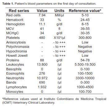
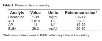
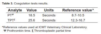
At clinical examination, a severe abdominal pain and bilateral pelvic limb paresis with tachycardia and tachypnea were found. Superficial and deep sensibility was negative. Rectal temperature was 38.8 °C. At neurological evaluation, depression was present without neural reflexes. The patient presented Babiski's signs in pelvic limbs and a severe contracture of gastrocnemius muscles. Subcutaneous emphysema and cold limbs were observed. After the clinical exam was complete, a blood sample was taken for measuring blood parameters (Table 1), blood chemistry values (Table 2), and coagulation (Table 3). The patient was given a hydration schedule. Evidences of thyroid pathologies were not found.
Therapeutic approach
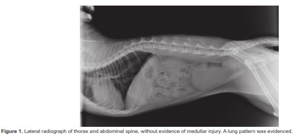
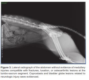
First, the cat was given a hydration schedule consisting of 20 drops/min intravenous saline (0.9% NaCl). Analgesic therapy was established with tramadol (2 mg/kg, IV) every 8 h; methylprednisolone succinate (18 mg total) every 6 h; dymethylsulfoxyde (0.36 mg diluted in NaCl) every 6 h, IV; and ketoprofen (2 mg/kg) every 24 h (Abbott, 2010; August, 2006). After 24 hours the cat had not responded and cold limbs were observed. A thorax and lumbar spine radiography was taken (Figures 1 to 3). The finding of valentine's heart-like shape is indicative of dilated cardiomiopathy (Nelson, 1999) and findings in lung suggested aortic thromboembolism. After two days of hospitalization, an ATE diagnosis was established (Figure 2).
The therapeutic schedule was then completed with furosemide (5 mg/kg) given twice a day, and hydration was established with 5% dextrose. Additionally, angiotensine converting enzime inhibitors (ACEI) antihypertensives (0.5 mg/kg) twice a day p.o., and heparine infusion (10 UI/kg/h) were administered. Cephalotine (20 mg/kg twice a day, IV) was given to prevent sepsis. The cat did not show any response to this therapeutic schedule. On the contrary, friable and cyanotic pelvic limbs and an emphysematous process with partial skin loss and follicular fragility were found. At neurological examination, superficial or deep sensibility was not found, and a completely relaxed anal sphincter and a lack of tail motor response were observed. The owner subsequently signed the informed consent to authorize euthanasia.
Findings at Necropsy
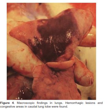
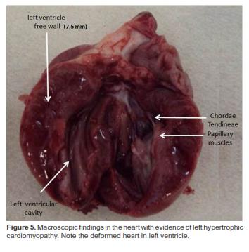
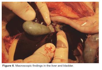
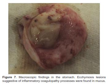
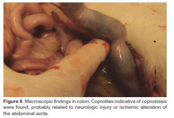
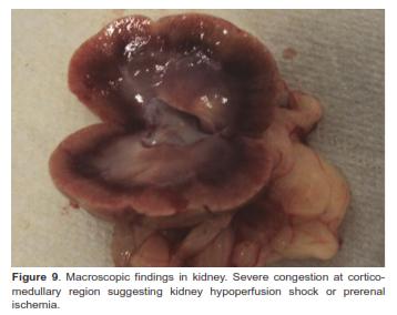
Congestive lesions were found in caudal and intermediate lung lobes at necropsy (Figure 4). Lung congestion and/or hemorrhagic lesions suggestive of cardiogenic-type congestion were evident. Increased size of mediastinum at left ventricle was found (Figure 5), and myocardium wall was thickened (7.5 mm), compatible with HCM (Abbott, 2010). Liver and bladder were congestive (Figure 6). Petechial and hemorrhages were found in stomach mucus (Figure 7). Coprolites were the most relevant finding in colon (Figure 8). Local hemorrhagic lesions and wide congestive areas suggesting a severe ischemic infarction were found in kidneys (Figure 9).
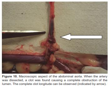
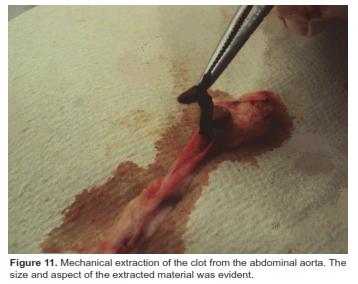
Several ecchymosis lesions were found at the sub lumbar abdominal muscles. After dissection of the abdominal aorta, a 2 cm long clot was found at the bifurcation of the common iliac arteries (Figures 10 and 11). This finding confirmed the diagnosis of feline aortic thromboembolism.
Discussion
In this study, the first case of feline thromboembolism is reported in Colombia. Clinical signs of our patient can overlap with those of medullar compression. Survival rate of cats with partial or complete episodes of feline thromboembolism is 15% and 93%, respectively. Global survival rate is 36% to 39% (Baty, 2004). Mortality rate of cats at 36 hours after the beginning of the episode can reach 28%. However, cats could receive palliative treatment for pain amelioration (Baty, 2004). Hypertrophic cardiomyopathy (HCM) was considered the cause of thromboembolic disease in our patient because the normal value of the ventricular septal thickness in cats is 6 mm (Abbott, 2010; August, 2006).
The results of blood parameter in our patient did show slight leukocytosis with bands and increased coagulation times (PT and PTT). These findings could be associated to deficiency of extrinsic and common coagulation factors, acquired vitamin K deficiency, intravascular disseminated coagulopathy (IDC) (a clinical entity that could end in coagulopathy by consumption), and with liver/biliary insufficiencies. Once liver dysfunction and toxic entities were discarded, three probable entities must be considered in our patient (Stockham and Scott, 2008): IDC, local consumption coagulopathy, and increased PTT of unknown cause.
ALT and creatinine plasma values were increased in our patient probably as a consequence of muscle injuries caused by ischemia and necrosis associated to the thrombus (Bonagura, 2009). These findings are in agreement with the results of a retrospective study carried out with 127 cats with ATE, where more than 70% of the patients had increased ALT, AST, and creatinine plasma levels (Smith et al., 2003).
The prophylaxis for thrombus formation in cats at risk of ATE has not been defined (Smith and Tobias, 2004). In our patient, clinical signs of heart or previous cardiogenic anomalies were not previously reported. For this reason, no treatment for avoiding thrombus formation had been defined. The recommended treatment for ATE in cats does not include thrombolytic therapy or omega-3 fatty acid supplementation. On the contrary, aspirin (5mg/kg/72 h) and non-fractionated L-heparin (NFH) (250 to 300 U/kg/8 h s.c.) are prescribed (Lunsford and Mackin, 2007). Clopidogrel could be used as an alternative to aspirin for long-term control of the disease (Lunsford and Mackin, 2007).
ATE with subjacent HCM was confirmed in our case by macroscopic findings in abdominal aorta. Hypertrophy could be symmetric or asymmetric, located in the subaortic region (Haggstrom, 2003; Cesta et al., 2005). Primary HCM includes a genetic cause, but could be secondary to hyperthyroidism, systemic arterial hypertension, acromegaly, and myocardial tumor inflammatory infiltration (Chetboul and Biourge, 2009). Prognosis of cats affected by ATE is reserved and the starting time for therapy depends on speed of diagnosis. The lesions found in the pelvic limbs in our patient at day three after consultation are in agreement with a previous report (Smith et al., 2003).
The radiographic finding compatible with lung edema (Figure 1) and tortuous lung veins are frequent in HCM (Nelson, 1999). Tsujino et al. (2005) reported chronic suppurative pyelo-nephrosis and liver and spleen congestion supporting the preventive use of antibiotics in early ATE diagnosis (Tsujino et al., 2005).
Conclusion
In conclusion, the acute phase of the signs in our patient, the severe pain he exhibited, the cold temperature of his pelvic limbs, and the lack of bleeding when a deep nail cut (including nail chorion) was performed, are clinical signs highly suggestive of feline thromboembolism.
Acknowledgements
INCA-CES research group activities are funded by University CES (Medellín, Colombia). Authors thank Dr. Juan Maldonado-Estrada (University of Antioquia) for critical review of this manuscript.
References
1. Abbott JA. Feline hypertrophic cardiomyopathy: an update. Vet Clinics North Am Small Anim Pract 2010; 40:685-700. [ Links ]
2. August J. Consultations in feline internal medicine. Vol 5. London: Elsevier Saunders; 2006. p. 341-55 [ Links ]
3. Baty C. Feline hypertrophic cardiomyopathy: an update. Vet Clinics North Am Small Anim Pract 2004; 34:1227-1234. [ Links ]
4. Birchard S. Manual clínico de pequeñas especies. 1st ed. México: McGraw-Hill Interamericana; 1996. [ Links ]
5. Bonagura J. Kirk's current veterinary therapy. 14th ed. London: Elsevier Saunders; 2009. [ Links ]
6. Cesta MF, Baty CJ, Keene BW, Smoak IW, Malarkey DE. Pathology of end-stage remodeling in a family of cats with hypertrophic cardiomyopathy. Vet Pathol 2005; 42: 458-467. [ Links ]
7. Chandler E, British Small Animal Veterinary Association. Feline medicine and therapeutics. 3rd ed. Ames Iowa: Blackwell Pub, Iowa State Press; 2004. [ Links ]
8. Chetboul V, Biourge V. Enfermedades cardiovasculares adquiridas en el gato: influencia de la nutrición. In: Enciclopedia de la nutrición clínica felina. 1st ed. Aimargues (Francia): Royal Canin; 2009. p. 323-54. [ Links ]
9. Ettinger S. Tratado de medicina interna veterinaria: enfermedades del perro y el gato. 6th ed. Madrid: Elsevier; 2007. [ Links ]
10. Haggstrom J. Hypertrophic cardiomyopathy in cats-it used to be so simple!. J Fel Med Surg 2003; 5:139-141. [ Links ]
11. Kirk RW, Bistner SI, Ford RB, Raffe MR. Manual de terapéutica y procedimientos de urgencia en pequeñas especies. México: McGraw-Hill Interamericana; 2002. [ Links ]
12. Lunsford KV, Mackin AJ. Thromboembolic therapies in dogs and cats: an evidence-based approach. Vet Clin North Am Small Anim Prac 2007; 37:579-609. [ Links ]
13. Nelson R. Small animal internal medicine. 2nd ed. St. Louis: Mosby; 1998. [ Links ]
14. Nelson R. Manual of small animal internal medicine. 1st ed. St. Louis: Mosby; 1999. [ Links ]
15. Schaer M. Medicina clínica del perro y el gato. Barcelona: Masson; 2006. [ Links ]
16. Smith SA, Tobias AH, Jacob KA, Fine DM, Grumbles PL. Arterial thromboembolism in cats: acute crisis in 127 cases (1992-2001) and long-term management with low-dose aspirin in 24 cases. J Vet Int Med 2003; 17:73-83. [ Links ]
17. Smith S, Tobias A. Feline arterial thromboembolism: an update. Vet Clin North Am Small Anim Prac 2004; 34:1245-1271. [ Links ]
18. Sodikoff C. Perfiles de laboratorio en las enfermedades de los pequeños animales: guía para el diagnostico de laboratorio. Buenos Aires: Inter-Medica; 1988. [ Links ]
19. Stockham SL, Scott MA. Fundamentals of veterinary clinical pathology. Ames, Iowa: Blackwell Pub; 2008. [ Links ]
20. Tsujino, Kumiko, Yoshiaki Hikasa, Saburo Minami, Yoshiharu Okamoto, Takehito Morita, Akinori Shimada. Chronic myocardial infarction due to arteriosclerosis of coronary arteries followed by acute thromboembolism of caudal abdominal aorta in a cat. J Vet Med Sci 2005; 67:631-634. [ Links ]
Notas al pie
¤ To cite this article: Molina VM, Estrada J, Salas SA, González MS. Feline aortic thromboembolism: first case reported in Colombia. Rev Colomb Cienc Pecu 2012; 25:639-645.
