


Services on Demand
Journal
Article
 Article in xml format
 Article references
 Send this article by e-mail
Indicators
-
 Cited by SciELO
 -
 Access statistics
Related links
-
 Cited by Google
 -
 Similars in
SciELO
Similars in
SciELO -
 Similars in Google
Share

 Permalink
Revista Colombiana de Ciencias Pecuarias
Print version ISSN 0120-0690
Rev Colom Cienc Pecua vol.26 no.2 Medellín Apr./June 2013
CLINICAL CASE
Osteomyelitis and periosteal reaction in a red brocket deer (Mazama americana)¤
Osteomielitis y reacción perióstica en un venado soche (Mazama americana)
Osteomelite e reação periosteal em um veado (Mazama americana)
Ana M Henao-Duque1*, DVM; Juliana Peña-Stadlin2, DVM; Dave Wehdeking-Hernández2, DVM.
* Corresponding author: Ana María Henao Duque, Programa de Ofidismo/Escorpionismo, Universidad de Antioquia, Carrera 50A N° 63-65, Tel. 2631914, Medellín, Colombia. e-mail: anahe1989@hotmail.com
1 Programa de Ofidismo/Escorpionismo, Universidad de Antioquia, AA 1226, Medellín, Colombia.
2 Unidad de Bienestar Animal, Fundación Zoológica de Cali, AA 760045, Cali, Colombia.
(Received: June 24, 2012; accepted: January 28, 2013)
Summary
Anamnesis and treatment approach: a female red brocket deer (Mazama americana, Erxleben 1777) presented a post-traumatic abscess in the left-carpometacarpal joint. The deer was treated with enrofloxacin (5 mg/kg) and ivermectin (0.2 mg/kg) with no response. The animal underwent two surgical procedures to remove purulent material and perform adequate antisepsis as well as several antimicrobial therapies, with satisfactory results in a period of 68 days. Clinical and laboratory findings: according to the radiological and laboratory tests the animal developed a metacarpal bone osteomyelitis with periosteal reaction due to a beta-hemolytic Streptococcus abscess. Conclusion: to our knowledge, this is the first report of medical procedures in this species. The most relevant findings and treatment schedule are discussed.
Key words: antimicrobial therapy, cervid, post-traumatic abscess.
Resumen
Anamnesis y aproximación terapéutica: una hembra de venado soche (Mazama americana), que presentó un absceso postraumático en la articulación carpo-metacarpiana del miembro anterior izquierdo fue tratada con enrofloxacina (5 mg/kg) e ivermectina (0,2 mg/kg), sin responder al tratamiento. Se sometió a dos procedimientos quirúrgicos con el fin de extraer el material purulento y realizar una antisepsia adecuada, además de varias terapias antibióticas, con resultados satisfactorios en un término de 68 días. Hallazgos clínicos y de laboratorio: de acuerdo a los resultados radiológicos y de laboratorio el animal desarrolló osteomielitis con reacción perióstica metacarpiana, debido a un absceso por Streptococcus beta-hemolítico. Conclusión: este es el primer reporte sobre procedimientos médicos en esta especie, se discuten los hallazgos clínicos y el esquema de tratamiento más relevante.
Palabras clave: absceso postraumático, cérvido, terapia antimicrobiana.
Resumo
História e tratamento: uma fêmea de veado (Mazama americana) apresentou um abscesso póstraumático na articulação do carpo-metacarpo do membro esquerdo. Foi tratada com enrofloxacino (5 mg/kg) e ivermectina (0,2 mg/kg), sem responder ao tratamento. Logo foi submetida a dois procedimentos cirúrgicos para remover o pus e fazer limpeza com antisséptico apropriado, além de várias terapias antibióticas, com resultados satisfatórios em um período de 68 dias. Resultados clínicos e laboratoriais: de acordo com os resultados dos exames radiológicos e de laboratório, o animal desenvolveu osteomielite no metacarpo com reação periosteal devido a um abscesso por Streptococcus beta-hemolítico. Conclusão: este é o primeiro relatório sobre procedimentos médicos nesta espécie, discutimos os achados clínicos e o sistema de tratamento adequado.
Palavras chave: abscesso pós-traumático, cervídeo, terapia antimicrobiana.
Introduction
The red brocket deer (Mazama americana) is native to South America and is included in the International Union for Conservation of Nature (IUCN) Red List of Threatened Species. This species is the largest of the genus Mazama, reaching 30 to 40 kg body weight and 65 cm height. The species inhabits dense forests and riverbanks covered with vegetation (Mayor et al., 2011; Abril et al., 2010). The IUCN classifies this species as ''data deficient'' (IUCN, 2008) and the conservation status of its wild populations is unknown.
The most common infectious diseases on cervids are usually caused by Yersinia pseudotuberculosis, Corynebacterium (Actinomyces) pyogenes, Escherichia coli, Pasteurella multocida, Streptococcus spp., Staphylococcus spp., Fusobacterium necrophorum, and Pseudomonas spp. (Barbanti et al., 2001).
According to reports and studies on wild cervids around the world, mandibular osteomyelitis is a common post-mortem finding (Konjevic et al., 2011; Flueck and Smith-Flueck, 2008). Konjevic et al. (2011) reported a Slovenian deer (Capreolus capreolus) with chronic pyogranulomatous mandibular osteomyelitis. Osteomyelitis has been observed only in the jaw of wild elk and reindeer in North America (Hoefs and Bunch, 2001). Pathogenicity and mortality caused by this disease is unknown in wild cervids. Some authors suggest that this infectious process could be subclinical. According to a study of Patagonian huemules (Hippocamelus bisulcus) the most common bone diseases are secondary alveolar chronic osteomyelitis and osteoarthritis (Flueck and Smith- Flueck, 2008).
Although cervid pathologies are well known, currently there are no protocols for their treatment. For this reason, it is necessary to extrapolate treatments described for domestic mammals such as horses. The previously established treatment principles for septic arthritis and osteomyelitis in horses include systemic broadspectrum antimicrobial drugs that reach effective concentrations in bone (Hardy, 2006).
However, medical procedures for captive cervids are limited because of their nervous behavior and incidence of post-capture myopathy syndrome, a problem that is easily developed and potentially fatal. This situation makes the procedures recommended in the literature impractical.
Patient examination
Anamnesis
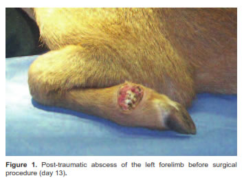
A female red brocket deer was found with superficial wounds, caused by intra-specific aggression. The deer presented 1/4 lameness in the left forelimb (Flo et al., 2002), without superficial injuries. The treatment assigned was oral ivermectin as a preventive therapy against myasis, and enrofloxacin for 5 days (Table 1). An abscess compromising the fifth phalanx of the left forelimb was found in the carpometacarpal joint (Figure 1). Enrofloxacin and meloxicam therapy was initiated (Table 1). As the animal had a 2/4 lameness (Flo et al., 2002) on day 13, a chemical restrain with previous fasting was initiated using Zoletil 50® (Tiletamin/Zolacepam) and Rompun 2%® (Xylazine hydrochloride) intramuscular (Table 1). During removal of the abscess the animal was anesthetized and given oxygen (6 L/m).
Clinical findings
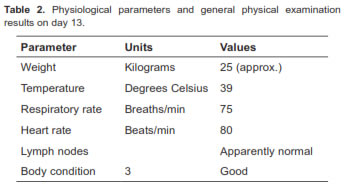
The animal was in adequate body condition (3/5), and presented recent rostral abrasions and a post-traumatic abscess (Figure 1; Table 2).
Therapeutic approach
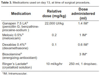
A surgical procedure was performed to extract caseous and necrotic material of the fifth phalanx. A deep digital flexor tendon resection was observed and could not be fixed. The healthy phalanx was fixed to fibrous connective tissue with a tension suture. The wound was left open, with a wick impregnated with healing ointment and an elastic bandage. Medications and doses used during surgery are described in table 3. The animal was treated with antimicrobial drugs for 7 days and continued with analgesic therapy. A probiotic and antioxidant therapy with vitamin E was started on day 16. On day 17 the animal had diarrhea. Consequently, oral antimicrobial therapy was suspended.
Monitoring
Analgesic and antioxidant therapy was restarted. Prophylactic antifungal therapy was also administered (Table 1). Lameness increased to 4/4 (Flo et al., 2002) on day 30, with a very swollen forelimb and a fistula. Antimicrobial therapy was restarted. A second surgical procedure was scheduled.
Second surgical procedure
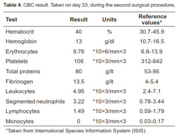
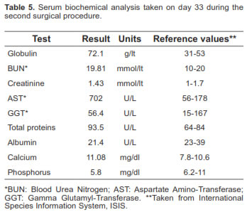
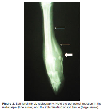
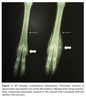
Surgery was performed on day 33 day using the same anesthetic protocol (Table 1). Blood samples were taken from the saphenous vein during surgery for Complete Blood Count (CBC, Table 4) and blood chemistry (Table 5). Additionally, a deep swab was taken for culture as well as two radiographs: 1) Latero-lateral (LL) view of the carpometacarpal joint; 2) Antero-posterior (AP) view comparing both forelimbs.
At the surgical procedure, the wound and the surrounding tissue were swollen with exposure and avulsion of the deep digital flexor tendon. A deep wash with Harris solution was performed. The necrotic and infected tissues were removed. A tension suture was made. Physiological parameters were maintained as normal and oxygen saturation ranged between 77% and 94%. The animal was left in an outdoor enclosure with plenty of hay for recovery.
The CBC results showed marked thrombocytopenia associated with platelet aggregation. The biochemistry serum result showed aspartate aminotransferase (AST) increased 3.9 times due to the muscle trauma. Hyperproteinemia, hyperglobulinemia, and hypoalbuminemia were correlated with a chronic inflammatory process. This was also correlated with fibrinogen increase. Additionally, hypercalcemia caused by septic osteomyelitis was found. Hypophosphatemia is usually associated with hypercalcemia (Sodikoff, 1996).
Monitoring
The radiographic findings demonstrated osteomyelitis in the carpometacarpal joint, with metacarpal periosteal reaction (Figures 2 and 3). Figure 4 shows the wound on day 34.
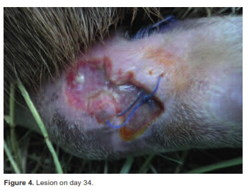
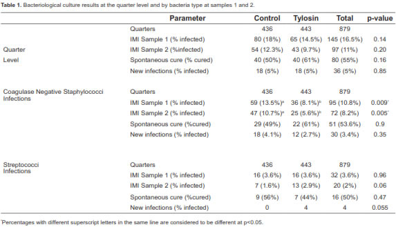
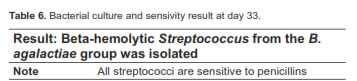
Treatment was altered after receiving bacterial culture and sensivity testing results (Table 6). Enrofloxacin was exchanged for Ganapen LA 7.5®, a long-acting penicillin (Table 1). The animal had a guarded prognosis.
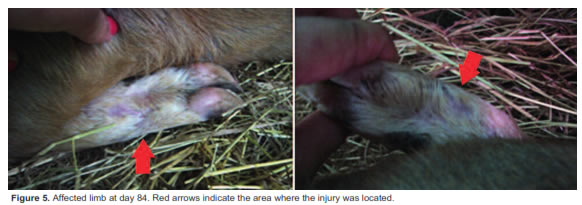
Antimicrobial systemic therapy was suspended at day 53 because the animal showed clinical improvement. Analgesic and antioxidant therapy continued. Figure 5 shows the lesion and the affected limb at day 84, at full recovery.
Discussion
Beta-hemolytic Streptococcus was isolated at the culture testing. This bacterium is included among commonly infectious etiologic agents in cervids. This is an important finding because osteomyelitis has been reported in wild cervids around the world (Konjevic et al., 2011; Flueck and Smith-Flueck, 2008), Actinomyces pyogenes being the most common etiologic agent for abscesses (Wobeser, 2001; Turnquist and Fales, 1998). Pathogens such as Actinobacillus sp., Escherichia coli, Klebsiella spp., Pseudomonas spp., Salmonella spp. and Streptococcus spp. have been described in horses with septic arthritis and osteomyelitis (Hardy, 2006).
Radiographs are essential diagnostic tools for traumas unresponsive to therapy. Therefore, diagnosis should not be limited to a single radiograph. In the case of osteomyelitis or arthritisaffected horses, radiographs should be taken on a weekly basis until clinical signs of deterioration or resolution are present (Hardy, 2006).
In the case presented here, it is important to highlight that therapy was successful because of the docile temperament of this particular animal, which allowed a favorable medical management. It is also important to ensure appropriate antimicrobial therapy from the beginning. Additionally, the presence of osteomyelitis suggests antimicrobial drug combinations should be used to reach effective concentrations in bones and achieve a synergistic effect (Hardy, 2006). An aggressive therapy with long-action penicillin was implemented after betahemolytic Streptococcus was isolated. Rifamycin was administered as a local antimicrobial drug. Rifamycin combined with other drugs is effective in treating osteomyelitis and septic arthritis, however is not recommended to be administered by itself because it can rapidly generate bacterial resistance (Hardy, 2006).
Conclusion
Although the patient was a cervid, this case had a positive outcome because its docility and human acceptance facilitated handling and medical treatment. It is the important to emphasize animal conditioning programs for medical procedures in captive wildlife in order to facilitate successful medical therapy.
Literature reports about interventions and medical procedures with these animals in captivity are scarce. It is recommended to refer to data from similar species, which allow extrapolating therapies and medical handling.
Notes
¤ To cite this article: Henao-Duque AM; Peña-Stadlin J, Wehdeking-Hernández D. Osteomyelitis and periosteal reaction in a red brocket deer (Mazama americana). Rev Colomb Cienc Pecu 2013; 26:137-143.
Acknowledgements
The research and medical treatment completed in this report were made possible with the support of the Animal Welfare Unit of the Cali Zoo Foundation, especially with the help of zookeeper Rodrigo Campo. We gratefully acknowledge the contributions made by Camilo Londoño in the preparation and review of this manuscript.
References
Abril VV, Carnelossi EAG, González S, Duarte JMB. Elucidating the Evolution of the Red Brocket Deer Mazama americana Complex (Artiodactyla; Cervidae). Cytogenet Genome Res 2010; 128:177-187. [ Links ]
Barbanti JM, Merino M, Gonzalez S, Veloso AL, Mansano J, Juán MP, Pandolfi JR, Gouveia I, Do Nascimento AA, Zacarias R, Pessoa J, Catao JL, Werther K, García JE, Da Silva RJ, Reiko E. Order Artiodactyla, family Cervidae (deer). In: Fowler EM, Cubas SZ, editors. Biology, Medicine and Surgery of South American Wild Animals. Iowa State: University Press; 2001. p.402-22. [ Links ]
The IUCN Red List of Threatened Species. Version 2011.2. [Accessed November 19, 2011] URL: http://www.iucnredlist.org/apps/redlist/details/29619/0 [ Links ]
Flo GL, Dyson SJ, Radostits OM. Exploración del sistema músculo esquelético. In: Radostits OM, editor. Examen y diagnóstico clínico en veterinaria. Madrid: ELSEVIER, Ediciones Halcourt SA; 2002. p.573-659. [ Links ]
Hardy J. Etiology, diagnosis, and treatment of septic arthritis, osteitis, and osteomyelitis in foals. Clin Tech Equine Pract 2006; 5:309-317. [ Links ]
Hoefs M, Bunch TD. Lumpy jaw in wild sheep and its evolutionary implications. J Wildlife D 2001; 37:39-48. [ Links ]
Konjevic D, Jelenko I, Severin K, Policnik H, Janicki Z, Slavica A, Njemirovskij V, Stanin D, Pokorny B. Prevalence of Mandibular Osteomyelitis In Roe Deer (Capreolus capreolus) in Slovenia. J of Wildlife Diseases 2011; 47:393-400. [ Links ]
Kraus BM. Lacerations involving synovial structures: Initial treatment and novel therapy for infectious arthritis/ tenosynovitis. Clin Tech Equine Pract 2006; 5:82-92. [ Links ]
Mayor P, Bodmer RE, López-Béjar M, López-Plana C. Reproductive biology of the wild red brocket deer (Mazama americana) female in the Peruvian Amazon. An Repro Sci 2011; 128:123-128. [ Links ]
Sodikoff CH. Pruebas diagnósticas y de laboratorio en las enfermedades de pequeños animales. 2nd edition. Madrid: Harcourt Brace; 1996. [ Links ]
Turnquist SE, Fales WH. Disseminated Actinomyces pyogenes infection in a free-ranging white-tailed deer. J Vet Diagn Invest 1998; 10:86-89. [ Links ]
Flueck WT, Smith-Flueck JAM. Age-independent osteopathology in skeletons of a South American cervid, the Patagonian huemul (Hippocamelus bisulcus). J of Wildlife D 2008; 44:636-648. [ Links ]
Wobeser G. Staphylococcus infection. In: Williams ES, Barker IK, editors. Infectious diseases of wild mammals. 3th ed. Iowa State: University Press/Ames; 2001. p.509-10. [ Links ]
