


Services on Demand
Journal
Article
 English (pdf)
 Article in xml format
 Article references
 Send this article by e-mail
Indicators
-
 Cited by SciELO
 -
 Access statistics
Related links
-
 Cited by Google
 -
 Similars in
SciELO
Similars in
SciELO -
 Similars in Google
Share

 Permalink
Revista Colombiana de Ciencias Pecuarias
Print version ISSN 0120-0690
Rev Colom Cienc Pecua vol.27 no.3 Medellín July/Sept. 2014
CLINICAL CASE
Idiopathic stringhalt in a Colombian Creole horse*
Arpeo idiopático en un caballo Criollo Colombiano
Harpejamento idiopático em um cavalo Crioulo Colombiano
Diego Duque1, MV; Valentina Velasquez2, MVZ (est.); Laura Espinosa2, MVZ (est.); Maria P Arias2,1†, MV, MS, PhD.
1 Centro de Medicina Veterinaria y Zootecnia, Universidad CES, Medellín, Colombia.
2 Facultad de Medicina Veterinaria y Zootecnia, Universidad CES, Medellín, Colombia.
† Corresponding author: Maria P Arias. Facultad de Medicina Veterinaria y Zootecnia. Universidad CES. Calle 10 A # 22 – 04. El Poblado, Medellín, Colombia. Email: marias@ces.edu.co.
(Received:June 20, 2013; accepted:January 28, 2014)
Summary
Anamnesis: an adult horse that showed hind limb hyperflexion was examined. Clinical and laboratory findings: at locomotion examination bilateral hyperflexion was observed; the right hind limb was more severely affected than the left. Electromyographic and histopathological examination revealed neural denervation and muscular atrophy supporting the idiopathic stringhalt diagnosis. Treatment approach: a lateral digital extensor tenectomy and partial myectomy was practiced in both hind limbs, accompanied by medical treatment and implementation of a mild exercise plan. The effectiveness of surgery is still controversial in these cases; however, this patient evidenced slow improvement after surgery and exercise seemed to be instrumental in the recovery of his normal locomotion. Conclusion: to our knowledge, this is the first report of a clinical case compatible with idiopathic stringhalt in Colombian Creole horses, but further studies are necessary to clarify the etiology and pathogenesis of stringhalt in Colombia.
Key words: axonopathy, denervation, electromyography, hyperflexion.
Resumen
Anamnesis: se examinó un caballo adulto que mostraba hiperflexión de ambos miembros posteriores. Hallazgos clínicos y de laboratorio: al examen locomotor se observó hiperflexión de ambos miembros posteriores pero el miembro posterior derecho parecía estar más afectado. El examen histopatológico y la electromiografía revelaron denervación neural y atrofia muscular soportando el diagnóstico de arpeo idiopático. Abordaje terapéutico: se practicó tenectomía y miectomía parcial del extensor digital lateral en ambos miembros posteriores, acompañada de tratamiento médico con la implementación de un plan de ejercicio ligero. La eficacia de la cirugía es controversial aún, sin embargo, en este caso, una lenta recuperación fue evidente y el ejercicio pareció ser un factor clave. Conclusión: el presente caso clínico es para nuestro conocimiento el primero compatible con arpeo idiopático en el Caballo Criollo Colombiano, pero se deben realizar más estudios para clarificar la etiología y patogenia del arpeo en Colombia.
Palabras clave: axonopatía, denervación, electromiografía, hiperflexión.
Resumo
Anamnese: foi examinado um cavalo adulto que mostrava hiperflexão dos dois membros posteriores. Achados Clínicos e de laboratorio: o exame foi observado hiperflexão de ambos os membros posteriores, estando mais afectado o membro posterior direito. O exame histopatológico e eletromiografia revelou denervação neural e atrofia muscular levando ao diagnóstico de harpejamento idiopático. Abordagem terapêutica: foi realizada tenectomia e miectomia parcial do músculo extensor digital lateral em ambos os membros posteriores, acompanhada de tratamento médico com a implementação de um plano de exercícios leves. A eficácia da cirurgia é ainda controversa, no entanto, neste caso foi evidente uma recuperação lenta e o exercício pareceu ser um fator fundamental. Conclusão: este relato de caso é, a nosso conhecimento, o primeiro compatível com harpejamento idiopático, mas devem ser realizados mais estudos para esclarecer a etiologia e patogenia do harpejamento no Cavalo Crioulo Colombiano.
Palavras chave: axonopatia, denervação, eletromiografia, hiperflexão.
Introduction
Stringhalt in horses–recently renamed Equine reflex hypertonia–consists of an intermittent hyperflexion of the tarsocrural joint, and may affect one or both hind limbs (Furr et al., 2011). It is considered a form of spasticity due to upper motor neuron dysfunction or hyperexcitability of motor neurons (Crabill et al., 1994). Stringhalt has long been recognized in conventional (idiopathic stringhalt) and epidemic (Australian stringhalt) forms (Slocombe et al., 1992) but it has not been determined which stringhalt form affects Colombian Creole horses. The aim of this report is to describe a case of the idiopathic stringhalt form in a Colombian Creole horse.
Patient examination
Anamnesis
A 5-year-old Colombian Creole horse was presented to the Veterinary Clinic of CES University in Medellin (Colombia) with a two-month history of a sudden, progressive bilateral abnormal gait and exaggerated hock flexion. The horse had lived for one year in the same pasture with other horses that had not developed clinical signs consistent with stringhalt. There was no history of recent trauma or previous medical problems.
Clinical findings
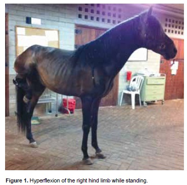
The horse weighed 316 kg on arrival, the patient was excited, very nervous and reluctant to walk. Physical parameters were normal. Hind limb hyperflexion was evident at clinical examination. He showed an exaggerated flexion of both pelvic limbs when he was obligated to walk. Hyperflexion was bilateral but the right hind limb seemed to be more affected than the left. He kept his hind limbs hyperflexed while standing for several seconds until he relaxed and was unable to move backwards more than a few steps. Excitement triggered hyperflexion (Figure 1). The clinical diagnosis was bilateral stringhalt grade V/V, according to the Huntington scale (Huntington et al., 1989).
Diagnostic aids used
Chemistry and routine hematological values were normal. Electromyographic (EMG) examination of pelvic limb muscles with a concentric electrode (Neurodiagnostics LBmII®) inserted into the lateral digital extensor and long digital extensor muscles was recorded at a sampling rate of 10 milliseconds and 100 μV amplitude, following the Van Wessum protocol (1999). The horse was sedated with Xylazine IV., at a dose of 0.1 mg/Kg. Insertional activity, motor neurons and muscle activity were evaluated at rest and during reflex contraction. Insertional activity was measured 3 times and then each muscle was observed for spontaneous activity at rest (Wijnberg, 2003). EMG showed an exaggerated electrical activity of the lateral digital extensor muscle and a disorganized spontaneous electrical activity in both hind limbs. Decreased voluntary activity compatible with denervation of the hindquarters' lateral digital extensor muscles was observed.
Biopsies of the lateral digital extensor muscle and superficial peroneal nerve were collected during a surgical approach, immersed in 10% phosphatebuffered formalin solution for 24 h and shipped to the Veterinary Pathology Laboratory of the Universidad Nacional de Colombia. Tissues were dehydrated, paraffin-embedded and 3 μm sections were cut for conventional paraffin-embedded light microscopy examination. Samples of skeletal muscle and peripheral nerve were stained with haematoxylineosin and Masson's Trichrome (Raimondo, 2009). Histopathological examination showed multifocal irregularity in shape and size of some muscle fascicules. The superficial peroneal nerve presented vacuolization of some nerve fibers, and axonal degeneration with evidence of regeneration. Different levels of myelin degradation and connective tissue fragmentation were also found. The pattern was consistent with acute denervation (Figure 2).
Treatment approach
Medical therapy
The initial therapy focused on reducing any secondary inflammatory damage and restoring motion range. It included: Flunixin meglumine (1.1 mg/kg IV, every 12 h during 5 days) and 100 mL DMSO at 10% diluted in Hartman (IV, every 24 h during 3 days, followed by half of this dose for 3 additional days). Ranitidine (8.8 mg/kg) was administered for 7 days every 8 h to protect the gastric mucosa. Phenytoin sodium was administered orally (15 mg/kg body weight twice a day for three weeks) to decrease hyperflexia, since it has been reported as effective in some stringhalt cases (Huntington, 1991; Furr, 2011), Vitamin E (6000 UI, each 24 h PO) and Thiamine (10 mg/kg) were also prescribed throughout the hospitalization period. This initial treatment failed to provide any improvement of the condition.
Surgical procedure
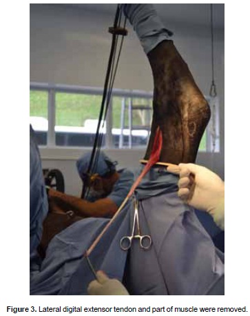
One month after admission, the patient had lost body condition and no clinical improvement was observed, therefore, the animal underwent a lateral digital extensor myotenectomy. The patient was premedicated with Xylazine (0.5 mg/kg IV), Diazepam (0.05 mg/kg IV) and Ketamine (0.5 mg/kg IV) and put under general anesthesia using isofluorane. The horse was on dorsal recumbence with limb extension throughout the procedure. A 1 cm incision was made over the lateral digital extensor tendon, just proximal to its junction with the long digital extensor tendon, which was exposed by blunt dissection. Then, a 7 cm longitudinal incision was done directly over the muscle, in the lateral aspect of the limb, above the lateral malleolus of the tibia. The muscle belly was exposed by blunt dissection. The tendon was severed in the distal incision and through the proximal incision by traction and the muscle was also severed at the proximal aspect (Figure 3). Superficial peroneal nerve was localized by blunt dissection and a 2 cm partial longitudinal section was removed. Fascia and subcutaneous tissue were closed with a simple continuous pattern using 2 - 0 absorbable suture. The skin of the proximal incision was closed with simple-continuous pattern using N.0 synthetic non-absorbable material. The distal incision was closed with skin sutures. The procedure was repeated in the contralateral limb. Finally, both limbs were bandaged. Penicillin (15,000 IU/kg IM, a single dose) and Phenilbutazone (4.4 mg/kg IV each 24 h during 8 days) were administered.
Biopsies of superficial peroneal nerve and lateral digital extensor muscle were taken, immersed in 10% buffered formalin and shipped to the Veterinary Pathology Laboratory of the Universidad Nacional de Colombia.
Clinical management and evolution
One day after surgery the horse was alert, but excitement or walking on hard floor triggered signs of stringhalt again. A slow improvement was seen when the horse was on pasture. However, hyperflexion became apparent when he returned to the box, although this behavior was not constant. The bandage was changed every day after surgery.
The patient remained hospitalized for three months while treatment with Thiamine and Vitamin E continued. As mild exercise is recommended, this horse was exercised three times a day by walking slowly on soft ground for 10 minutes and remaining outside for 3 h daily as part of the therapeutic management. The owner was instructed to hand-walk the horse when discharged, following the same postoperative exercise program for three additional months, which resulted in notorious locomotion improvement. A progressive recovery of the normal gait was observed several months after the surgery.
Discussion
Australian stringhalt is caused by neurotoxins produced by Hypochoeris radicata in response to drought stress (MacKay, 2013). This condition was described for the first time in Australia in 1848 (Domange, 2013), although several cases have been reported in other countries (Araya, 1998; Araujo, 2008; Rodrigues, 2008). Idiopathic stringhalt is a mechanical lameness condition for which different possible causes have been proposed, such as articular lesions of the hock, nervous lesions, spasmodic contractions of the metatarsus flexor muscle due to reflex irritation and degeneration of the sciatic or peroneal nerves and affections of the spinal cord (Aleman, 2011; Dixon, 1969). The stringhalt form has not yet been defined in Colombian Creole horses, although it is a frequent condition in this breed.
Stringhalt occurs in all breeds and ages of horses. The affected horse displays hind limbs spasmodic flexion and abnormal gait during progression because of a distal axonopathy with a skeletal muscle denervation and atrophy (Domange, 2010). In this patient, the main manifestation was a spasmodic and excessively rapid flexion of hind limbs, more marked when the horse was required to move.
Histopathological changes associated with stringhalt in muscle and nerve plays an essential role in the diagnosis of neuromuscular disorders by providing information on whether a disease process affects muscle, nerve, or both (Aleman, 2008). Nonspecific histological alterations include neurogenic muscle atrophy seen as anguloid myofiber shape or marked size differences, axonal degeneration and myelin splitting and degeneration (Summers, 1995). In this case, the histopathological alterations found were axonal degeneration with evidence of regeneration and vacuolization of some neural fibers and neurogenic muscle atrophy evidenced by different size and shape of myofibers.
EMG provides essential information (Wijnberg, 2004) and may support or refute the diagnosis (Wijnberg, 2000), so it should always be considered in the examination of a horse with signs of stringhalt in order to make the best clinical decisions (Van Wessum, 1999). Abundant denervation with decreased voluntary activity in the lateral digital extensor muscle was observed in this animal. Findings revealed the presence of a peripheral neuropathy with axonal damage, predominantly distal, and corroborate a histopathological diagnosis of neural denervation.
Sometimes it is necessary to give a minimum dose of sedatives to impatient horses during EMG examination (Wijnberg, 2004). In this patient, we used a low dose of Xylazine, just enough to put the electrodes on the muscles.
Identification and classification of a specific neuromuscular dysfunction require EMG and histopathological analyses of muscle and nerve (Stevens, 2009). In this patient, there was no history of nervous episodes or traumatic events, and the initial cause could not be clearly established. EMG and histopathological findings were compatible with a distal axonopathy, while epidemiological and clinical evidence support the diagnosis of idiopathic stringhalt since other horses living in the same conditions were not affected; no toxic agents were found in the pasture.
The use of muscle relaxants such as phenytoin in oral doses of 15-25 mg/kg from two (Furr, 2011) to three weeks (Wijnberg, 2000) has shown to be effective in horses suffering from Australian stringhalt. Pharmacological effects involve neuronal stabilization and control of the electrical activity of peripheral nerves and skeletal muscle membrane (Huntington, 1991). In this patient, medical treatment with phenytoin sodium was not useful in reducing the clinical signs of stringhalt.
The effectiveness of the surgery remains controversial. According to Torre (2005), symptoms do not always disappear after surgery. Armengou (2010) reported that rapid clinical improvement after surgery is unusual. Hahn (2008) reported that surgical removal of a section of the myotendinous region containing the Golgi tendon organ of the lateral digital extensor muscle relieves the clinical signs in many cases. In this case, one month post-operation a slow improvement was evident, and hyperflexion was only observed when the horse was taken out of its stall or became stressed.
As mild exercise is recommended, this horse was exercised three times a day by walking slowly on soft ground for 10 minutes and remained outside for 3 h daily as part of the therapeutic management. Hyperflexion diminished markedly with time and the exercise plan. A progressive recovery to normal gait was observed. One year after the surgery, the condition resolved almost completely, suggesting that exercise and time were very useful for the full recovery of this patient.
Conclusion
This case is to our knowledge the first compatible with idiopathic stringhalt in a Colombian Creole Horse but further studies are necessary to clarify stringhalt etiology in Colombia.
Acknowledgments
Special thanks to Dr. Jorge Arias for his assistance.
Notes
* To cite this article: Duque D, Velasquez V, Espinosa L, Arias MP. Idiopathic stringhalt in a Colombian Creole horse. Rev Colomb Cienc Pecu 2014; 27:227-233.
References
Aleman M. A review of equine muscle disorders. Neuromuscular Disorders 2008; 18:277-287. [ Links ]
Armengou L, Añor S, Climent F, Shelton GD, Monreal L. Antemortem diagnosis of a distal axonopathy causing severe stringhalt in a horse. J Vet Intern Med 2010; 24:220-223. [ Links ]
Araujo JAS. Stringhalt in Brazilian horses caused by Hypochaeris radicata. Toxicon 2008; 52:190-193. [ Links ]
Araya O, Krause A, Solis de Ovando M. Outbreaks of stringhalt in southern Chile. Vet Rec 1998; 142:462-3. [ Links ]
Crabill JI, Goulden BE, Jolly DR. Stringhalt in horses: a distal axonopathy. Neuropath Appl Neuro 1986; 12:459-475. [ Links ]
Dixon RT, Stewart GA. Clinical and pharmacological observations in a case of equine Strnghalt. Aust Vet J 1969; 45:127. [ Links ]
Domange C, Casteignau A, Collignon G, Pumarola M, Priymenk N. Longitudinal study of Australian stringhalt cases in France. J Anim Physiol Anim Nutr 2010; 94:712-720. [ Links ]
Domange C, Schroeder H, Violle N, Peiffer J, Canlet C, Paris A, Priymenko N. Mining the brain metabolome to understand behavioural disruptions induced in mouse fed Hypochoeris radicata (L.), a neurotoxic plant for horse. NeuroToxicology 2013; 38:74-83. [ Links ]
Furr M, Reed S, Hahn C. Miscellaneous Movement Disorders. In: Equine Neurology. Iowa, U.S.A: Blackwell Publishing; 2011. p.365-373. [ Links ]
Hahn C. Common peripheral nerve disorders in the horse. In Practice 2008; 30:322-329. [ Links ]
Huntington PJ, Jeffcott LB, Friend SCE, Luff AR, Finkelstein DI, Flynn RJ. Australian stringhalt – epidemiological, clinical and neurological investigations. Equine Vet. J 1989; 21:266-273. [ Links ]
Huntington PJ, Seneque S, Slocombe RF, Jeffcott LB, McLean A, Luff AR. Use of phenytoin to treat horses with Australian stringhalt. Aust Vet J 1991; 68:221-4. [ Links ]
MacKay RJ, Wyera S, Gilmour A, Kongara K, Harding DR, Clark S, Mayhewa IG, Thomson CE. Cytotoxic activity of extracts from Hypochaeris radicata. Toxicon 2013; 70:194-203. [ Links ]
Raimondo S, Fornaro M, Di Scipio F, Ronchi G, Giacobini M, Geuna S. Chapter 5: Methods and protocols in peripheral nerve regeneration experimental research: part II-morphological techniques. Int Rev Neurobiol 2009;87:81-103. [ Links ]
Rodrigues A, De La Corte FD, Graça DL, Rissi RR, Schild AD, Kommers CD, Barros C. Harpejamento em eqüinos no Rio Grande do Sul. Pesq. Vet. 2008; 28:23-28. [ Links ]
Slocombe RF, Huntington PJ, Friend SC, Jeffcott LB, Luff AR, Finkelstein DK. Pathological aspects of Australian stringhalt. Equine Vet J 1992; 24:174-183. [ Links ]
Stevens RD, Marshall SA, Cornblath DR, Hoke A, Needham DM, De Jonghe B, Ali NA, Sharshar T. A framework for diagnosing and classifying intensive care unit-acquired weakness. Crit Care Med 2009; 37:S309-15. [ Links ]
Summers BA, Cummings JF, de Lahunta A. Disease of the peripheral nervous system. In: Summers BA, Cummings JF, de Lahunta A, editors. Veterinary Neuropathology. St Louis, MO: Mosby; 1995. p.451-493. [ Links ]
Torre F. Clinical diagnosis and results of surgical treatment of 13 cases of acquired bilateral stringhalt (1991–2003). Equine Vet J 2005; 37:181-183. [ Links ]
Van Wessum R, Van Oldruitenborgh-Oosterbaan MM, Clayton HM. Electromyography in the horse in veterinary medicine and in veterinary research: a review. Vet Quart 1999; 21:3-7. [ Links ]
Wijnberg ID, Back W, Van der Kolk JH. The use of electromyographic examination as a diagnostic tool and phenytoin sodium as treatment in a case of classic stringhalt in a Dutch warmblood horse. Tijdschr Diergeneeskd 2000; 15:743-747. [ Links ]
Wijnberg ID, Van Der Kolk JH, Franssen H, Breukink HJ. Needle electromyography in the horse compared with its principles in man: a review. Equine vet. J 2003; 35:9-17. [ Links ]
Wijnberg ID, Back W, De Jong M, Zuidhof MC, Van den Belt AJ, Van der Kolk JH. The role of electromyography in clinical diagnosis of neuromuscular locomotor problems in the horse. Equine Vet J. 2004; 36:718-22. [ Links ]
