SciELO - Scientific Electronic Library Online

 
vol.31 issue1PROCESO EDITORIAL (cuarta entrega): Funciones y responsabilidades de los evaluadores (1a parte)Cross-sectional study of 13 Leptospira serovars in cows in a Colombian dairy region author indexsubject indexarticles search
Home Pagealphabetic serial listing  

Services on Demand

Journal

Article

Indicators

Related links

  • On index processCited by Google
  • Have no similar articlesSimilars in SciELO
  • On index processSimilars in Google

Share


Revista Colombiana de Ciencias Pecuarias

Print version ISSN 0120-0690

Rev Colom Cienc Pecua vol.31 no.1 Medellín Jan./Mar. 2018

https://doi.org/10.17533/udea.rccp.v31n1a01 

Original Articles

Comparison of three sampling procedures for evaluating intestinal villi: A swine model**

Comparación de tres procedimientos de muestreo para evaluar vellosidades intestinales: Modelo porcino

Comparação de três métodos de amostragem para a avaliação das vilosidades intestinais: Modelo suíno

Mateo Itza-Ortiz1 

Andrés Quezada-Casasola1  * 

Yamicela Castillo-Castillo1 

Elizabeth Rodríguez-Galindo2 

José María Carrera-Chávez1 

Ubicelio Martín-Orozco1 

Esaúl Jaramillo-López1 

Ana Calzada-Nieves1 

1Departamento de Ciencias Veterinarias, Universidad Autónoma de Ciudad Juárez, Ciudad Juárez, Chihuahua, México.

2Investigación Aplicada S.A. de C.V. Tehuacán, Puebla, México.


Abstract

Background:

Villi morphology and function affect the absorption capacity of the small intestine. Most tissues are fragile and their morphology may change with excessive manipulation and inadequate sampling techniques. Intestinal sampling includes methodologies such as cutting longitudinally or transversely, keeping the intestinal content in it and preserving all in a 10% formalin solution; washing the intestinal sample in saline solution while emptying it by pressing downwards with two fingers, conserving the sample in a 10% formalin solution and knotting both ends of the sample, introducing 10% formalin into it and preserving it in the same solution.

Objective:

To compare height, area and desquamation caused by washing, pressing, and knotting used in sampling and conservation techniques of small intestine villi of pigs.

Methods:

Samples (n = 270) from duodenum, jejunum and ileum of 30 Landrace × Yorkshire crossed pigs, aged 7 to 8 months were randomly subjected to washing, soft pressing or knotting procedures, fixed in 10% formalin solution, embedded in paraffin, and stained with eosin and hematoxylin. Intestinal villi in each slide were observed to determine height, surface area and cellular desquamation of each villus.

Results:

Villi height from duodenum and ileum knotted samples was higher (p<0.05) compared with samples from the other procedures in the same anatomical portion, which were similar to each other (p>0.05). Villi from knotted jejunum samples were the shortest (p<0.05) compared to the other two procedures, which were similar to each other (p>0.05). Knotted samples from ileum had larger villi area compared with the rest of the procedures and intestinal portions (p<0.05). Villi desquamation was similar among procedures and portions of the intestine (p>0.05).

Conclusion:

Knotting is the recommended procedure for intestinal cell morphometry evaluation, as values of villi height and area are higher. Desquamation in the three procedures may be related to epithelial restoration processes.

Keywords: histopathology; knotting; sampling; tissue conservation

Resumen

Antecedentes:

La morfología y función de las vellosidades afectan la capacidad de absorción del intestino delgado. La mayoría de los tejidos son frágiles y su morfología puede cambiar con una manipulación excesiva y técnicas de muestreo inadecuadas. El muestreo intestinal incluye metodologías tales como el corte longitudinal o transversal, conservando el contenido intestinal y conservando todo en una solución de formol al 10%; lavado de la muestra de intestino en solución salina mientras se vacía, presionándola hacia abajo con dos dedos, conservando la muestra en solución de formol al 10% y anudando ambos extremos de la muestra, introduciendo en ella formol al 10% y preservándola en la misma solución.

Objetivo:

Comparar la altura, área y descamación causada por el lavado, presión y anudamiento utilizados en la toma de muestras y técnicas de conservación utilizadas en vellosidades intestinales en cerdos.

Métodos:

Se obtuvieron 270 muestras de duodeno, yeyuno e íleon de 30 cerdos cruzados Landrace × Yorkshire de 7 a 8 meses de edad, y se sometieron aleatoriamente a procedimientos de lavado, prensado suave o anudado y fueron fijados en solución de formol al 10%, procesados por inclusión en parafina y teñidos con eosina y hematoxilina. Se observaron las vellosidades intestinales de cada muestra para determinar su altura, la superficie de cada vellosidad y la descamación celular.

Resultados:

La altura de las vellosidades de las muestras anudadas de duodeno e íleon fue mayor (p<0,05) que las muestras de los otros procedimientos en la misma porción anatómica, las cuales fueron similares entre sí (p>0,05). Las vellosidades procedentes de muestras de nudos de yeyuno fueron las más cortas (p<0,05) en comparación con los otros dos procedimientos, que fueron similares entre sí (p>0,05). Las muestras anudadas del íleon presentaron mayor área de vellosidades que el resto de los procedimientos y porciones intestinales (p<0,05). La descamación de las vellosidades fue similar en todos los procedimientos y porciones del intestino (p>0,05).

Conclusión:

El procedimiento de anudamiento es el recomendado para la evaluación morfométrica de células intestinales, considerando que los valores de altura y área de las vellosidades son mayores. La observación de la descamación en los tres procedimientos puede estar relacionada con un proceso de restauración epitelial.

Palabras clave: anudamiento; conservación tisular; histopatología; muestreo

Resumo

Antecedentes:

A morfologia e função das vilosidades afeta a capacidade de absorção no intestino delgado. A maioria dos tecidos são frágeis e podem mudar sua morfologia com um manuseio excessivo e técnicas de amostragem inadequada. A amostragem de intestino inclui metodologias tais como corte longitudinal ou transversal, conservando o conteúdo intestinal e mantendo tudo em uma solução de formalina a 10%. A amostra do intestino é lavada em solução salina enquanto se esvazia pressionando para baixo com dois dedos, após isso a amostra é submergida em uma solução de formalina a 10% e atam-se as duas extremidades do intestino, mais formalina 10% é introduzida no mesmo e é preservado na mesma solução.

Objetivo:

Comparar a altura, área e descamação nas vilosidades do intestino delgado em porcos, causada pela lavagem, pressão e colocação do nó y pelas técnicas de conservação.

Métodos:

Duzentos e setentaa mostras de duodeno, jejuno e íleo de 30 porcos de cruzamento de raças Landrace e Yorkshire de sete a oito meses de idade foram submetidos aleatoriamente a procedimentos de lavagem, prensagem suave ou colocação de nós e foram fixados em solução de formalina a 10%, processados para inclusão em parafina e corados com hematoxilina e eosina. Foram observadas as vilosidades de cada amostra para determinar a sua altura, a superfície de cada célula vilosidade e descamação celular.

Resultados:

A altura das vilosidades nas amostras de duodeno e íleo com nó foi maior (p<0,05) do que as amostras de outros procedimentos na mesma parte anatómica, as quais foram semelhantes entre si (p>0,05). Amostras de jejunos com nó apresentaram vilosidades menores (p<0,05) em comparação com os outros dois procedimentos, que foram semelhantes entre si (p>0,05). Amostras de íleo com nó apresentaram maior área nas vilosidades do que outros procedimentos e outras porções do intestino (p<0,05). A descamação das vilosidades foi semelhante em todos os procedimentos e porções do intestino (p>0,05).

Conclusão:

O procedimento de colocação do nó é o recomendado para a avaliação morfométrica das células intestinais, já que os valores de altura e área das vilosidades são mais elevados. A observação da descamação nos três procedimentos poderia estar relacionada com um processo de restauração epitelial.

Palavras chave: amostragem; colocação de nó; histopatologia; preservação de tecido

Introduction

The study of intestinal function allows to understand growth and maintenance of the digestive tract, as these factors are associated with efficient digestion (Marchini et al., 2011; Marchini, 2005). The absorption of nutrients in the small intestine depends on length, density and placement of intestinal villi, as well as size and density of enterocyte microvilli (Velasco et al., 2010; Roa and Meruane, 2012). Shortening of intestinal villi is associated to pathogens or chemicals that modify the intestinal morphology, which decreases nutrient absorption (Assis et al., 2010).

The study of intestinal villi involves histological procedures, and the first and crucial step is the collection of samples (Sisson and Grossman, 2002). Most tissues are fragile and their morphology may change with excessive manipulation and inadequate sampling techniques. The next and most important step is the fixation of the sampled tissue (Wick, 2008).

Sampling of the intestine is probably the most delicate procedure because of the presence of normal bacteria; therefore, autolysis begins immediately after an animal dies (Sisson and Grossman, 2002), and the ideal sample must come from a recently deceased animal. Usually two or three samples are taken from duodenum, jejunum and ileum (Schweer et al., 2016). In pigs, the recommended sample length is four to five cm (Sisson and Grossman, 2002; Hedemann et al., 2005). To avoid autolysis, it is important to consider that these tissues must be fixed in formalin immediately after sampling (Segalés and Domingo, 2003).

In studies involving intestinal villi, several sampling methodologies have been reported: 1) by cutting the intestine, keeping its content inside and preserving it all in a 10% formalin solution; 2) the pressure technique, by washing the intestine sample in saline solution while pressing it downwards with the thumb and index fingers to empty the content, and further conservation of the sample in a 10% formalin solution; 3) the knotting technique, using nylon suture to knot both ends of the sample, introducing 10% formalin into it and preserving it in the same solution. In other studies, combinations of these techniques have been used (Arce et al., 2008; Itzá-Ortiz et al., 2008; Assis et al., 2010; Velasco et al., 2010).

The aim of the present study was to compare the height, area, and morphological changes caused by washing, pressing and knotting sampling, and conservation techniques of small intestine villi in finishing pigs.

Materials and methods

Ethical considerations

All procedures involving animals were conducted following guidelines approved in official techniques of animal care and health in México (Ley federal de sanidad animal; articles 19 to 22), NOM-051-ZOO-1995: Humanitarian care of animals during mobilization, and NOM-033-ZOO-1995: Humanitarian slaughter of domestic and wild animals, and the International guiding principles for biomedical research involving animals by the Council for International Organizations of Medical Sciences (CIOMS).

Experimental location and animals

The present study was carried out at Departamento de Ciencias Veterinarias of Universidad Autónoma de Ciudad Juárez, Mexico. Samples were obtained from 30 Landrace and Yorkshire-crossed finishing pigs, with 74.5 (± 10.8) Kg average body weight. Pigs were slaughtered at the municipal abattoir in Ciudad Juárez (Industrializadora Agropecuaria de Ciudad Juárez, Chihuahua, México) following conventional recommended procedures.

Sampling and evaluation procedures

All samples were taken by the same technician. Individual gastrointestinal tracts were obtained, and samples of each of the three portions of the small intestine were taken by transversally cutting approximately five cm of tubular tissue (Wick, 2008). Samples were taken from the ascending portion of the duodenum, middle portion of jejunum, and ileum portion adjacently prior to the ileocecal valve.

Immediately after evisceration, three samples were taken from the three portions of the intestine and the same amount of different procedures were assigned for each sample: 1) washing the samples: Tissue samples were introduced in a plastic container with % saline solution. Afterwards, samples were held at one end with soft tissue surgical forceps and the intestinal lumen was washed using a 5 mL syringe, removing all contents; 2) applying soft pressure: Samples were placed between the thumb and index finger and a downward soft pressure was applied to eliminate intestinal contents; 3) knotting the samples: Samples were treated by knotting each end with nylon suture, and injecting a 10% formalin solution into the lumen without removing intestinal contents. Immediately after the three procedures, samples were placed in sterile containers with 10% formalin solution for tissue fixation and conservation during 24 to 48 h (Wick, 2008). Afterwards, tissues were processed at the pathology lab at Investigación Aplicada, S. A. de C. V. (IASA) by conventional paraffin embedding, obtaining samples of 4 µm standard thickness, followed by eosin and hematoxylin staining on glass slides for further microscopic morphometric observation and determination of possible morphological changes.

Slides were evaluated using a Leica DM3000 microscope connected to a processor with imaging software (LAS Interactive Measurement ES, LEICA license; Leica Microsystems AG, Wetzlar, Germany). Evaluations were made by measuring 100 intestinal villi per slide, obtaining the average of the following variables:

  1. Villi height (µm), measured from the apex to the base of each villus.

  2. Villus surface area (µm2; Figure 1).

Additionally, cellular desquamation of the villi was classified as mild, moderate or severe, as described by Neog et al. (2011), and cellular autolysis was determined following the description by Jensen et al. (2010).

Statistical analysis

A total of 270 slides were evaluated and randomly distributed in three treatments, according with the procedure (washing, soft pressure, or knotting), as follows: 30 samples per treatment per intestinal portion (270 samples). Villi height and area were analyzed by ANOVA under a completely randomized block design, in which the block factor included the portion of the intestine from which samples were obtained (duodenum, jejunum, or ileum) using proc GLM of SAS program (Version 9.4; SAS Inst. Inc, Cary, NC, USA, 2013).

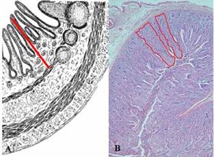
Figure 1 Schematic representation of measurements of (A) height (µm) and (B) area (µm2) of villi from duodenum, jejunum, and ileum in pigs (adapted and modified from Gartner, 2002). 

Results are presented as mean values ± SD. Mean comparisons were performed with Tukey tests and differences in mean value comparisons were considered as statistically significant at p ≤ 0.05. Proportions of morphologic alterations related to desquamation and presence of autolysis signs in samples of each treatment were compared using chi- square tests at p<0.05 using the GENMOD procedure of SAS.

Results

Intestinal villi measurements after sampling procedures are shown in Table 1. Villi height values of knotted samples from duodenum and ileum were higher (p<0.05) compared to the other procedures in the same anatomical portions, which were similar (p>0.05). Also, villi from knotted jejunum samples were the shortest (p<0.05) compared to the other two procedures. Regarding villi area, knotted samples from ileum had higher values compared to the rest of the procedures and intestinal portions (p<0.05).

Table 1 Morphometric characteristics of small intestine villi of pigs subjected to sample procedures of washing, soft pressure, and knotting. 

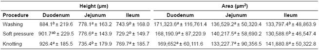
a, b Values with different superscripts indicate significant differences (p<0.05).

Table 2 Desquamation of small intestine villi in pig samples subjected to washing, soft pressure, and knotting. 

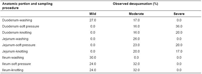
Villi desquamation as a result of sampling procedure did not show differences among procedures in the three intestinal portions (p>0.05); however, most samples showed some degree of desquamation (Table 2). In addition, 15 (5.5%) washed, 10 (3.7%) mild-pressed and 14 (5.2%) knotted samples (14.4% overall; p>0.05) showed similar signs of autolysis on the apical portion of the villi.

Discussion

The observation of intestinal villi allows evaluating the factors associated to intestinal structure and functional integrity, including enterocytes and mucosa membranes involved in digestive processes, and is widely used to explain weight gain during pig growth (Marchini, 2005; Skrzypek et al., 2010; Jung and Saif, 2017).

Our results showed differences in height and area of intestinal villi among sampling procedures and should not be considered a response variable of an experimental factor. Regarding the height observed in knotted samples, it has been reported that repair mechanisms after a lesion involving epithelial integrity may be involved in intestinal segments with shorter, thinner and fewer villi. These findings related to villi morphometry are associated to digestive disorders caused mainly by bacteria (Escherichia coli) or parasites (Eimeria spp and nematodes) that can activate humoral or cellular immune mechanisms and may cause changes in villi structure (length or area) and mature enterocytes numbers (Gartner, 2002). These mechanisms tend to reintegrate the epithelial tissue slowly and progressively. On the other hand, villus size and the relationship between villi number and epithelial area tends to diminish as age increases (Tsukahara et al., 2012), which may be the case of the present study, as animals may have gone thru different intestinal tissue repair procedures before the experiment. The knotting procedure is thought to be less “aggressive” to the intestinal villi, as it consists of knotting both ends of the intestine and introducing formalin solution, without any additional manipulation that could deteriorate the tissue. The washing and pressing procedures involve mechanical manipulation as the pressure wash or the sweep made with the fingers, which may have affected the tissue structure, possibly explaining the lower height of villi in washed and softly-pressured samples. Villi height and area were higher and wider in the duodenum, and tended to decrease towards the caudal portions of the intestine. Other authors reported variations in villi length among species and gastrointestinal physiology; in particular, villi in the cranial portions of the intestine are larger, more uniform and have a larger area, which optimizes digestion and absorption processes (Tsukahara et al., 2012). In swine, villi height increases notably since the first day of life (Huygelen et al., 2014) as their area increases gradually with age (Arce et al., 2008; Yunusova et al., 2013; Horn et al., 2014). Alterations in intestinal villi due to histologic cutting procedures have been reported, which is known as histologic artifacts. These artifacts represent unwanted changes that may occur during the cutting and mounting of a sample on a glass slide or may be due to inadequate sample processing (Narváez, 2015).

It is worth mentioning that the tissue cutting method used during the sample mounting process in the present study had an adequate quality control, thus minimizing potential artifact effects and other alterations. Therefore, the morphologic alterations observed are considered to be randomly-occurring in the three experimental procedures. Other authors (Bravo, 2011; Venne et al., 2014) have pointed out that some histologic artifacts may induce changes such as autolysis on cells and tissues by using an inadequate fixation technique, prolonged time of fixation, the use of extremely small amounts of the fixing agent -such as 10% solution of formalin, incomplete inclusion of the tissue, use of poorly sharpened blades, incorrect placing of the blade, presence of paraffin residues, folding of the tissue, poor elimination of stains used, presence of air bubbles, among others. Wick (2008) and Bravo (2011) recommend appropriate identification of the predictable changes in the sample due to improper processing or handling. On the other hand, Narváez (2015) points out that these artifacts may be effectively identified and have no influence on the study.

Autolysis or post-mortem degeneration observed in some samples of the present study may be due to improper fixation. According to Rubio et al., (2010) and Bravo (2011), when the tissue is not fixed rapidly or when the volume of fixative compound is not sufficient for the size of the sample, autolysis may begin, with the subsequent loss of observable detail and morphology of cells. Nevertheless, these can be considered permissible changes in a study that involves intestinal villi and are easily identified with common microscopic observation (Bravo, 2011). It is important to mention that, even though these changes occurred in similar proportions in all three sampling procedures, the knotting procedure showed to be less “aggressive” to the villi. Also, this technique may take longer to carry out since it requires other person to help with the knotting and injection of formalin solution. In conclusion, although none of the procedures in the present study can be considered better, the knotting procedure is recommended when intestinal cell morphometry -especially villi height and area - is to be evaluated. Additionally, artifacts can be found indistinctly in the washing, pressing and knotting procedures, and they may be related with epithelial restoration processes.

Acknowledgements

The authors wish to show their appreciation to the personnel of Industrializadora Agropecuaria de Ciudad Juárez, specially MVZ Fernando Corella and Franco Gamboa Rubio for their support during the sampling process and to Dr. Efrén Díaz Aparicio (INIFAP) for reviewing the manuscript.

References

Arce MJ, Ávila GE, Lopez CC. Comportamiento productivo y cambios morfológicos en vellosidades intestinales del pollo de engorda a 21 días de edad con el uso de paredes celulares del Saccharomyces cerevisiae. Vet Méx 2008; 39(2):223-228. [ Links ]

Assis RCL, Luns FD, Beletti ME, Assis RL, Nasser NM, Faria EM, Cury MC. Histomorphometry and macroscopic intestinal lesions in broilers infected with Eimeria acervulina. Vet parasitol 2010; 168(3):185-189. [ Links ]

Bravo GM. Manual de procedimientos y técnicas histopatológicas. Facultad de Medicina Veterinaria y Zootecnia-Universidad Michoacana de San Nicolás de Hidalgo. Morelia, Michoacán, México. 2011. [ Links ]

Gartner LP, Hiatt JL. Texto Atlas de Histología. 2nd ed. McGraw Hill-Interamericana. 2002. [ Links ]

Hedemann MS, Mikkelsen LL, Naughton PJ, Jensen BB. Effect of feed particle size and feed processing on morphological characteristics in the small and large intestine of pigs and on adhesión of Salmonella entérica serovar Typhimuriumm DT12 in the ileum in vitro. J Anim Sci 2005; 83:1554-1562. [ Links ]

Horn N, Ruch F, Miller G, Ajuwon KM, Adeola O. Impact of acute water and feed deprivation events on growth performance, intestinal characteristics, and serum stress markers in weaned pigs. J Anim Sci 2014; 92(10):4407-4416. [ Links ]

Huygelen V, De Vos M, Willemen S, Fransen E, Casteleyn C, Van Cruchten S, Van Ginneken C. Age-related differences in mucosal barrier function and morphology of the small intestine in low and normal birth weight piglets. J Anim Sci 2014; 92(8):3398-3406. [ Links ]

Itza-Ortiz MF, Lopez-Coello C,Avila-Gonzalez E, Gomez-Rosales S, Arce-Menocal J, Velazquez-Madrazo P. Efecto de la fuente energética y el nivel de energía sobre la longitud de vellosidades intestinales, la respuesta inmune y el rendimiento productivo en pollos de engorda. Vet Mexico 2008; 39(4):357-376. [ Links ]

Jensen TK, Boensen HT, Vigre H, Boye M. Detection of Lawsonia intracellularis in formalin-fixed porcine intestinal tissue samples: Comparison of immunofluorescence and in-situ hybridization, and evaluation of the effects of controlled autolysis. J Comp Path 2010; 142:1-8. [ Links ]

Jung K, Saif LJ. Goblet cell depletion in small intestinal villous and crypt epithelium of conventional nursing and weaned pigs infected with porcine epidemic diarrhea virus. Res Vet Sci 2017; 110:12-15. [ Links ]

Marchini CFP. Efeito da temperatura ambiente cíclica elevada sobre os parâmetros produtivos, fisiológicos, morfométricos e proliferação celular da mucosa intestinal de frango de corte. Vet Noticias 2005; 12(2):63-64. [ Links ]

Marchini CFP, Lourenço da Silva P, Bueno de Mattos Nascimento MA. Body weight, intestinal morphometry and cell proliferation of broiler chickens submitted to cyclic heat stress. Int J Poult Sci 2011; 10(6):455-460. [ Links ]

Narváez ADJ. La microscopía: Herramienta para estudiar células y tejidos [Access date: March 3, 2015]. URL: URL: http://www.medic.ula.ve/histologia/anexos/microscopweb/MONOWEB/anexos/MIcrografiasInterpretacion3D/artificios.htmLinks ]

Neog BK, Barman NN, Bora DP, Dey SC, Chakraborty A. Experimental infection of pigs with group A rotavirus and enterotoxigenic Escherichia coli in India: Gross, histopathological and immunopathological study. Vet Ital 2011; 47(2):117-128. [ Links ]

Roa I, Meruane M. Desarrollo del Aparato Digestivo. Int J Morph 2012; 30(4):1285-1294. [ Links ]

Rubio LA, Ruiz R, Peinado MJ, Echeverri A. Morphology and enzymatic activity of the small intestinal mucosa of Iberian pigs as compared with a lean pig strain. J Anim Sci 2010; 88:3590-3597. [ Links ]

SAS. 2013. SAS/STAT User’s guide (Version 9.4). Cary, NC: SAS Institute Inc. [ Links ]

Schweer WP, Pearce SC, Burrough ER, Schwartz K, Yoon KJ, Sparks JC, Gabler NK. The effect of porcine reproductive and respiratory syndrome virus and porcine epidemic diarrhea virus challenge on growing pigs II: Intestinal integrity and function. J Anim Sci 2016; 94(2):523-532. [ Links ]

Segalés J, Domingo M. La necropsia en el ganado porcino; Diagnóstico anatomopatológico y toma de muestras. 1st ed. Madrid, Spain: Boehringer Ingelheim; 2003. [ Links ]

Sisson S, Grossman JD. Anatomía de los animales domésticos. 5th ed. Barcelona, Spain: Salvat Editors; 2002. [ Links ]

Skrzypek T, Valverde-Piedra JL, Skrzypek H, Kazimierczak W, Szymanczyk SE, & Zabielski R. Changes in pig small intestinal absorptive area during the first 14 days of life. Livest Sci 2010; 133:53-56. [ Links ]

Tsukahara T, Kishino E, Inoue R, Nakanishi N, Nakayama K, Ito T, Ushida K. Correlation between villous height and the disaccharidase activity in the small intestine of piglets from nursing to growing. Anim Sci J 2012; 84(1):54-59. [ Links ]

Velasco S, Alzueta MC, Rodríguez ML, Rebolé A, Ortiz LT. Los prebióticos tipo inulina en alimentación aviar I: características y efectos a nivel intestinal. Rev Complutense C Vet 2010; 4(2):87-104. [ Links ]

Venne P, John-Baptiste A, Vitsky A. Post-mortem histological artifacts created by poor tissue handling during necropsy. J Histotech 2014; 37(2):43-47. [ Links ]

Wick MR. Diagnostic Histochemistry. 1st ed. New York, NY. Cambridge University Press. 2008. [ Links ]

Yunusova RD, Neville TL, Vonnahme KA, Hammer CJ, Reed CJ, Taylor JB, Redmer DA, Reynolds LP, Caton JS. Impacts of maternal selenium supply and nutritional plane on visceral tissues and intestinal biology in 180-day-old offspring in sheep. J Anim Sci 2013; 91(5):2229-2242. [ Links ]

**To cite this article: Itza OM, Quezada CA, Castillo CY, Rodríguez GE, Carrera CJM, Martín OU, Jaramillo LE, Calzada NA. Comparison of three sampling procedures for evaluating intestinal villi: A swine model. Rev Colomb Cienc Pecu 2017; 31(1):3-9.

Conflicts of interest The authors declare they have no conflict of interest regarding the work presented in this report.

Received: March 02, 2017; Accepted: November 21, 2017

* Corresponding author: Andrés Quezada Casasola. Departamento de Ciencias Veterinarias, Instituto de Ciencias Biomédicas. Universidad Autónoma de Ciudad Juárez. Anillo envolvente y Estocolmo s/n Cd. Juárez, Chihuahua, México. C.P. 32310. E-mail: aquezada@uacj.mx

Creative Commons License This is an open-access article distributed under the terms of the Creative Commons Attribution License