


 English (pdf)
 Article in xml format
 Article references
 Send this article by e-mail
 Cited by SciELO
  Cited by Google
  Similars in
SciELO
Similars in
SciELO  Similars in Google
 Permalink
Introduction
Veterinary ocular pathology is an emerging area, with reports dating from the beginning of the 20th century (Gelatt, 2008). In Colombia, this discipline is still modest when compared to ophthalmology, the specialty from which it derives. The increasing number of veterinary pathology laboratories and institutions offering ophthalmology services will soon depend on the development of this field.
Neoplasms are especially important in clinical practice for being the most frequent cause of death in dogs (47.0%; Withrow et al., 2013). Primary eye neoplasms are relatively uncommon in domestic animals. Frequencies in dogs can be as low as 0.9 (Withrow et al., 2013) to 39.0% (Dubielzig et al., 2010) of all neoplasms.
Eye-related neoplasms are important since they have a significant impact on visual ability, comfort, and longevity, and can lead to tissue destruction and metastasis (Labelle and Labelle, 2013). Therapeutic options and prognosis for life and for the eye will depend on neoplasm type and its anatomical location (Dubielzig, 2017).
It is known that disease prevalence varies largely between countries and between regions within a country. Although reports of ocular diseases in specific animal species and etiological entities are available, there is still the need for data about general frequency and distribution of eye-related neoplasms in our country. Absence of such data does not bias the diagnosis of lesions submitted to pathology labs, but unavailability somehow forces local pathologists and veterinary students to confront their findings with those reported in the international literature. In addition, understanding the type and frequencies of eye-related neoplasms in dogs would provide information on epidemiological background and potentially help to identify tendencies over time and even risk factors.
The aim of the present report was to describe the frequency and distribution of eye-related neoplasms in dogs using diagnostic records collected during 13 years at the Animal Pathology Laboratory of Universidad de Antioquia (Colombia).
Materials and methods
Ethical considerations
The study involved the analysis of information obtained during regular histopathological diagnosis. Approval from an ethical committee on animal experimentation was not necessary.
Study population, histopathological analysis and data collection
A retrospective search of reports on eye-related neoplasms in dogs was performed. Experienced veterinary pathologists at the Animal Pathology Laboratory of Universidad de Antioquia (Colombia) collected cases between January 2005 and December 2017 from routine histopathological examination. Samples were formalin-fixed, 2-4 μm-paraffin sectioned, and finally, hematoxylin and eosin-stained. Inclusion criteria considered neoplasms affecting eyelid, ocular globe, and third eyelid in dogs during the period. Data extracted included year of case report, information on the affect ed dog (i.e., age, sex, breed) and neoplasms characteristics (i.e. location, histological type, cellular origin). Nomenclature of the eye-related neoplasms was adopted in conformity with the World Health Organization’s Histological Classification of Ocular and Otic Tumors of Domestic Animals (Wilcock et al., 2002).
Data analysis
Data from the records was entered in Excel worksheets (Microsoft Corp., Redmond, WA, USA). Descriptive statistics was presented as percentages and 95% confidence intervals (CI) based on the total number of eye-related neoplasms. Statistical analyses were conducted using Stata14.0 (StataCorp 2015, Texas, USA).
Results
A total of 250 eye-related-neoplasm reports were found in 246 dogs (one report/animal, with the exception of four animals with both eyes simultaneously affected by the same type of neoplasm). Eye samples were submitted from 40 veterinary clinics located in Aburrá valley (Antioquia province, Colombia) and surrounding areas. Reports included dogs from 33 breeds (including mixed-breeds), males and females, aged between 6 months and 18 years. Data regarding distribution of eye-related neoplasms by age, sex, and breed are listed in Table 1.
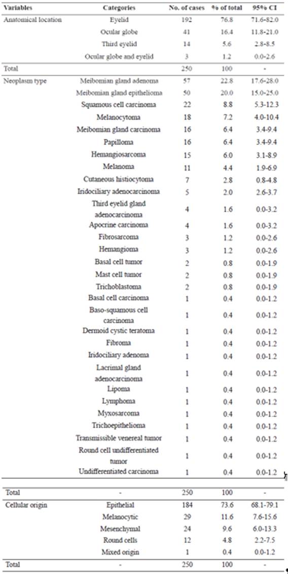
Data on distribution of eye-related neoplasms by location, neoplasm type, and cellular origin are listed in Table 2. The 250 cases were classified in 30 types, in three locations, both eyes, and three cellular origins.
Table 1 Distribution of eye-related neoplasms by age, sex, and breed of dogs reported at the Animal Pathology Laboratory of Universidad de Antioquia (Colombia) between 2005 and 2017.

CI: Confidence interval.
Table 2 Distribution of eye-related neoplasms by anatomical location, type, and cellular origin, at the Animal Pathology Laboratory of Universidad de Antioquia (Colombia) between 2005 and 2017.
CI: Confidence interval.
The following text describes the findings for eye-related neoplasms, grouped according to the international classification system reported by Dubielzig (2017) and mentioned according to frequency as a report group in this study (from most to least frequent). Details for each description refers to significant findings and others to details reported due to interest of the authors.
Meibomian gland neoplasms
Neoplasms of the Meibomian glands were the most frequently found in this study (49.2%; 123/250). These neoplasms, classified as adenoma, epithelioma, and carcinoma, presented an epithelial origin. Meibomian adenoma (Figure 1A) was the most commonly found Meibomian gland neoplasm, as well as the most frequently reported in this study. Labrador retriever (17/57) and Poodle (10/57) were the two most commonly affected breeds. No sex predilection was found. Only one case was classified as ductal adenoma in a 10-year-old female Labrador retriever. Meibomian epithelioma (Figure 1B) was the second most frequently reported neoplasm in this study and it was restricted to the eyelid. A 60.0% (30/50) of the cases occurred in male dogs. Labrador retriever (12/50), Poodle (7/57), mixed-breed (7/57), and Schnauzer (7/57) were the most frequently affected breeds. Three of these cases had ductal differentiation (3/50). After the squamous cell carcinoma, the Meibomian carcinoma (Figure 1C) was the most commonly found malignant neoplasm. A 62.5% (10/16) of the affected dogs were males. Mixed-breed dogs (5/16) and Labrador retrievers (4/16) were the most frequently affected breeds.
Surface epithelium neoplasms
Papilloma was the most commonly benign epidermal neoplasm found. The eyelid was the location involved in most of the cases (81.3%; 13/16). The bulbar conjunctiva was affected twice (12.5%; 2/16) and only one case was reported affecting the third eyelid. Bulldog (3/16) and mixed-breed dogs (3/16) were the most frequently affected breeds. Squamous cell carcinoma (Figure 1D) was the most commonly malignant neoplasm found in this study and it was reported individually in all anatomical locations (eyelid, third eyelid, and ocular globe). Only one neoplasm case affected two anatomical locations at the same time (third eyelid and ocular globe). The eyelid was involved in most of the cases (59.1%; 13/22), the ocular globe in six cases (27.3%), and the third eyelid, twice (9.1%). The average age for this neoplasm was 8.7 years, and breeds mostly affected were Boxer (7/22) and Rottweiler (3/22). Basal cell neoplasms were present four times in the eyelid, two of them corresponding to benign basal cell neoplasms, found in two females Poodle and Labrador retriever. Basal cell carcinoma was reported in a 10-year-old male Boxer. Likewise, only one case of baso-squamous cell carcinoma was found affecting the eyelid of a 7-year-old male Schnauzer.
Iridociliary neoplasms
Iridociliary neoplasms (6/250) were restricted to the ocular globe. One of them was an adenoma and it was reported in an 8-year-old female Labrador retriever. The remaining were adenocarcinomas affecting three dogs. The age range for iridociliary adenocarcinoma was 5-11 years, with an average of 9.1. No breed tendency was found.
Melanocytic neoplasms
Melanocytoma (Figure 2A) was the most common melanocytic neoplasm (62.1%; 18/29). The age range for this neoplasm was 4-16 years, with an average of 8.7. This neoplasm had higher frequency in males (72.2%; 13/18). The eyelid was the anatomical site most commonly affected by melanocytoma (77.8%; 14/16). The other four cases were intraocular (22.2%). There was no breed predilection in regard to the ocular globe. Melanoma (Figure 2 B) was slightly more frequent in males (54.5%; 6/11). The ocular globe and the third eyelid were affected each one, twice. There was not breed predilection, but three of the four dogs were males. Eyelid melanoma (7/11) had an age range of diagnosis between 7-11 years, with Pitbull (2/7) and Labrador retriever (2/7) as the most commonly affected breeds.
Vascular neoplasms
Eighteen vascular neoplasms (7.2%; 18/250) were found. Three cases corresponded to hemangioma (3/18) involving individually the three anatomical locations reported herein. The age range for this neoplasm was 4-14 years, and the breeds affected were Dalmatian, Bull terrier, and mixed-breed dogs. Hemangiosarcoma (15/18) was found in the ocular globe and in the third eyelid (Figure 2C). The ocular globe was affected in 80.0% (12/15) of the cases, and was restricted to the third eyelid in three of the cases. The age range for this neoplasm was 5-13 years, with an average of 8.7. Mixed-breed dogs (6/15) and Dalmatian (2/15) were the most commonly affected breeds. The ocular hemangiosarcoma mostly affected males (66.7%; 8/12).
Fibrous tissue neoplasms
A fibroma was reported in the eyelid of a 13-year-old male Cocker Spaniel. A fibrosarcoma was found in the eyelid of two male Labrador retrievers, and in one case, the neoplasm affected the ocular globe of an 11-years-old female Poodle. A myxosarcoma was reported in the third eyelid of a 4-year-old male Weimaraner.
Round cell neoplasms
Twelve round cell tumors were found (4.8%; 12/250). One of them was an undifferentiated tumor in the eyelid of a 4-year-old male Pug and another one was a transmissible venereal tumor (TVT) in a 2- year-old male mixed-breed dog. Cutaneous histiocytoma was restricted to the eyelid of dogs between 2-8 years of age. Males were most commonly affected (71.4%; 5/7) as well as Labrador retrievers (2/7). A lymphoma was found affecting the eyelid of an 8-year-old male Poodle. Two mast cell tumors (0.8%; 2/250) were reported as, a high-grade mast cell neoplasm affecting the ocular globe of a 12-year-old female Poodle, and a low-grade mast cell neoplasm affecting the ocular globe of a 5-year-old male Golden retriever.
Third eyelid gland adenocarcinoma
This malignant neoplasm, derived from the apocrine third eyelid gland, was found four times in our study, three of them affecting only the third eyelid and one the ocular globe as well. The age range for this neoplasm was 6-15 years. This neoplasm affected a male Beagle, Boxer, and Fox terrier, and a female Weimaraner.
Apocrine neoplasms
Four reports for apocrine carcinoma were found, and two of them had ductal proliferation (ductal apocrine carcinoma). All the animals were males aged 7-10 years. This neoplasm was found twice in Boxers, once in a Poodle and a Golden retriever. The neoplasm affected the eyelid and extended to the ocular globe of an 8-year-old male Boxer. The other three apocrine neoplasms were restricted to the eyelid.
Hair follicle neoplasms
Three benign hair follicle neoplasms were found, corresponding to two trichoblastomas and a trichoepithelioma, all of them located in the eyelid. Trichoblastomas were reported in an 11-year-old male Siberian husky and in a 12-year-old female Poodle. A trichoepithelioma was reported in a 12-year-old male Poodle.
Other eye-related neoplasms
The eyelid of a 14-year-old male Poodle was affected by a lacrimal gland adenocarcinoma (Figure 2D). Additionally, a dermoid cystic teratoma was found in the cornea of a young female Chihuahua.
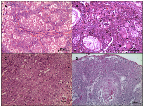
Figure 1 Histopathological features. (A) Meibomian gland adenoma in the upper eyelid of a 7- year-old Labrador retriever (mass size = 0.5 cm). (B) Meibomian gland epithelioma in the upper eyelid of an adult Beagle (note the predominant presence of reserve cells). (C) Meibomian gland carcinoma in the upper eyelid of an 8-year-old Golden retriever. (D) Squamous cell carcinoma affecting the cornea of an 8-year-old male Pug (Hematoxylin & Eosin stain).

Figure 2 Histopathological features. (A) Melanocytoma in the lower eyelid of a 9-year-old female Yorkshire terrier (note the diffuse presence of melanocytes affecting the dermis). (B) Intraocular infiltrating melanoma in the iris of a 10-year-old male Siberian huskie. The neoplasm extended to both ocular chambers (a magnification of the same lesion after melanin bleaching, presenting pleomorphism and mitotic figures is shown in the inset figure). (C) Hemangiosarcoma in a 5-year-old Dalmatian. Note the presence of dilated structures containing red blood cells affecting the vascular tunic; additionally, this neoplasm extended to the cornea (a greater formation of vascular structures with erythrocytes inside, typical of this neoplasm, is shown in the inset figure). (D) Lacrimal gland adenocarcinoma affecting the palpebral conjunctiva of a 14-year-old male Poodle (Hematoxylin & Eosin stain).
Discussion
Many neoplasm types can affect the eye and adnexal structures in domestic animals. When considering neoplasms, thorough evaluation of the health of the eye and patient is essential to determine the biologic behavior of the condition. Fernandez et al.(2013) analyzed a considerable number of ocular neoplasms (n = 173) finding numerous cases in dogs over 10 (43.2%) and 6-10 years (40.6%), and few cases in those under 5 years of age (9.9%). Several studies have evaluated ocular neoplasms, observing an age predilection, mainly in dogs between 8 and 11 years of age (Abarca et al., 2002; Heath et al., 2003; Laus et al., 2008; Conceição et al., 2010; Fernandez, 2013; Werner et al., 2017). These reports are in agreement with our findings, where the age group with the highest incidence (8-11 years of age) presented significant difference when compared to the other age groups. Even though histiocytoma is common in young dogs (with an average age of 3 years; Conceição et al., 2010; Fernandez, 2013) the age range in the present study was 2-8 years.
Fernandez (2013) reported a distribution of ocular neoplasms by sex, with a higher presentation in male dogs (56.2%) compared to females (43.8%). In addition, Silva et al.(2016) reported a very similar distribution by sex (55.6 vs. 43.4%), which is consistent with our findings.
Labrador retriever and Poodle dogs were the most commonly affected breeds in our study, similar to Silva et al.(2016) who reported that mixed-breed (26.6%), Labrador retriever (16.1%) and Poodle (11.2%) were the breeds most commonly affected by ocular neoplasms. Other researchers reported that Labrador retriever and Poodle breeds are predisposed to ocular neoplasms -mostly adenomas, adenocarcinomas, and melanomas (Conceição et al., 2010; Fernandez, 2013; Dees et al., 2015).
The eyelid was the most frequently affected structure. Consistently with the literature, the most important neoplasms were those affecting the Meibomian glands (Dees et al., 2015). We hypothesize that this could be due to the direct contact of this structure with the environment, increasing the chances of damage and further tissue adaptation to injuries.
The most commonly affected structure in our study was the Meibomian gland, agreeing with the literature (Dubielzig, 2017). Werner et al. (2017) reported 9.4 years as the average age for Meibomian neoplasms, which is consistent with our findings (9.9 years). Silva et al.(2016) reported the epithelioma (24.3%) and adenoma (15.4%) as the most common neoplasms affecting the eye and adnexa structures in dogs, similar to our study. The only difference is that adenoma was slightly more frequent compared to epithelioma in our study. Werner et al.(2017) reported that Labrador retrievers was the breed most commonly affected by adenoma and epithelioma of the Meibomian glands, which is consistent with what is reported herein.
The literature reports that Meibomian adenocarcinoma is a rare neoplasm of the eyelid, accounting for less than 1.0% of all eyelid neoplasms, with no gender or breed susceptibility (Wilcock et al., 1998; Tavasoli et al., 2012). We found that this neoplasm was one of the lowest occurrence, but it was not as low as reported in the literature.
Eye-related squamous cell carcinoma frequently affected Boxer and Rottweiler in our study. Similar findings about presentation of squamous cell carcinoma in brachycephalic dogs have been reported (Takiyama et al., 2010). This could be due to chronic exposition to excessive sunlight in this kind of dogs (Busse et al., 2008). Nevertheless, Dreyfus et al.(2011) reported lower incidence of these neoplasms in brachycephalic breeds. Conceição et al.(2010) reported that squamous cell carcinoma was the most common eyelid neoplasm in dogs older than 10 years of age. We found no sex predilection for this neoplasm as it has been reported by the literature (Goldschmidt and Goldschmidt, 2017).
Literature reports that iridociliary neoplasms represent 12.5% of the eye-related neoplasms in dogs, which is consistent with our findings -representing the 14.6% of intraocular neoplasms. Retriever dogs are considered the most commonly affected by these kind of neoplasms (Petterino et al., 2014; Okawauchi et al., 2016). Nevertheless, our study did not find such tendency. We found an average presentation of 8.5 years of age for iridociliary neoplasms, which is consistent with previous reports (Fernandez, 2013).
In a retrospective study by Teixeira et al. (2010), melanoma was the most common melanocytic eye-related neoplasm found in dogs, but none of them related to eyelid. We found 14 cases of melanocytoma in the eyelid; similar to what was reported by Wang and Kern (2015) who outlined melanocytic neoplasms as mainly benign, except in the conjunctiva, where the malignant form is more commonly seen. In our study, the malignant form was frequently observed in the ocular globe. The frequency of this neoplasm was higher in males in our study as well as reported by Wang and Kern (2015).
Soft tissue sarcomas are a heterogeneous group of neoplasms of mesenchymal origin, representing 15% of all skin neoplasms in dogs (Campos et al., 2015). The literature reports that canine eyelid sarcomas are infrequent and predisposition to fibrosarcoma in Golden retrievers could exist (Fernandez, 2013; Nordio et al., 2017). In our study, Labrador retriever was the most commonly affected breed, genetically related to the breed reported in the literature. Hendrix et al.(2000) reported five cases of fibrosarcoma out of 44 cases of ocular neoplasms, with no predilection by age, sex, or breed.
Histiocytoma is the most common neoplasm seen in young dogs, and the eyelid is a typical site of occurrence (Conceição et al., 2010; Lew et al., 2010). It has been reported that ocular histiocytoma is more frequently found in dogs younger than three years of age (Fernandez, 2013). Nevertheless, we found it in 2 to 8-year-old dogs, averaging five years.
Although there are few reports of TVT in the eye of dogs (a 1-year-old mixed-breed male and a 4-year-old male Labrador retriever), this diagnosis should be taken into account in sexually active young dogs (Milo and Snead, 2017).
According to the literature, adenocarcinoma is more commonly found in the third eyelid gland, compared to adenoma (Fernandez, 2013; Labelle and Labelle, 2013; Dees et al., 2015).
Dees et al.(2015) reported an age range of 3-16 years and a male predilection (60.9%), and the breeds most commonly affected by these neoplasms were mixed-breed (20.2%) followed by Labrador retriever (7.5%). The literature also reports a predisposition in geriatric dogs, with an average age of 11 years (Conceição et al., 2010). We found four cases in different breeds, with a male tendency (75%) and the age range was 6-15 years.
Apocrine sweat gland adenocarcinomas account for 0.7 to 2.2% of all skin-associated tumors in dogs (Stern and Eisele, 2011). Simko et al.(2003) reported a breed tendency (27.8% in mixed-breeds) with 9 years average age in a study of 44 dogs with apocrine skin neoplasms. The literature reports only two cases of apocrine carcinoma in the eyelid of dogs, corresponding to a 13-year-old male Shetland sheepdog and a 12-year-old female Miniature pinscher (Hirai et al., 1997; Stern and Eisele, 2011). In agreement with Simko et al.(2003), we found a tendency in males (100%) and a similar age of presentation (7-10 years), although the breeds affected in our study were different.
As with many retrospective studies, there are difficulties in veterinary medicine reports. This can be due to lack of information in the analysis request form and/or medical records when samples are submitted to the pathology laboratory. Similarly, information is scarce in our country as well as in those sharing similar cultural and ecological conditions. To our knowledge, this is the first retrospective report aimed to eye-related neoplasms in dogs published in Colombia.
In conclusion, the behavior of eye-related neoplasms affecting dogs is similar to that reported in the literature, with variations between countries and regions. Labrador retriever and Poodle dogs were overrepresented in this study, which can be explained by their great presence in Colombia. More complete clinical information at sample submission is required for valuable epidemiological information to make specific and useful pathological diagnoses. We should increase our understanding of the epidemiological behavior of eye-related neoplasms in dogs during the clinical practice. The present study provides useful epidemiological data for the diagnosis of ocular neoplastic disease by pathologists and clinicians. Further retrospective epidemiological studies are needed to understand the current situation in the country.

