SciELO - Scientific Electronic Library Online

 
vol.34 issue2Estimating biological reference intervals for cell blood counts in a dog populationReducing Salmonella enterica serovar Enteritidis contamination in food: lytic bacteriophages in a homemade mayonnaise-like matrix author indexsubject indexarticles search
Home Pagealphabetic serial listing  

Services on Demand

Journal

Article

Indicators

Related links

  • On index processCited by Google
  • Have no similar articlesSimilars in SciELO
  • On index processSimilars in Google

Share


Revista Colombiana de Ciencias Pecuarias

Print version ISSN 0120-0690On-line version ISSN 2256-2958

Rev Colom Cienc Pecua vol.34 no.2 Medellín Apr./June 2021  Epub June 23, 2022

https://doi.org/10.17533/udea.rccp.v34n2a03 

Brief communications

Serum concentration of thyroxine and triiodothyronine in newborn calves from cows supplemented with barium selenate**

Concentración sérica de tiroxina y triiodotironina en terneros recién nacidos de madres suplementadas con selenato de bario

Concentração de tiroxina e triiodotironina sérica em bezerros recém-nascidos de mães suplementadas com selenato de bário

Víctor R Leyán1  * 

Ricardo H Chihuailaf2 

Fernando G Wittwer2 

1Instituto de Inmunología, Facultad de Medicina, Universidad Austral de Chile, Valdivia, Chile.

2Instituto de Ciencias Clínicas Veterinarias, Facultad de Ciencias Veterinarias, Universidad Austral de Chile, Valdivia, Chile.


Abstract

Background:

Barium selenate is an inorganic source of selenium (Se) used in prolonged-release preparations to treat selenium deficiency in bovines.

Objective:

To evaluate serum concentrations of triiodothyronine (T3) and thyroxine (T4) hormones in newborn calves from mothers supplemented with barium selenate during prepartum.

Methods:

Six black Frisian pregnant cows were supplemented with barium selenate subcutaneously during the last two months of gestation, until calving. Six cows were used as controls. All cows were subjected to a low Se diet, consisting of hay from natural pasture and commercial concentrate lacking Se. The Se balance was measured through the activity of erythrocyte glutathione peroxidase (GPx). Serum concentration of T3 and T4 in calves was determined by electrochemiluminescence.

Results:

Se supplementation during prepartum increased GPx activity in cows from day 45 post-supplementation (p<0.05). Calves from supplemented mothers showed higher average serum Se concentration than calves from non-supplemented mothers. The average concentration of T3 in the calves from supplemented mothers was lower in the first hour of life (p<0.05) compared with calves from mothers of the non-supplemented group. A decrease (p<0.05) in T4 serum concentrations was observed in both groups at seven days of age.

Conclusions:

Administration of barium selenate to cows during prepartum generates a reduction in serum concentration of T3 in the first hour of life of calves.

Keywords: barium selenate; bovine; calves; cows; glutathione peroxidase; mineral nutrition; selenium; supplementation; thyroid hormones; thyroxine; triiodothyronine

Resumen

Antecedentes:

El selenato de bario es una fuente inorgánica de selenio (Se) utilizada en preparaciones de liberación prolongada para corregir el estado de carencia de Se en bovinos.

Objetivo:

Evaluar las concentraciones séricas de triyodotironina (T3) y tiroxina (T4) en terneros recién nacidos de madres suplementadas durante el preparto con selenato de bario.

Métodos:

Seis vacas frisón negro con 7 meses de gestación fueron suplementadas vía subcutánea con selenato de bario dos meses previos a la fecha de parto. Otras seis vacas permanecieron como controles. Todas las vacas se mantuvieron con una dieta cuyo aporte de Se fue inferior a los requerimientos y consistió en heno de pradera natural y concentrado comercial sin Se. El balance de Se se midió usando la actividad eritrocitaria de glutatión peroxidasa (GPx) y las concentraciones de T3 y T4 en terneros mediante electroquimioluminiscencia.

Resultados:

La suplementación con Se aumentó la actividad de GPx en vacas desde el día 45 post suplementación (p<0,05). Los terneros de madres suplementadas mostraron una concentración sérica promedio de Se mayor que los terneros de madres no suplementadas. La concentración promedio de T3 de terneros de madres suplementadas fue menor en la primera hora de vida (p<0,05) que en terneros de madres no suplementadas. A los 7 días de edad hubo una disminución (p<0,05) en las concentraciones séricas de T4 en ambos grupos.

Conclusión:

La administración de selenato de bario en vacas preparto genera una disminución en la concentración sérica de T3 en la primera hora de vida del ternero.

Palabras clave: bovino; hormonas tiroideas; nutrición mineral; selenato de bario; selenio; suplementación; terneros; tiroides; tiroxina; triyodotironina; vacas

Resumo

Antecedentes:

O selenato de bário é uma fonte inorgânica de selênio (Se) usada em preparações de liberação prolongada para corrigir o status de deficiência de Se em bovinos.

Objetivo:

Avaliar as concentrações séricas de triiodotironina (T3) e tiroxina (T4) em bezerros recém-nascidos de mães suplementadas durante o pré-parto com selenato de bário.

Métodos:

Seis vacas friesianas negras aos 7 meses de gestação foram suplementadas com selenato de bário por via subcutânea dois meses antes do parto. Seis outras vacas permaneceram como controle. Todas as vacas foram mantidas em uma dieta cuja contribuição de Se foi inferior aos requeridos e consistiram em feno natural da pradaria e concentrado comercial sem Se. O balanço de Se foi medido usando a atividade eritrocitária das concentrações de glutationa peroxidase (GPx) e T3 e T4 em bezerros por eletroquimiluminescência.

Resultados:

A suplementação com atividade de GPx aumentou em vacas a partir do dia 45 após a suplementação (p<0,05). Os bezerros de mães suplementadas apresentaram uma concentração sérica média de Se maior que os bezerros de mães não suplementadas. A concentração média de T3 dos bezerros das mães suplementadas foi menor na primeira hora de vida (p<0,05) do que nos bezerros das mães não suplementadas. Aos 7 dias de idade houve uma diminuição (p<0,05) nas concentrações séricas de T4 nos dois grupos.

Conclusão:

A administração de selenato de bário em vacas de parto gera uma diminuição na concentração sérica de T3 na primeira hora de vida do bezerro.

Palavras-chave: bezerros; bovino; hormônios da tireóide; nutrição mineral; selenato de bário; selênio; suplementação; triiodotironina; tireóide; tiroxina; vacas

Introduction

The hormones thyroxine (T4) and triiodothyronine (T3) are synthesized exclusively by the thyroid through a series of reactions linked to the histological organization of the gland (Carvalho and Dupuy, 2017). Thyroid hormones affect growth, development, differentiation, reproduction, and several routes of intermediary metabolism. Although T3 is the biologically active hormone, T4 is considered a pro-hormone that requires conversion to T3 (Suttle, 2010). The deiodination process is catalyzed by one of the three deiodinase enzymes currently described, distributed mainly in extrathyroidal tissues (Beckett and Arthur, 2005). All deiodinases contain a residue of selenocysteine in their active site (Stadtman, 2000); therefore, an adequate metabolic balance of selenium (Se) is essential for synthesis, activation, metabolism, and secretion of thyroid hormones. An insufficient supply of Se in ruminants compromises the metabolism of thyroid hormones affecting the conversion of T4 to T3 (Contreras et al., 2002; 2005; Voudouri et al., 2003; Rowntree et al., 2004). Calves born from mothers with negative metabolic balances of Se have a compromised Se status and neonatal synthesis of thyroid hormones (Awadeh et al., 1998; Guyot et al., 2011). To correct or control clinical deficiency it is common to resort to inorganic (e.g., sodium selenite) or organic Se sources (e.g., selenized yeasts; Davis et al., 2008). In this regard, Se metabolic path depends on the source employed (Ortman and Pehrson, 1999; Juniper et al., 2008; Slavik et al., 2008), with selenite being more easily metabolized to the immediate precursors of selenocysteine in comparison with organic forms of Se. Barium selenate (BaSeO4) is an inorganic source used in the formulation of prolonged-release preparations to provide an adequate Se status for at least 7 months (Leyán et al., 2004; Judson and Badidge, 2010), generating a high and sustained concentration of Se in plasma and liver compared to other inorganic forms (Davis et al., 2008).

The hypothesis of this study was that blood concentration of thyroid hormones in newborn calves from Se-deficient mothers can be improved by supplementing the mother with barium selenate as a source of Se. Therefore, this study aimed to evaluate the blood concentration of T3 and T4 in newborn calves from mothers supplemented with barium selenate during prepartum.

Materials and methods

Ethical considerations

The experiment was developed according to the bioethics protocols for the use of animals in research of Universidad Austral de Chile (Fondecyt 119-09939).

Animals

Twelve clinically healthy black Friesian cows were used for the study, conducted in Valdivia, Chile (39º48'LS; 73º15'LO). The animals were distributed in two groups of six cows each, homogeneous in terms of age (6.5 years), number of deliveries (3.5 deliveries), body weight (594 kg), gestational age (230 days), and milk production in the last lactation (6,406 L).

Experimental design

Throughout the experimental period, animals were housed in individual stalls on concrete floor and straw bed. All cows were fed a Se-deficient diet (Se <0.05 ppm) consisting of 9.5 kg of natural pasture hay [Se = 0.02 ppm of dry matter -DM), and 1 kg of commercial concentrate (Cosetan® Biomaster IANSA, Quepe, Chile; Se = 0.12 ppm DM) in individual feeders, and water ad libitum. One group of animals was selected at random and supplemented with Se (Se-S), and the other group remained as control without supplementation (Se- D). After calving, which occurred in August (late winter), the calves received 4 L colostrum from their mothers during the first 24 hours and stayed with them for three days. Afterward, they were placed in individual pens where they continued to receive whole milk (4 L/d).

Selenium supplementation

The Se-S group cows were supplemented subcutaneously with 1 mg Se/kg using barium selenate (Deposel®, Young Animal Health Ltd., New Zealand) in a single dose of 1 mL/50 kg/ BW, administered 60 days before the expected calving date.

Sampling

Blood samples from each cow were collected by coccygeal venipuncture into heparin-coated and noncoated tubes before supplementation (day 0) and then every 15 days for 90 days. Blood samples were collected from the calves into noncoated tubes immediately after birth, before colostrum ingestion, and at 7 days post- birth. Blood plasma and serum were maintained at -20 °C until analysis.

Selenium metabolic balance

The Se balance in cows was evaluated by blood activity of glutathione peroxidase (GPx) using a commercial reagent (Ransel®, Randox), according to Contreras et al. (2002). The activity of GPx was expressed in U/g of hemoglobin (Hb).

Serum T3 and T4 concentration in calves

Serum concentrations of T3 and T4 hormones was measured by electrochemiluminescence technique in an Elecsys 1010 device (Roche Diagnostics, Basel, Switzerland). The values were expressed in nmol/L. The serum T3 /T4 ratio was calculated by their quotient on serum concentration values.

Serum selenium concentration in calves

Serum selenium concentration in calves was quantified in an atomic absorption spectrophotometry device (Thermo® Series AA Solaar, Waltham, MA, USA), at 196 nm, with hydride generation in a (Thermo Scientific model VP100, Waltham, MA, USA), after acid digestion of the sample as indicated by Muñiz-Naveiro et al. (2005). The values are expressed in mg/L.

Statistical analysis

Data are expressed as means and standard deviation. The Shapiro-Wilk test was used to establish normality of the data. Significance of differences in Se-balance between supplemented and non-supplemented cows was evaluated using a variance analysis of repeated measures followed by Tukey's multiple comparisons test. Differences in concentration of thyroid hormones in the calves were assessed by Student's "t" test. The analysis was performed with the GraphPad© Prism 3 software (GraphPad Software, San Diego, CA, USA), and it was considered significant at p<0.05.

Results

Mean initial values of GPx activity in the Se-D and Se-S groups were 111 ± 52 U/g Hb and 118 ± 29 U/g Hb (p>0.05), respectively. Subsequent measurements in the Se-D group showed a reduction in blood activity of GPx up to values considered marginal (<100 U/g Hb) at day 30 and the following days (Figure 1). In contrast, in the Se-S group, an increase was observed in blood activity of GPx with respect to the initial value. The differences between groups were detected from day 45 until the end of the experiment (p<0.05).

The average T3 concentration was lower in the first hour of life in calves from the Se-S mothers compared with calves from Se-D mothers (p<0.05; Table 1). The Se-D values presented on day 7 were similar between groups (p>0.05). The T4 concentrations during the first hour of life were also similar between groups (p>0.05). On day 7, both groups presented a decrease that was significantly lower in the Se-S group (p<0.05). The T3:T4 ratio was similar between groups in the first hour of life (p>0.05) and on day 7. However, the ratio between periods was higher in the Se-D group (p<0.05; Table 1).

Discussion

The Se requirement for dairy cattle is 0.3 ppm according to the NRC (2001). The diet used in this experiment contained 0.04 ppm Se; thus, the non-supplemented cows were in negative metabolic balance for this mineral. Accordingly, blood activity of GPx in the Se-D cows remained within a low-marginal range (<100 U/g Hb; Ceballos and Wittwer, 1996). Although dietary minimum Se concentration for grazing cattle should be greater than 0.05 ppm to prevent alterations in health and productivity (Suttle, 2010), no clinical signs of Se deficiency were observed during the trial period in the present study. Supplementation with barium selenate significantly increased GPx activity of cows in the Se-S group from day 45 post-supplementation, reaching values higher than 200 U/g Hb during delivery, which are considered appropriate for lactating cows (Ceballos and Wittwer, 1996). This result agrees with reports by other researchers using different Se sources (Awadeh et al., 1998; Guyot et al., 2011). According to Gunter et al. (2013), the increase in blood Se in calves of supplemented mothers depends on the Se source.

The effect of organic and inorganic Se supplementation on different systems, including the function of the thyroid gland in cattle, has been reported by various researchers (Awadeh et al., 1998; Rowntree et al., 2004; Koenig and Beauchemin, 2009; Guyot et al., 2011; Rose et al., 2012); however, few studies refer to the effect in newborns from mothers supplemented with Se. In the present study, the average concentration of T3 was lower in the first hour of life (p<0.05) in calves from Se-S mothers (Table 1).

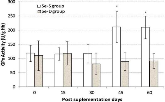
Figure 1 Blood activity of GPx (mean ± SD in U/g Hb) of cows maintained fed ration with low selenium, supplemented during prepartum (day 0) with barium selenate (Se-S) and non-supplemented (Se-D). Parturition period between 45 and 60 days. Asterisks (*) indicate significant differences (p<0.05) between groups. 

Table 1 Serum concentrations (mean ± SD in nmol/L) of T3, T4, and T3:T4 ratio in calves at 1 hour (h) and 7 days after birth from cows fed a low-selenium diet supplemented with barium selenate (Se-S) and non- supplemented (Se-D) during prepartum. 

1 h T3 7-day 1 h T4 7-day T3:T4 ratio 1 h 7-day
Se-S 5.92 ± 0.6 4.64 ± 0.94 181 ± 51 101 ± 21 0.0347 0.0464
Se-D 8.64 ± 2.46* 4.23 ± 1.16 188 ± 31 88 ± 31 0.0472 0.0488

* p<0.05 between groups; ≠ p<0.05 between periods.

Although no studies conducted under similar conditions were found in the literature, other researchers have reported values of thyroid hormones in the newborn by applying experimental models different from those in this study (Davicco et al., 1982; Stojić et al., 2002; Kirovski et al., 2008). These differences can be attributed not only to the hormone determination methodologies but also to the potential influence of the environment on the animals subjected to experimentation. In this regard, Stanko et al. (1991) indicated that calves born during the winter period have higher concentrations of T3 and T4 than those born in other seasons because cold stimulates synthesis and secretion of these hormones in the newborn. Colostrum intake is another factor related to the concentrations of thyroid hormones in the neonate (Grongnet et al., 1985). In the present study, the calves received colostrum ad libitum from their mothers, which presented different metabolic Se status (Figure 1); however, some studies have indicated that iodine transferred transplacentally from the mother to the fetus would be more relevant than iodine transferred by colostrum (Davicco et al., 1982; Guyot et al., 2011).

The decrease in hormone concentration after birth is consistent with other reports (Awadeh et al., 1998; Takahashi et al., 2001; Stojić et al., 2002; Guyot et al., 2011; Rose et al., 2012). In this sense, the high initial T3 concentration in the newborn is related to the synthesis of thermogenin, a protein necessary for heat generation in the brown adipose tissue (Carstens, 1994). When analyzing Se between groups, T3 concentration was lower in the calves from Se-supplemented mothers compared with the non-supplemented. This unexpected effect could be attributed to the administration of barium selenite; a low-soluble salt formulated for subcutaneous deposition of Se to be released over a one year period. The uncontrolled release can reach values that, in the conditions of this test, could be considered supra-nutritional, leading to lower conversion of T4 to T3 in newborn calves. With a similar Se source, this paradoxical effect has been observed in heifers supplemented with barium selenate. The erythrocyte GPx activity exceeded by far metabolically adequate values, decreasing the cellular immune response (Leyán et al., 2006). This hypothetical effect associated with Se formulation is supported by studies conducted with different Se sources. In this regard, Awadeh et al. (1998) reported that T3 concentration in calves from mothers supplemented with 60 ppm Se, in the form of organic Se, was higher than in calves from mothers supplemented with the same amount of inorganic Se, with the highest T3:T4 ratio in calves from mothers who received salts with 120 ppm Se. However, in other trials, no effect on the concentration of thyroid hormones in newborns was observed by supplementing prepartum cows with selenized yeast or selenite (Rowentree et al., 2004; Koenig and Beauchemin, 2009).

There was also no effect on thyroid hormone concentrations of newborns when using intra- ruminal boluses formulated on the basis of Se, iodine and cobalt, and supplied two months before delivery (Rose et al., 2012). The effect of different Se formulations (selenite and selenium yeast) on thyroid hormones concentration in adult animals has also been studied and, although they increase GPx activity and blood Se concentration, they do not cause differences in T3, T4 concentration and T3:T4 ratio with respect to non-supplemented animals (Gunter et al., 2013). These results were attributed to a compensatory response of non-supplemented animals, in which the expression of the enzyme iodothyronine deiodinase type I in the thyroid would be increased.

The studies conducted on Se effects on the thyroid gland function have used organic Se, selenite salts and elemental selenium with varied results. To the best of our knowledge, this is the first study using slow-release barium selenate in cows during prepartum to assess its effect on the concentration of thyroid hormones in the calve, which results do not allow for establishing criteria for practical use. The results of this study raise questions about the adequate balance of Se in the prepartum cow and the consequences of supranutritional contributions that could cause negative metabolic responses in the newborn. Accordingly, further studies are required on this subject.

References

Awadeh FT, Kincaid RL, Johnson KA. Effecto flevel and source of dietary selenium on concentrations of thyroid and immunoglobulins in beef cows and calves. J Anim Sci 1998; 76(4):1204-1215. DOI: https://doi.org/10.2527/1998.7641204xLinks ]

Beckett GJ, Arthur JR. Selenium and endocrine systems. J Endocrinol 2005;184(3):455-465. DOI: https://doi.org/10.1677/joe.1.05971Links ]

Carstens G. Cold thermoregulation in the newborn calf. Vet Clin North Am Food Anim Pract 1994; 10:69-106. DOI: https://doi.org/10.1677/joe.1.05971Links ]

Carvalho DP, Dupuy C. Thyroid hormone biosynthesis and release. Mol Cel Endocrinol 2017; 458(1):6-15. DOI: https://doi.org/10.1016/j. mce.2017.01.038Links ]

Ceballos A, Wittwer F. Metabolismo del selenio en rumiantes. Arch Med Vet 1996; 28(2):5-18. Available at: https://www.researchgate.net/profil/FernandoWittwer/publication/287436421_Selenium_metabolism_in_ruminants/links/569d48d808aed27a702f9e6c/Selenium-metabolism-in-ruminants.pdfLinks ]

Contreras PA, Matamoros R, Monroy R, Kruze J, Leyán V, Andaur M, Böhmwald H, Wittwer F. Effect of a Selenium deficient diet on blood values of T3 and T4 in cows. Comp Clin Path 2002; 11(2): 65-70. DOI: https://doi.org/10.1007/s005800200000Links ]

Contreras PA, Wittwer F, Matamoros R, Mayorga IM, van Schaik G. Effect of grazing pasture with a low selenium content on the concentrations of triiodothyronine and thyroxine in serum, and GSH-Px activity in erythrocytes in cows in Chile. N Z Vet J 2005(1); 53:77-80. DOI: https://doi.org/10.1080/00480169.2005.36472Links ]

Davicco MJ, Vigouroux E, Dardillat C, Barlett JP. Thyroxine, triiodothyronine and iodide in different breeds of newborn calves. Reprod Nutr Dévelop 1982; 22(2):352-362. DOI: https://doi.org/10.1051/rnd:19820306Links ]

Davis PA, McDowell LR, vanAlstyne R, Marshall TT, Buergelt CD, Weldon RN, Wilkinson NS. Effects of form of parenteral or dietary selenium supplementation on body weight and blood, liver, and milk concentrations in beef cows. Prof Anim Sci 2008(1); 24:52-59. DOI: https://doi.org/10.15232/S1080-7446(15)30810-XLinks ]

Grongnet JF, Grongnet-Pinchon E, Witowski A. Neonatal levels of plasma thyroxine in male and female calves fed a colostrum or immunoglobulin diet or fasted for the first 28 hours of life. Reprod Nutr Dévelop 1985; 25(3):537-543. DOI: https://doi.org/10.1051/rnd:19850406Links ]

Gunter SA, Beck PA, Hallford DM. Effects of supplementary selenium source on the blood parameters in beef cow and their nursing calves. Biol Trace Elem Res 2013; 152(2):204-211. DOI: https://doi.org/10.1007/s12011-013-9620-0Links ]

Guyot H, de Oliveira LA, Ramery E, Beckers JF, Rollin F. Effect of a combined iodine and selenium supplementation on I and Se status of cows and their calves. J Trace Elem Med Biol 2011; 25(2):118-124. DOI: https://doi.org/10.1016/j.jtemb.2011.02.003Links ]

Judson GJ, Babidge PJ. Depot injection of barium selenate for long-term prevention of selenium in adequacy in beef cattle. Aust Vet J 2010; 88(4):154-155. DOI: https://doi.org/10.1111/j.1751-0813.2010.00555.xLinks ]

Juniper DT, Phipps RH, Ramos-Morales E, Bertin G. Effect of dietary supplementation with selenium-enriched yeast or sodium selenite on selenium tissue distribution and meat quality in beef cattle. J Anim Sci 2008(11); 86:3100-3109. DOI: https://doi.org/10.2527/jas.2007-0595Links ]

Kirovski D, Lazarević M, Baričević-Jones I, Nedić O, Masnikosa R, Nikolić JA. Effects of peroral insulin and glucose on circulating insulin-like growth factor-I, its binding proteins and thyroid hormones in neonatal calves. Can J Vet Res 2008; 72(3):253-258. Available at: https://www.ncbi.nlm.nih.gov/pmc/articles/PMC2327243/pdf/cjvr72_pg253.pdfLinks ]

Koenig KM, Beauchemin KA. Supplementing selenium yeast to diets with adequate concentrations of selenium: Selenium status, thyroid hormone concentrations and passive transfer of immunoglobulins in dairy cows and calves. Can J Anim Sci 2009; 89(1):111-122. DOI: https://doi.org/10.4141Links ]

Leyán V, Wittwer F, Contreras PA, Kruze J. Concentraciones de inmunoglobulinas séricas y calostrales de vacas selenio deficientes y en el suero sanguíneo de sus terneros. Arch Med Vet 2004; 36(2):155-162. DOI: http://dx.doi. org/10.4067/S0301-732X2004000200006Links ]

Leyán V, Wittwer F, Contreras PA, Schurig G. Efecto de una dieta con bajo aporte de selenio sobre la respuesta inmune a la vacuna Brucella abortus Cepa RB51 en vacas lecheras. Arch Med Vet 2006; 38(2):129-135. DOI: http://dx.doi.org/10.4067/S0301-732X2006000200006Links ]

Muñiz-Naveiro O, Domínguez-González R, Bermejo-Barrera A, Cocho JA, Fraga JM, Bermejo-Barrera P. Determination of total selenium and selenium distribution in the milk phases in commercial cow’s milk by HG-AAS. Anal Bioanal Chem 2005; 381(6):1145-1151. DOI: https://doi.org/10.1021/jf050155wLinks ]

NRC-National Research Council. Nutrient Requirements of Dairy Cattle. 7th ed. Washington DC: Natl. Acad. Press; 2001. p.141-43. DOI: https://doi.org/10.17226/9825Links ]

Ortman K, Pehrson B. Effect of selenate as a feed supplement to dairy cows in comparison to selenite and selenium yeast. J Anim Sci 1999; 77(12):3365-3370. DOI: https://doi.org/10.2527/1999.77123365xLinks ]

Rose M, Pearson S, Cratchley T. Effect of iodine, selenium and cobalt rumen boluses given to dry dairy cows on the immunoglobulin and thyroid hormone status of calves. Anim Sci J 2012; 83(7):543-548. DOI: https://doi.org/10.1111/j.1740-0929.2011.00991.xLinks ]

Rowntree JE, Hill GM, Hawkins DR, Link JE, Rincker MJ, Bednar GW, Kreft Jr RA. Effect of Se on selenoprotein activity and thyroid hormone metabolism in beef and dairy cows and calves. J Anim Sci 2004; 82(10):2995-3005. DOI: https://doi.org/10.2527/2004.82102995xLinks ]

Slavik P, Illek J, Brix M, Hlavicova J, Rajmon R, Jilek F. Influence of organic versus inorganic dietary selenium supplementation on the concentration of selenium in colostrum, milk and blood of beef cows. Acta Vet Scand 2008; 50:43-48. DOI: https://doi.org/10.1186/1751-0147-50-43Links ]

Stadtman TC. Selenium biochemistry. Mammalian selenoenzymes. Ann N Y Acad Sci 2000; 899(1):399-402. DOI: https://doi.org/10.1111/j.1749-6632.2000.tb06203.xLinks ]

Stanko RL, Guthrie MJ, Randel RD. Response to environmental temperatures in Brahman calves during the first compared to the second day after birth. J Anim Sci 1991; 69(11):4419-4427. DOI: https://doi.org/10.2527/1991.69114419xLinks ]

Stojić V, Nikolić JA, Huszenicza GY, Šamanc H, Vozdić G, Kirovski D. Plasma levels of triiodothyronine, thyroxine and cortisol in newborn calves. Acta Vet-Beograd 2002; 52:85-96. DOI: https://doi.org/10.2298/AVB0203085SLinks ]

Suttle N. The mineral nutrition of livestock. 4th ed. Oxfordshire (UK): MPG Books Group; 2010. p.306-33. DOI: http://dx.doi.org/10.1079/9781845934729.0000Links ]

Takahashi K, Takahashi E, Ducusin RJ, Tanabe S, Uzuka Y, Sarashina T. Changes in serum thyroid hormone levels in newborn calves as a diagnostic index of endemic goiter. J Vet Med Sci 2001; 63(2):175-178. DOI: https://doi. org/10.1292/jvms.63.175Links ]

Voudouri AE, Chadio SE, Menegatos JG, Zervas GP, Nicol F, Arthur JR. Selenoenzyme Activities in Selenium-and Iodine-Deficient Sheep. Biol Trace Elem Res 2003; 94(3):213-224. DOI: https://doi.org/10.1385/BTER:94:3:213Links ]

** To cite this article: Leyán VR, Chihuailaf RH, Wittwer FG. Thyroxine and triiodothyronine serum concentration in newborn calves from cows supplemented with barium selenate. Rev Colomb Cienc Pecu 2021; 34(2): 145-153. DOI: https://doi.org/10.17533/udea.rccp.v34n2a03

Funding Fondecyt (Nº 119-0993).

Received: May 10, 2020; Accepted: July 10, 2020

*Corresponding author. Instituto de Inmunología, Facultad de Medicina, Universidad Austral de Chile, Valdivia, Chile. E-mail: vleyan@uach.cl

Conflicts of interest

The authors declare they have no conflicts of interest with regard to the work presented in this report.

Author contributions

Víctor R. Leyán: Design of the study, sample processing, and manuscript writing. Ricardo H. Chihuailaf: Data processing and manuscript writing. Fernando G. Wittwer: Design of the study, sample processing, and manuscript reviewing and edition.

Creative Commons License This is an open-access article distributed under the terms of the Creative Commons Attribution License