SciELO - Scientific Electronic Library Online

 
vol.45 issue1Heparin and insulin in the management of acute hypertriglyceridemic pancreatitisAdenomegaly or adenophaty? author indexsubject indexarticles search
Home Pagealphabetic serial listing  

Services on Demand

Journal

Article

Indicators

Related links

  • On index processCited by Google
  • Have no similar articlesSimilars in SciELO
  • On index processSimilars in Google

Share


Acta Medica Colombiana

Print version ISSN 0120-2448

Acta Med Colomb vol.45 no.1 Bogotá Jan./Mar. 2020  Epub Sep 15, 2020

https://doi.org/10.36104/amc.2020.1420 

Images in internal medicine

Fibrothorax. Remembering the tragedy

Óscar Mateo Pardo-Rodrígueza  * 

Humberto Carlo Parra-Bonillab 

a Médico Cirujano Universidad Nacional de Colombia, Médico Hospitalario Servicio de Cirugía General E.S.E. Hospital Universitario La Samaritana. Bogotá D.C. (Colombia).

b Médico Internista Pontificia Universidad Javeriana. Médico Internista E.S.E. Hospital Universitaio La Samaritana Bogotá. Especialista en Epidemiología Colegio Mayor de Nuestra Señora del Rosario. Bogotá D.C. (Colombia).


A 69-year-old male with high blood pressure, 50% LVEF heart failure, oxygen-dependent chronic obstructive pulmonary disease, a heavy smoking history and treated pulmonary tuberculosis was admitted to the emergency room due to mMRC 4/4 dyspnea, productive cough with yellow sputum, non-quantified fever, and diminished breath sounds. A chest x-ray showed left-sided diffuse heterogenous radiopacity and a high-resolution CT revealed left fibrothorax.

Fibrothorax is defined as the sequela of intense pleural inflammation, causing thickening and fibrosis 1,2. Transforming growth factor-beta (TGF-|3) plays the most important role 3. It occurs most often as a complication of empyema and hemothorax, but also in pulmonary tuberculosis, connective tissue diseases, uremia, paragonimiasis, radiation therapy, asbestosis and with medications such as ergot alkaloids 2-4. It is diagnosed by imaging. Treatment may be pharmacological, using systemic corticosteroids, or surgical with decortication 2,5. It has been associated with lung cancer 6.

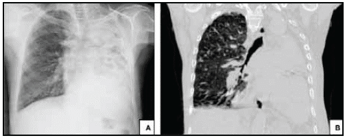
Figure 1 (A) AP chest x-ray showing diffuse heterogenous radiopacity of the left chest with associated ipsilateral loss of volume. Radiografía de tórax en proyección P-A. Evidencia radiopacidad difusa heterogénea del hemitorax izquierdo asociada perdida de volumen ipsilateral. (B) Coronal high-resolution CT in lung window projection showing loss of volume in the left lung with consolidation and varicose bronchiectases. 

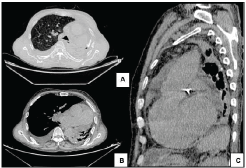
Figure 2 Chest computerized tomography image using an axial lung window. (A) and a mediastinal window; (B) sagittal mediastinal window; (C) showing left pleural thickening and calcifications. Free right pleural effusion. Mediastinal adenopathies. Cardiomegaly and precapillary pulmonary hypertension showing loss of left lung volume with consolidation and varicose bronchiectases. 

Referencias

1. Sulaiman S, Fasanya A, and Thirumala R. Extensive Calcified Fibrothorax. Am J Respir Crit Care Med Vol 195, Iss 4, pp e25-e26, Feb 15, 2017 [ Links ]

2. Light RW, Lee YCG. Pneumothorax, chylothorax. hemothorax, and fibrothorax. In: Broaddus CV, Mason RJ, Ernst JD, King TE Jr, Lazarus SC, Murray JF, Nadel JA, Slutsky A, Gotway M, editors. Murray & Nadel's textbook of respiratory medicine, 6th ed. Philadelphia, PA: Elsevier Saunders; 2015:1439-1460 [ Links ]

3. Michael A. Jantz, Veena B. Antony, Pleural Fibrosis. Clinics in Chest Medicine, Volume 27, Issue 2, 181 - 19 [ Links ]

4. Nachiappan, A. Et Al. Pulmonary Tuberculosis: Role of Radiology in Diagnosis and Management. RadioGraphics 2017; 37:52-72. Jan -Feb, 2017 [ Links ]

5. Hanane, Asri H & Zegmout, A. Historic sequelae of lung tuberculosis. Pan African Medical Journal. 2018;30:210. June, 2018. [ Links ]

6. Xu H, Koo HJ, Lee HN, Lim S, Lee JW, Choi CM, Kim MY.Lung Cancer in Patients With Tuberculous Fibrothorax and Empyema: Computed Tomography and 18F-Fluorodeoxyglucose Positron Emission Tomography Findings. Journal Comput Assist Tomogr. 2017 Sep/Oct;41(5):772-778. [ Links ]

Received: August 15, 2019; Accepted: February 11, 2020

* Correspondence: Dr. Oscar Mateo Pardo-Rodríguez. Bogotá, D.C. (Colombia). E-mail:ompardor@unal.edu.co

Creative Commons License This is an open-access article distributed under the terms of the Creative Commons Attribution License