SciELO - Scientific Electronic Library Online

 
vol.45 número4Teaching and learning anatomy. Pedagogical methods, history, the present and tendenciesAcute myocardial infarction associated with cocaine use índice de autoresíndice de assuntospesquisa de artigos
Home Pagelista alfabética de periódicos  

Serviços Personalizados

Journal

Artigo

Indicadores

Links relacionados

  • Em processo de indexaçãoCitado por Google
  • Não possue artigos similaresSimilares em SciELO
  • Em processo de indexaçãoSimilares em Google

Compartilhar


Acta Medica Colombiana

versão impressa ISSN 0120-2448

Acta Med Colomb vol.45 no.4 Bogotá out./dez. 2020  Epub 12-Jun-2021

https://doi.org/10.36104/amc.2020.1562 

Case presentation

Pharyngeal tuberculosis. Concerning a case in Peru

Jesús Neyra-León1  * 

Enike Gerecz-Cornejo1 

Janish Cabrejos-Valderrama2 

Pedro Antonio-Travezan3 

1 Estudiantes de Medicina. Escuela Profesional de Medicina Humana, Universidad Privada San Juan Bautista. Asociación de Investigación y Ciencia de los Estudiantes de Medicina de la UPSJB. Ica (Perú).

2 Médico Otorrinolaringólogo. Escuela profesional de Medicina Humana, Universidad Privada San Juan Bautista, Hospital Regional. Ica (Perú).

3 Médico Anatomopatólogo. Escuela Profesional de Medicina Humana, Universidad Privada San Juan Bautista. Ica (Perú).


Abstract

Pharyngeal tuberculosis is an extrapulmonary form of this disease which makes up less than 1% of cases, most of which are secondary. In Peru, there are no case reports of this form of extra-pulmonary tuberculosis.

We present the case of a 42-year-old man who consulted due to a two-month history of sore throat and a pharyngeal lump, with no epidemiological or medical history. A biopsy of the lesion and Ziehl-Neelsen staining yielded results compatible with pharyngeal tuberculosis. A chest x-ray showed bilateral miliary lesions, confirming a secondary form of disseminated tuberculosis.

This highlights the importance of ruling out tuberculosis in all cases of chronic pharyngitis which are unresponsive to conventional treatment.

Key words: pharyngeal tuberculosis; chronic pharyngitis; tuberculosis; pharynx

Resumen

La tuberculosis faríngea es una forma extrapulmonar de esta enfermedad que corresponde a menos de 1% de casos, mayormente de carácter secundario. En Perú, no hay datos de reportes de casos en esta forma de tuberculosis extrapulmonar.

Presentamos el caso de un varón de cuarenta y dos años que consultó por dolor de garganta y tumoración en faringe de dos meses de evolución, sin antecedentes epidemiológico o patológico. Se realiza biopsia de la lesión y coloración Ziehl Neelsen dando compatible a tuberculosis faríngea. En la radiografía de tórax se encontró lesiones pulmonares diseminadas bilaterales de tipo miliar, confirmando forma secundaria a tuberculosis diseminada.

Por ello, se resalta la importancia de descartar tuberculosis ante toda faringitis crónica sin respuesta al tratamiento convencional.

Palabras clave: tuberculosis faríngea; faringitis crónica; tuberculosis; faringe. (fuente: DeCS BIREME)

Introduction

Peru is one of the few countries in the world in which a variety of tuberculosis forms abound. In 25% of tuberculosis cases there is extrapulmonary involvement 1,2. The Peruvian Ministry of Health (MINSA) has an operational report from 2015 containing a total of 20,203 new cases of tuberculosis, among which 16,342 (80.8%) were pulmonary tuberculosis (PTB) and 3,861 (19.2%) were extrapulmonary tuberculosis (EPTB). Within the EPTB cases, males were predominant, with a higher number of cases between 15 and 44 years 3.

The extrapulmonary forms include central nervous system, peritoneal, lymph node, and osteoarticular forms, among others. However, there are very uncommon forms which are rarely seen or are not reported due to an unsuspected diagnosis 1,4,5.

Pharyngeal tuberculosis is an extrapulmonary manifestation of this disease which accounts for less than 1% of cases 6,7. There are few case reports of this type worldwide, and the nasal airway has been suggested as the principal means of transmission for the infection 8. There are no known case reports of this type in Peru.

We present the case of a 42-year-old patient who was seen due to chronic refractory pharyngitis. A series of exams were ordered, with a positive result for pharyngeal tuberculosis.

Case report

A 42-year-old male patient with no significant epidemiological or medical history, a native of Ica, was seen for a sore throat and pharyngeal lump. He reported having begun to experience a sore throat approximately two months before, and had been seen at the health center and treated for chronic pharyngitis with painkillers and antibiotics.

He continued to have discomfort, and began to notice a pharyngeal lump which made swallowing food difficult, associated with a 10 kg weight loss, and therefore was seen by an otorhinolaryngologist. On physical exam, a small, whitish lump on an erythematous base was seen on the left oropharynx (Figure 1).

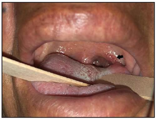
Figure 1 Shows a small, whitish lump on an erythematous base on the left oropharynx (arrow) with periodic secretion, as described by the patient. 

Figure 2 Disseminated bilateral milary pulmonary lesions seen on the chest x-ray. 

Laryngoscopy was ordered which did not show involvement of upper airways other than that previously described: a granulomatous lesion extending to the palate, from which a biopsy was taken. The sample was sent for analysis to rule out a specific chronic process. The biopsy results showed multiple granulomatous formations, some with central caseous necrosis and Langhans multinucleated giant cells (Figure 3). Furthermore, when the sample underwent Zielh Neelsen staining, it was positive for Koch bacilli (Figure 4), giving the correct histopathological picture for pharyngeal tuberculosis.

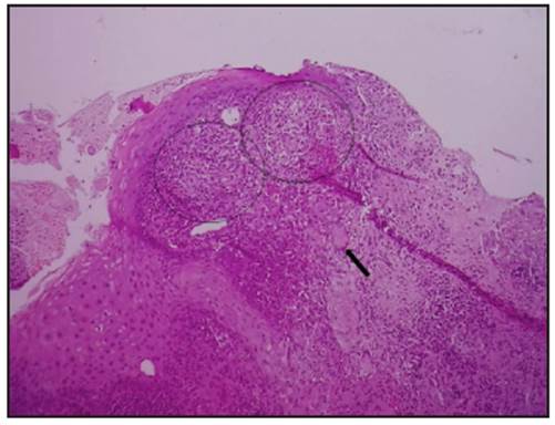
Figure 3 This sample shows multple granulomatous formations, some with central caseous necrosis (circles) and Langhans multinucleate giant cells (arrow). Staining method: hematoxylin-eosin. Magnification: 100x. 

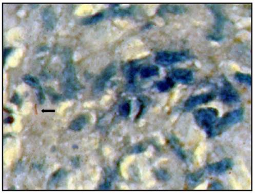
Figure 4 Exposure of the sample to Zielh Neelsen staining was positive, showing Koch bacilli, providing the correct histopathological picture for pharyngeal tuberculosis. Magnification: 1,000x. Staining method: Zielh-Neelson staining. 

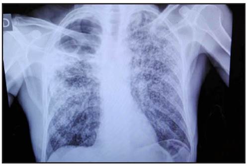
Tests were run seeking the primary focus, with disseminated bilateral miliary pulmonary lesions seen on the chest x-ray (Figure 2), confirming a form secondary to systemic tuberculosis. The patient was informed of the diagnosis and referred to pulmonology to begin treatment. Currently, the patient is receiving 2HREZ/4H3R3 medical treatment and is progressing well with no complications. We obtained the patient's informed consent to write this article.

Discussion

Globally, tuberculosis continues to be a public health problem. It is a preventable and curable infectious disease, which may attack any part of the body 9. In Peru, almost 27,000 new cases of active disease are reported per year, and we have one of the highest rates of tuberculosis among American countries, with it being the 15th cause of death, nationally 3. Ica is a very high-risk zone, with an incidence of 78/100,000 inhabitants per year.

Extrapulmonary tuberculosis is the new case in which tuberculosis is diagnosed in one or more organs other than the lung, using bacteriological, clinical, histological or other criteria 3. The preferred sites are the lymph nodes and pleura; on the other hand, in otorrhynolaryngology, the most common form of tuberculosis is that of the lymph nodes, followed by the larynx 10,11, with the pharyngeal form being very rare 6,12-14.

Pharyngeal tuberculosis has been described in few cases, with odynophagia being the main chief complaint 6. In most cases, it results from direct dissemination (through expectoration and inhalation of bacilli) or through the bloodstream. The first form was the case for our patient, as described in the literature 15. Bacilloscopy tends to be negative, with anatomical pathology being essential for diagnosis 6,12.

The recommended treatment regimen is the one used in our patient, as no resistance has been reported. An immediate diagnosis favors adequate resolution of the disease and prevents drug resistance 1,2,6.

Conclusion

Tuberculosis is common in otorhinolaryngology, as long as there is a diagnostic suspicion of it. Thus, we conclude that tuberculosis must be ruled out in any case of chronic pharyngitis that does not respond to conventional treatment.

Referencias

1. Ramírez M, Menéndez A, Noguerado A. Tuberculosis extrapulmonar, una revisión. Rev Esp Sanid Penit 2015; 17: 3-11. [ Links ]

2. Arciniegas W, Orjuela DL. Tuberculosis extrapulmonar: revisión de 102 casos en el Hospital Universitario San Jorge de Pereira, 2000-2004. Biomédica 2006; 26:71-80. [ Links ]

3. Alarcón V, et al. Tuberculosis en el Perú: situación epidemiológica, avances y desafíos para su control. Revista Peruana de Medicina Experimental y Salud Publica. 2017; 34(2): 299-310. [ Links ]

4. Ketata W, Rekik WK, Ayadi H, Kammoun S. Tuberculosis extrapulmonar. Rev Pneumol Clin. 2015 abril-junio; 71 (2-3): 83-92. [ Links ]

5. Sánchez L, et al. Tuberculosis extrapulmonar. Revista Argentina de Diagnóstico por Imágenes. 2016; 5(14): 25-37. [ Links ]

6. Mouhsinea A, et al. Tuberculosis faríngea: 5 casos clínicos. Acta Otorrinolaringol Esp. 2016;67(3):162-166. [ Links ]

7. Paredes A. Tuberculosis en Otorrinolaringología. Rev otorrinolaringol cir cab-cuello. 2001; 61: 87-94. [ Links ]

8. Vergara E, et al. ¿Es la vía aérea nasal el principal medio de transmisión para la tuberculosis faríngea? Acta Otorrinolaringol Esp . 2017 Jan - Feb;68(1):66-67. [ Links ]

9. Cardona P. Patogénesis de la tuberculosis y otras micobacteriosis. Enferm Infecc Microbiol Clin. 2018;36(1):38-46. [ Links ]

10. Chiesa CM, et al. Tuberculosis de cabeza y cuello: estudio retrospectivo de 6 años. Acta Otorrinolaringol Esp . 2015; 67(1): 9-14. [ Links ]

11. Paredes A. Tuberculosis en Otorrinolaringología. Rev otorrinolaringol cir cab-cuello . 2001; 61: 87-94. [ Links ]

12. Spini RG, et al. Tuberculosis faríngea. Caso clínico. Arch Argent Pediatr 2015;113(4): e230-e233. [ Links ]

13. Alcalá T, et al. Tuberculosis en faringe y laringe: presentación de un caso. Revista Habanera de Ciencias Médicas 2010:9(4) 545-552. [ Links ]

14. Gallardo A, et al. La tuberculosis faríngea: un caso clínico. Acta Otorrinolaringol Esp . 1998 Oct; 49 (7): 587-90. [ Links ]

15. Vergara E, et al. ¿Es la vía aérea nasal el principal medio de transmisión para la tuberculosis faríngea? Acta Otorrinolaringol Esp . 2017 Jan - Feb;68(1):66-67. [ Links ]

Received: June 06, 2019; Accepted: April 13, 2020

* Correspondencia: Jesús Neyra-León, Enike Gerecz-Cornejo. Ica (Perú). E-mail: jesus.24.47.1996@gmail.com

Creative Commons License This is an open-access article distributed under the terms of the Creative Commons Attribution License