text in
  English (pdf)
 Article in xml format
 Article references
 Send this article by e-mail
 Cited by SciELO
  Cited by Google
  Similars in
SciELO
Similars in
SciELO  Similars in Google
 Permalink
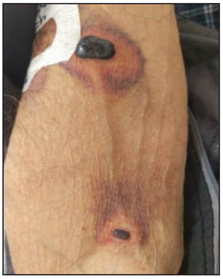
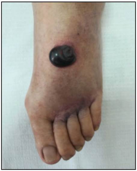
A 72-year-old man was hospitalized for acute heart failure. He had a history of arterial hypertension being treated with enalapril and hydrochlorothiazide. Thromboprophylaxis was begun at admission with 40 mg of subcutaneous enoxaparin daily, in the abdomen. Three days later, tense, blood-filled bullous skin lesions with a 5-15 mm diameter appeared. The lesions were on the right arm (Figure 1) and the left foot (Fig ure 2); they had an erythematous halo and were not painful. Clotting, C-reactive protein, ESR and complete blood count were normal. A skin biopsy was performed, which showed a subepidermal lesion with hemorrhagic material in the papillary and reticular dermis with scant neutrophils and no vasculitis or thrombosis. These findings confirm the diagnosis of bullous hemorrhagic dermatosis at distant sites related to enoxaparin. This is an uncommon and self-resolving adverse effect of heparins, unrelated to the dose and at sites distant from their application. This patient was treated by discontinu ing the medication, and the lesions resolved seven days later with no recurrence after three months of follow-up.