


 text in
  English (pdf)
 Article in xml format
 Article references
 Send this article by e-mail
 Cited by SciELO
  Cited by Google
  Similars in
SciELO
Similars in
SciELO  Similars in Google
 Permalink
A 67-year-old male with a history of chronic obstructive pul monary disease and heavy smoking consulted due to progressive worsening of his dyspnea over the previous week along with a dry cough and unquantified fever at home. A chest x-ray showed bilateral calcified micronodules. A high-resolution computerized tomography confirmed these findings. The diagnosis was confirmed by the histological results of a bronchoalveolar lavage showing characteristic laminar microliths.
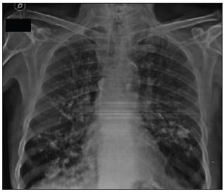
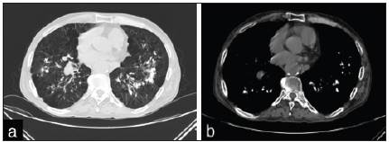
Figure 2. Axial high-resolution computerized tomography of the chest: A. Mediastinal window showing multiple bilateral calci fied densitites in the lower lobes. B. Pulmonary window showing bilateral calcified interlobular micronodular hyperdensities.
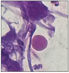
Figure 3. Hematoxylin and eosin staining of bronchoalveolar lavage liquid (100x) showing the presence of calcospherites
Pulmonary alveolar microlithiasis is a rare hereditary pul monary disease characterized by microcalcifications within the alveolar spaces 1. It is caused by mutations of the SCL34A2 gene which encodes the phosphate cotransporter in type II alveolar cells. This results in increased phosphate and calcium in lung surfactant, which leads to the formation and deposition of microliths within the alveoli as well as in other parts of the body 2. The definitive diagnosis requires histological identification of the microliths 3.

