


 text in
  English (pdf)
 Article in xml format
 Article references
 Send this article by e-mail
 Cited by SciELO
  Cited by Google
  Similars in
SciELO
Similars in
SciELO  Similars in Google
 Permalink
Introduction
Chronic kidney disease (CKD) is a significant cause of morbidity and mortality in the global population, and its prevalence is increasing due to increased life expectancy and a higher incidence of chronic illnesses including diabetes, high blood pressure, obesity, coronary disease, cerebrovas cular disease, systemic atherosclerosis and cancer, among others 1. In advanced stages of CKD, bone mineral metabo lism and calcium, phosphorus, vitamin D, fibroblast growth factor 23 (FGF23) and parathyroid hormone (PTH) balance disorders occur 2, which increases the risk of osteodystro phy, fibrous osteitis, osteomalacia or adynamic bone disease 3. All of these diseases may cause different complications such as uremic calciphylaxis. Below we present the case of a patient with penile uremic calciphylaxis facilitated by a state of shock and the use of vasopressors.
Case report
This was a 65-year-old man with a history of stage 5 or end-stage renal disease (ESRD) of unknown origin who had been on hemodialysis for five years, complicated in the last year with refractory hyperparathyroidism and consequently persistent hyperphosphatemia, being treated with cinacalcet 60 mg/day, calcium carbonate 1,500 mg three times a day, and aluminum hydroxide 230 mg three times a day. He had also already received calcitriol and paricalcitol. As comorbidities, he had coronary disease, dyslipidemia, high blood pressure, and non-alcoholic cirrhosis of the liver, which is why parathyroidectomy had not been able to be performed.
He presented to the emergency room in shock due to gastrointestinal bleeding related to bleeding esophageal varices, which were ligated. He received a transfusion of three units of red blood cells and required two 1 mg doses of terlipressin. The next day he developed intense pain in his hands, legs and external genitalia. The glans was cyanotic, progressing to frank necrosis (Figure 1). A penile Doppler was performed along with pelvic and extremity x-rays which showed calciphylaxis (6/8 Adragao index) (Figure 2). As an added complication, the patient developed rhabdomyolysis. Complementary studies showed hyperphosphatemia, hyper calcemia, and suppressed PTH for his CKD stage (Table 1). Treatment was begun with wound care, analgesia, sodium thiosulfate, and phosphate chelators, with which he was stabilized. One month later the patient had another massive bleed from the esophageal varices and died.
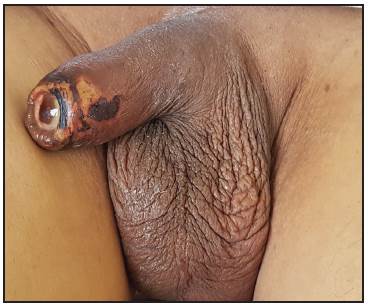
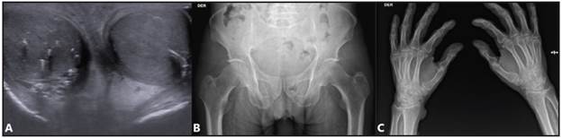
Figure 2 A: Testicular ultrasound showing multiple calcifications. B: Pelvic x-ray. C: Hand x-ray showing extensive calcification of the femoral, radial, ulnar and interdigital arteries. Adragao index: 6/8.
Table 1 Laboratory tests.
| Blood tests | Kidney tests | Coagulation profile | |||
|---|---|---|---|---|---|
| Hemoglobin | 6.2 g/dL | Creatinine | 6.5 mg/dL | PTT | 41.5 sec |
| Hematocrit | 18% | BUN | 85 mg/dL | PT | 18.5 sec |
| Leukocytes | 6,200 mm3 | - Serum electrolytes | INR | 1.60 | |
| Neutrophils | 4,400 mm3 | Sodium | 140 mEq/L | Endocrine studies | |
| Lymphocytes | 900 mm3 | Chloride | 99.9 mEq/L | PTH | 107.5 pg/mL |
| Platelets | 105 000 mm3 | Potassium | 5.02 mEq/L | Immunology profile | |
| MCV | 88.8 Fl | Calcium | 6.94 mg/dL | ANAs | Negative |
| Liver studies | Phosphate | 7.2 mg/dL | Others | ||
| AST | 46 u/L | Corrected serum calcium | 10.5 mg/dL | Albumin | 3.0 gr/dL |
| ALT | 28 u/L | Estudios infecciosos | CPK | 12,384 U/L | |
| Total bilirubin | 1.83 mg/dL | Hepatitis C Ab | Nonreactive | ||
| Direct bilirubin | 0.94 mg/dL | Hepatitis B sAg | Nonreactive | ||
| Alkaline phosphatase | 570 u/L | Hepatitis B anti core IgM | Nonreactive | ||
| GGT | 979.7 u/L | Anti-HBsAg | >799 mUI/mL | ||
| VDRL | Nonreactive | ||||
| MCV: mean corpuscular volume; ALT: alanine transaminase; AST: aspartate transaminase; GGT: gamma-glutamyl transferase; BUN: blood urea nitrogen; Ab: antibodies, Anti-HBsAg: Antibodies against hepatitis B surface antigen; PTH: parathyroid hormone; ANAs: antinuclear antibodies; CPK: creatinine phosphokinase. | |||||
Discussion
Uremic calciphylaxis (UC), also known as calcific uremic arteriolopathy (CUA) is a serious complication of bone mineral metabolism disorders in patients with CKD4. It occurs in 1-4% of patients with ESRD on dialysis5. It affects elderly, female, white, and diabetic patients more frequently, as well as those who are human immuno deficiency virus positive. It is important to know that other non-uremic causes of calciphylaxis have been reported, such as primary hyperparathyroidism, malignancies, and alcoholic liver disease 6.
In the pathogenesis of this complication, calcification of the middle layer of the arteries and large and medium muscular arterioles, as a result of phenotypic changes of vascular smooth muscle cells to osteoblast-like cells, has been reported. This change occurs secondary to hyperphos phatemia, hypercalcemia and high concentrations of PTH. Hyperphosphatemia increases the activity of the PiT-1 and PiT-2 calcium-dependent cotransporters, which regulate the genes associated with matrix mineralization. Hypercalcemia and hyperphosphatemia increase the release of vesicles from the matrix, resulting in hydroxyapatite deposition 7,8, which is why it is more common in patients receiving excess calcium and vitamin D analogs. Other possible risk factors involved are protein C and S deficiency, the use of vitamin K antagonists (warfarin), heart valves, high blood pressure, and dyslipidemia 4. The reported patient, for example, had refractory hyperparathyroidism, without the possibility of a parathyroidectomy, and was being treated with high doses of calcium and vitamin D analogs.
Uremic calciphylaxis also has the characteristic of tunica intima calcification and fibrosis in small and medium caliber arteries, resulting in ischemia and necrosis of the dermis and subcutaneous cell tissue 9. Penile calciphylaxis is espe cially rare and has been associated with a worse prognosis, with a mortality close to 69% and an average survival of 2.5 months after diagnosis 7. This was the case of the patient presented, who developed glans penis necrosis after having been in shock due to bleeding, which was also facilitated by the administration of terlipressin, which is a potent va soconstrictor.
Uremic calciphylaxis should be considered in patients with end-stage CKD who develop acute pain and necrotic lesions on an extremity or on the penis. The most common laboratory findings are hypercalcemia, hyperphosphatemia, and PTH disorders, but normal or low levels do not rule out the diagnosis. A biopsy is the gold standard for definitive diagnosis, showing calcification with intimal proliferation, fibrosis and, finally, thrombosis of the affected vessel, end ing in ischemia and necrosis of the tissue supplied 10. However, the advisability of performing a biopsy is debated, as it could worsen the extent of the lesion due to progres sion or superinfection 11. The patient in this case had the following risk factors for UC: bone mineral disease with refractory hyperphosphatemia which was treated for a long time with calcium and aluminum hydroxide-based chelating agents; he also had an exaggerated PTH suppression, which may have caused an adynamic bone disease which further favored the deposition of calcium and phosphorus in soft tissues and vascular tissue.
All the arteries, even the smallest arterioles, may be af fected; veins are more rarely affected. For example, the ra dial, ulnar and interdigital arteries are only muscular arteries, regulating blood flow with a thick tunica media of calcifiable smooth muscle. The iliac and femoral arteries (which are predominantly but not exclusively muscular) are also more susceptible to medial calcification. All are assessed using simple x-rays and are used for calculating the well-known Adragao index. On the other hand, the Kauppila index evaluates elastic arteries (abdominal aorta) which would be more susceptible to intimal calcification, since these elastic arteries (such as the subclavian and carotid) have a tunica media which contains more elastic fibers than muscle cells 12. In the patient discussed, the Kauppila index could not be calculated due to poor intestinal preparation, but the Adragao index had a score of six, which was compatible with vascular calcification.
The treatment of UC is controversial due to the rarity of the disease, and therefore the evidence is limited, and treatment is carried out according to the experiences or opinions of experts 13. More conservative approaches are recommended, using topical wound care, pain control and systemic therapy. To begin with, the discontinuation of calcium supplements and vitamin D analogs should be considered. Non-calcium phosphate chelators could be used, along with increased hemodialysis frequency using low-calcium dialysates 9. Sodium thiosulfate has emerged as a promising treatment in recent years as it favors calcium and phosphate chelation from the tis sues, although there are no randomized studies. However, two clinical trials are currently being conducted which are expected to produce promising results (controlled trial number ISRCTN73380053; and ClinicalTrials.gov number NCT03150420). The initial dose is 25 g in 100 milliliters of intravenous solution three times per week with hemodialysis. The length of treatment is uncertain at the moment 14. Its main adverse effects include volume overload, hypocalcemia, prolonged QT interval, hypoten sion and metabolic acidosis. These can be avoided through intralesional administration 15. The treatment of the presented patient was conservative, using wound care, sodium thiosulfate, phosphate chelation with sevelamer, and discontinuation of calcium and calcitriol. However, even so, the patient progressed poorly and died, which is in line with the literature and shows that calciphylaxis is a very late clinical manifestation, and when it occurs it indicates a very advanced stage of the disease with a very bad prognosis.
Efforts have been made to determine the efficacy of other treatments such as biophosphonates and even vitamin K1 supplements such as phylloquinone 16, showing a slower progression in pre-existing coronary calcifications, but with no clear effect in uremic calciphylaxis 17. Para-thyroidectomy has prevailed as a surgical treatment which could normalize calcium and phosphate homeostasis and is indicated in hyperparathyroidism cases in which PTH is not suppressed with medical treatment 9. Partial or radical penectomy is controversial and is not recommended as a first option due to the high rate of superinfection and increased necrotic tissue 11. The patient under discussion did not undergo parathyroidectomy due to his critical condition, and because his PTH was very suppressed. The penile lesions were treated with wound care.
Conclusions
Uremic calciphylaxis with penile involvement is a very rare condition which occurs as a complication of ESRD, reflecting a systemic involvement with generalized calciphylaxis, which is secondary to a long-term bone mineral disorder with chronic hyperphosphatemia and high exposure to calcium products and vitamin D analogs, leading to high morbidity and mortality. Prevention through appropriate treatment of bone mineral metabolism disorders is the basis for avoiding the occurrence of this irreversible condition with such a poor prognosis. However, when it does occur, early recognition, the application of conservative principles for managing the necrotic lesions, and optimization of the phosphate-calcium profile are the pillars of treatment.

