


 texto em
  Inglês (pdf)
 Artigo em XML
 Referências do artigo
 Enviar este artigo por email
 Citado por SciELO
  Citado por Google
  Similares em
SciELO
Similares em
SciELO  Similares em Google
 Permalink
This was a 35-year-old male of low socioeconomic status with no other significant history. He was admitted to the emergency room for sudden onset severe epigastric pain. On exam, his vital signs were within normal limits, and he had no signs of peritoneal irritation. Acute coronary syndrome and hollow organ perforation were ruled out. Studies showed elevated bilirubin, transaminases, alkaline phosphatase and, especially, amylase. He was diagnosed with acute pancreatitis (AP) with no evidence of multiple organ dysfunction. His studies were completed with an upper abdominal ultrasound showing no cholelithiasis, with collections in both paracolic gut ters. A double-contrast computed tomography of the abdomen (ab dominal CT) was ordered, which showed acute pancreatitis (Figure 1) and a tubular filling defect towards the duodenal papilla (Figure 2). An upper gastrointes tinal endoscopy (EGD) showed a single 25 cm parasite located in the duodenal papilla. After antibiotic cover age and deworming, the patient developed walled-off necrotizing pancreatitis in the fifth week of follow up, requiring a necrosectomy and collection drainage.

Figure 1 An axial view of a double-contrast computed tomography of the abdomen showing acute interstitial edematous pancre atitis (yellow pointer), with a tubular shape corresponding to ascaris (red pointere) and non-encapsulated peripancreatic fluid.
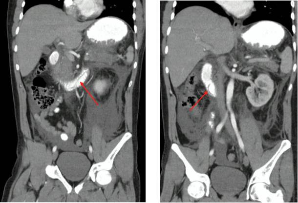
Figure 2 A coronal view of a double-contrast abdominal tomography showing an ascaris worm in the duodenal papilla, second and third part of the duodenum (red pointer).
Acute pancreatitis is an inflammatory condition usually caused by gallstones or excessive alcohol consumption. Khuroo et al. reported that 23% of the AP cases in India are caused by ascaris, associating it with poor hygiene conditions 1. The diagnosis is usually clinical, finding ascaris worms in the stool. Occasionally, the ultrasound shows indirect signs such as mobile figures within the hollow gut 2. In this exceptional case, there was no infestation; it was a single parasite, for which the double-contrast abdominal CT was very helpful. A prompt CT in the emergency room can provide the diagnosis or change treatment in close to 15% cases of AP 3.

