


 text in
  English (pdf)
 Article in xml format
 Article references
 Send this article by e-mail
 Cited by SciELO
  Cited by Google
  Similars in
SciELO
Similars in
SciELO  Similars in Google
 Permalink
This was a 42-year-old male who underwent surgery for transposition of the great arteries as a child. Twenty years later a catheterization through the right deep femoral artery was performed. Over the last five years he has had edema with a change in color of his right foot 50 seconds after going from a lying to a standing position (Figure 1). Computed tomography angiography corroborated the diagnosis of an arteriovenous fistula (AVF) in the right femoral system (Figures 2 and 3). He was referred to vas cular surgery for stent placement.

Figure 1 Sequential clinical images (A, B, C and D) of a 42-year-old male who had edema in the pelvic portion of the right lower extremity along with changes from a normal to a purplish color in the right ankle and foot over a span of 50 seconds.
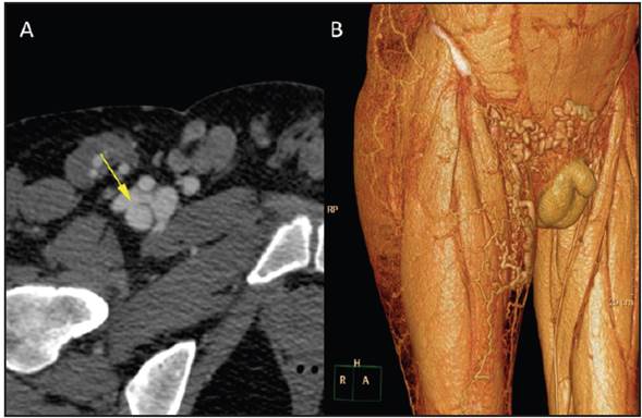
Figure 2 (A): an axial view of an iliofemoral computed tomography angiography of the connection between the anterior wall of the deep femoral artery and a blind venous sac connecting to the femoral vein. Note the mix of contrast in the venous territory in an arterial phase; (B): volumetric reconstruction of the iliofemoral computed tomography angiography in the venous phase, showing pre-pubic and right proximal thigh varicose bundles stemming from venous insufficiency driven by deep vein thrombosis.

Figure 3 Volumetric reconstruction of the iliofemoral computed tomography angiography (A): frontal plane; (B): sagittal plane. The arrow indicates the ante rior wall of the proximal third of the deep femoral artery, as well as a blind venous sac draining to the femoral vein, with aneurysmal dilation of the iliac vein.
Diagnostic and therapeutic percutaneous catheterization techniques entail a risk of vascular complications such as AVF, false aneurysms, hematomas, hemorrhages and arterial thromboses, with an incidence of 0.5% to 1% after diagnostic procedures; 0.0-9% after balloon angioplasty; 5.7-17% after stent implantation and 5.2-10% after percu taneous mitral vavuloplasty 1,2. Covered stents are used to resolve the AVFs 3.

