SciELO - Scientific Electronic Library Online

 
vol.47 issue3Simultaneous thromboses in patients with SARS-CoV-2 infectionAbsence of the posterior arch of the atlas author indexsubject indexarticles search
Home Pagealphabetic serial listing  

Services on Demand

Journal

Article

Indicators

Related links

  • On index processCited by Google
  • Have no similar articlesSimilars in SciELO
  • On index processSimilars in Google

Share


Acta Medica Colombiana

Print version ISSN 0120-2448

Acta Med Colomb vol.47 no.3 Bogotá July/Sept. 2022  Epub Mar 28, 2023

https://doi.org/10.36104/amc.2022.2328 

Images in internal medicine

Anemia and a kidney mass

EDWIN URIEL SUÁREZ-MERCHÁNa  * 

ADRIANA ORTEGA-LARRODÉb 

a Internista. Residente e Hematología, Departamento de Hematología, Hospital Universitario Fundación Jiménez Díaz; Madrid (España).

b Residente de Gastroenterología, Departamento de Gastroenterología, Hospital Universitario Fundación Jiménez Díaz. Madrid (España).


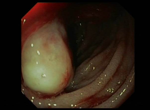
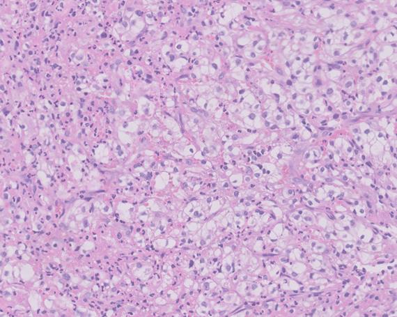
A 68-year-old woman, with no relevant medical history, was undergoing outpatient studies due to a small amount of painless gross hematuria and a kidney mass detected on ultrasound. She presented to the emergency room with a complaint of three months of asthenia, weight loss and dyspnea. Laboratory tests showed iron deficiency anemia (hemoglobin of 4.6 g/dL and ferritin of 11 ug/L). She underwent an upper GI endoscopy which revealed an eroded ampullary lesion suggestive of malignancy (Figure 1). Abdominal ultrasound confirmed the presence of an 8 cm kidney mass compatible with a kidney neoplasm (Figure 2) and a periampullary duodenal lesion with endoluminal infiltration. The histopathological study of the duodenal lesion showed ulcerated tissue infiltrated with renal carcinoma (Figure 3). In this case, the appropriate work up for iron deficiency anemia was able to adduce the duodenal lesion, which was compatible with metastasis. The ampulla of Vater is an unusual site for renal cell carcinoma metastasis and may present as GI bleeding, obstructive jaundice or iron deficiency anemia without obvious bleeding 1. The current treatment for renal carcinoma has improved its prognosis; it includes radical or partial nephrectomy with a curative intent, immunotherapy, and targeted therapy. Metastasis resection is available in some cases in which there are inoperable conditions 2,3.

Figure 1 Second duodenal portion: eroded ampullary lesion. 

Figure 2 Kidney mass in the lower pole of the right kidney (magnetic resonance, T2-HASTE). 

Figure 3 Fragment of ulcerated tissue infiltrated with clear cell carcinoma. 

Acknowledgements

To Teresa Reina Durán, M.D. (Pathologist) and Juan Porres Cubero, M.D. (Gastro-enterologist).

References

1. Ignatavicius P, Lizdenis P, Pranys D, Gulbinas A,Pundzius J, Barauskas G. Long-term Survival of Patient with Ampulla of Vater Metastasis of Renal Cell Carcinoma. Prague Med Rep. 2018;119(4):165-169. [ Links ]

2. Hsieh JJ, Purdue MP, Signoretti S, Swanton C, Albiges L, Schmidinger M, Heng DY, Larkin J, Ficarra V. Renal cell carcinoma. Nat Rev Dis Primers. 2017 Mar 9;3:17009. [ Links ]

3. Takagi T, Fukuda H, Ishihara H, Yoshida K, Kondo T, Kobayashi H, Iizuka J, Okumi M, Ishida H, Omae K, Tanabe K. Predictive factors for recurrence after complete metastasectomy in patients with metastatic renal cell carcinoma in the targeted therapy era. Urol Oncol. 2020 May;38(5):515-520. [ Links ]

Received: September 14, 2021; Accepted: October 10, 2021

* Correspondencia: Dr. Edwin Uriel Suárez-Merchán, Adriana Ortega-Larrodé. Madrid (España) E-Mail: edwin.suarez@quironsalud.es

Creative Commons License This is an open-access article distributed under the terms of the Creative Commons Attribution License